Transcription
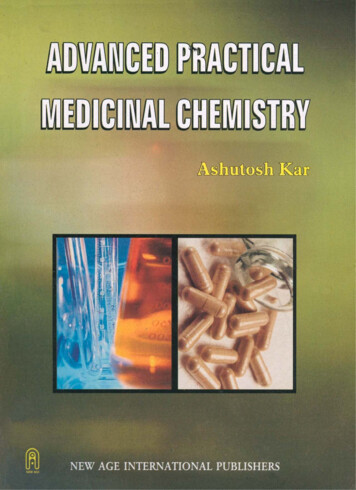
Fundamentals ofMedicinal ChemistryGareth ThomasUniversity of Portsmouth, UK
Fundamentals ofMedicinal Chemistry
Fundamentals ofMedicinal ChemistryGareth ThomasUniversity of Portsmouth, UK
Copyright # 2003 by John Wiley & Sons Ltd,The Atrium, Southern Gate, Chichester,West Sussex PO19 8SQ, EnglandNational01243 779777International (þ44) 1243 779777e-mail (for orders and customer service enquiries): cs-books@wiley.co.ukVisit our Home Page on http://www.wiley.co.uk or http://www.wiley.comAll rights reserved. No part of this publication may be reproduced, stored in a retrieval system, ortransmitted, in any form or by any means, electronic, mechanical, photocopying, recording,scanning or otherwise, except under the terms of the Copyright, Designs and Patents Act 1988 orunder the terms of a licence issued by the Copyright Licensing Agency, 90 Tottenham CourtRoad, London, UK W1P 9 HE, without the permission in writing of the publisher.Other Wiley Editorial OfficesJohn Wiley & Sons, Inc., 605 Third Avenue,New York, NY 10158–0012, USAWiley-VCH Verlag GmbH, Pappelallee 3,D-69469 Weinheim, GermanyJohn Wiley & Sons (Australia) Ltd, 33 Park Road, Milton,Queensland 4064, AustraliaJohn Wiley & Sons (Asia) Pte Ltd, 2 Clementi Loop #02–01,Jin Xing Distripark, Singapore 0512John Wiley & Sons (Canada) Ltd, 22 Worcester Road,Rexdale, Ontario M9W 1L1, CanadaWiley also publishes its books in a variety of electronic formats. Some content that appears inprint may not be available in electronic booksLibrary of Congress Cataloging-in-Publication DataThomas, Gareth, Dr.Fundamentals of medicinal chemistry / Gareth Thomas.p. cm.Includes bibliographical references and index.ISBN 0-470-84306-3 (cloth : alk. paper) ISBN 0-470-84307-1 (paper : alk. paper)1. Pharmaceutical chemistry. I. Title.RS403.T446 2003615’.19 — — dc212003014218British Library Cataloguing in Publication DataA catalogue record for this book is available from the British LibraryISBN 0-470 84306 3 (Hardback)ISBN 0-470 84307 1 (Paperback)Typeset in 11/14pt Times by Kolam Information Services Pvt. Ltd, Pondicherry, IndiaPrinted and bound in Great Britain by Antony Rowe LtdThis book is printed on acid-free paper responsibly manufactured from sustainable forestry,in which at least two trees are planted for each one used for paper production.
Acronymsxiii1 Biological Molecules1.1 Introduction1.2 Amino acids1111.2.1 Introduction1.2.2 Structure1.2.3 Nomenclature1331.3 Peptides and proteins41.3.1 Structure1.4 Carbohydrates1.4.11.4.21.4.31.4.41.4.51.4.6The structure of monosaccharidesThe nomenclature of monosaccharidesGlycosidesPolysaccharidesThe nomenclature of polysaccharidesNaturally occurring polysaccharides1.5 ionFatty acidsAcylglycerols ds1.6 Nucleic acids1.6.11.6.21.6.31.6.41.6.5IntroductionDNA, structure and replicationGenes and the human genome projectRNA, structure and transcriptionClassification and function of RNA1.7 Questions2 An Introduction to Drugs and their Action2.12.22.32.4IntroductionWhat are drugs and why do we need new ones?Drug discovery and design, a historical outlineSources of drugs and lead 33343737373943
viCONTENTS2.4.1 Natural sources2.4.2 Drug synthesis2.4.3 Market forces and ‘me-too drugs’2.5 Classification of drugs2.6 Routes of administration, the pharmaceutical phase2.7 Introduction to drug action2.7.1 The pharmacokinetic 2.7.2 Bioavailability of a drug2.7.3 The pharmacodynamic phase3454649494950515153532.8 Questions55An Introduction to Drug Discovery573.1 Introduction3.2 Stereochemistry and drug design57593.2.1 Structurally rigid groups3.2.2 Conformation3.2.3 Configuration3.3 Solubility and drug design3.3.1 The importance of water solubility3.4 Solubility and drug structure3.5 Salt formation3.6 The incorporation of water solubilizing groups in a structure3.6.13.6.23.6.33.6.44434545The type of groupReversibly and irreversibly attached groupsThe position of the water solubilizing groupMethods of introduction5960606162636465666667673.7 Questions70The SAR and QSAR Approaches to Drug Design714.1 Structure–activity relationships (SARs)4.2 Changing size and shape4.3 Introduction of new substituents7173734.3.1 The introduction of a group in an unsubstitutedposition4.3.2 The introduction of a group by replacing anexisting group4.4 Quantitative structure–activity relationships (QSARs)4.4.1 LipophilicityPartition coefficients (P)Lipophilic substitution constants (p)4.4.2 Electronic effectsThe Hammett constant (s)4.4.3 Steric effectsThe Taft steric parameter (Es)Molar refractivity (MR)Other parameters737678797980828283848485
CONTENTSvii4.4.4 Hansch analysisCraig plots85884.5 The Topliss decision tree4.6 Questions89925 Computer Aided Drug Design5.1 Introduction5.1.1 Molecular modelling methods5.1.2 Computer graphics5.2 Molecular mechanics5.2.1 Creating a molecular model using molecular mechanics5.3 Molecular dynamics5.3.1 Conformational analysis95959698981021041055.4 Quantum mechanics5.5 Docking5.6 Questions1051091106 Combinatorial Chemistry1136.1 Introduction6.1.1 The design of combinatorial syntheses6.1.2 The general techniques used in combinatorial synthesis6.2 The solid support method6.2.1 Parallel synthesis6.2.2 Furka’s mix and split technique6.3 Encoding methods6.3.1 Sequential chemical tagging methods6.3.2 Still’s binary code tag system6.3.3 Computerized tagging6.4 Combinatorial synthesis in solution6.5 Screening and deconvolution6.6 Questions7 Selected Examples of Drug Action at some Common Target Areas7.1 Introduction7.2 Examples of drugs that disrupt cell membranes and walls7.2.1 Antifungal agentsAzolesAllylaminesPhenols7.2.2 Antibacterial apentsIonophoric antibiotic actionCell wall synthesis inhibition7.3 Drugs that target enzymes7.3.1 Reversible inhibihors7.3.2 Irreversible inhibition7.3.3 Transition state inhibitors7.4 Drugs that target receptors7.4.1 Agonists7.4.2 Antagonists7.4.3 Partial 7
viiiCONTENTS7.5 Drugs that target nucleic Enzyme inhibitorsIntercalation agentsAlkylating agentsAntisense drugsChain cleaving agents7.6 Antiviral drugs7.6.1 Nucleic acid synthesis inhibitors7.6.2 Host cell penetration inhibitors7.6.3 Inhibitors of viral protein synthesis7.7 Questions8Pharmacokinetics8.1 Introduction to pharmacokinetics8.1.1 General classification of pharmacokinetic properties8.2 Pharmacokinetics and drug design8.3 Pharmacokinetic models8.4 Intravascular administration8.4.1 Intravenous injection (IV bolus)8.4.2 Clearance and its significance8.4.3 Intravenous infusion8.5 Extravascular administration8.5.1 Single oral dose8.5.2 The calculation of tmax and Cmax8.5.3 Repeated oral doses8.6 The use of pharmacokinetics in drug design8.7 Questions9Drug Metabolism9.1 Introduction9.1.19.1.29.1.39.1.4The stereochemistry of drug metabolismBiological factors affecting metabolismEnvironmental factors affecting metabolismSpecies and metabolism9.2 Secondary pharmacological implications of metabolism9.3 Sites of action9.4 Phase I metabolic ationReductionHydrolysisHydrationOther Phase I 83184184184184186186186189189189Phase II metabolic routesPharmacokinetics of metabolitesDrug metabolism and drug designProdrugs1901901931959.8.1 Bioprecursor prodrugs9.8.2 Carrier prodrugs195196
ixCONTENTS9.8.3 The design of prodrug systems for specific purposesImproving absorption and transportthrough membranesImproving patient acceptanceSlow releaseSite specificityMinimizing side effects9.9 Questions10 An Introduction to Lead and Analogue Syntheses10.1 Introduction10.2 Asymmetry in syntheses10.2.1 The use of non-stereoselective reactions toproduce stereospecific centres10.2.2 The use of stereoselective reactions toproduce stereospecific centres10.2.3 General methods of asymmetric synthesisMethods that use catalysts to obtainstereoselectivityMethods that do not use catalysts to obtainstereoselectivity10.3 Designing organic syntheses10.3.1 An introduction to the disconnection approach10.4 Questions11 Drug Development and Production11.1 Introduction11.2 Chemical 611.711.811.9Chemical engineering issuesChemical plant, health and safety considerationsSynthesis quality controlA case studyPharmacological and toxicological testingDrug metabolism and pharmacokineticsFormulation developmentProduction and quality controlPatent .5A.6A.7Sickle-cell anaemiaBacteriaCell membranesReceptorsTransfer through membranesRegression 35236237238239239240244246249250252
usesBlood–brain barrierEnzyme structure and species255256257259260Answers to Questions261Selected Further Reading273Index275
PrefaceThis book is written for second, and subsequent year undergraduates studyingfor degrees in medicinal chemistry, pharmaceutical chemistry, pharmacy,pharmacology and other related degrees. It is also intended for students whosedegree courses contain a limited reference to medicinal chemistry. The textassumes that the reader has a knowledge of chemistry at level one of a universitylife sciences degree. The text discusses the fundamental chemical principles usedfor drug discovery and design. A knowledge of physiology and biology isadvantageous but not essential. Appropriate relevant physiology and biologyis outlined in the appendices.Chapter 1 gives a brief review of the structures and nomenclature of the morecommon classes of naturally occurring compounds found in biological organisms. It is included for undergraduates who have little or no backgroundknowledge of natural product chemistry. For students who have studied naturalproduct chemistry it may be used as either a revision or a reference chapter.Chapter 2 attempts to give an overview of medicinal chemistry. The basicapproaches used to discover and design drugs are outlined in Chapters 3–6inclusive. Chapter 7 is intended to give the reader a taste of main line medicinalchemistry. It illustrates some of the strategies used, often within the approachesoutlined in previous chapters, to design new drugs. For a more encyclopediccoverage of the discovery and design of drugs for specific conditions, the readeris referred to appropriate texts such as some of those given under MedicinalChemistry in the Selected Further Reading section at the end of this book.Chapters 8 and 9 describe the pharmacokinetics and metabolism respectivelyof drugs and their effect on drug design. Chapter 10 attempts to give anintroductory overview of an area that is one of the principal objectives of themedicinal chemist. For a more in depth discussion, the reader is referred to themany specialized texts that are available on organic synthesis. Drug development from the research stage to marketing the final product is briefly outlined inChapter 11.The approach to medicinal chemistry is kept as simple as possible. The text issupported by a set of questions at the end of each chapter. Answers, sometimesin the form of references to sections of the book, are listed separately. A list ofrecommended further reading, classified according to subject, is also included.Gareth Thomas
AcknowledgementsI wish to thank all my colleagues, past and present without, whose help thisbook would have not been written. In particular, I would like to thank Dr. SArkle for access to his lecture notes and Dr. J Wong for his unstinted help andadvice, Dr. J Brown for again acting as a living pharmacology dictionary, Dr. PCox for the molecular model diagrams and his patience in explaining to me theintricacies of molecular modelling and Mr. A Barrow and Dr. D Brimage for thelibrary searches they conducted. I wish also to thank the following friends andcolleagues for proof-reading chapters and supplying information: Dr. L Adams,Dr. C Alexander, Dr. L Banting, Dr. D Brown, Dr. S Campbell, Dr. B Carpenter, Mr. P Clark, Dr. P Howard, Dr. A Hunt, Mrs. W Jones, Dr. T Mason, Dr. JMills, Dr. T Nevell, Dr. M Norris, Dr. J Smart, Professor D Thurston, Dr. GWhite and Mr. S Wills.Finally, I would like to thank my wife for her support whilst I was writing thetext.
denineAbequoseAngiotensin-converting enzymeAcetyl cholineAbsorption, distribution, metabolism and eliminationAdverse drug reactionAlanineArginineAspartateDeoxyadenosine triphosphateAdenosine triphosphateArea under the curveCCNSCoACYP-450CysCytosineCentral nervous systemCoenzyme ACytochrome P-450 familyCysteined.e.DHFDHFRDMPKDNADiastereoisometric excessDihydrofolic acidDihydrofolate reductaseDrug metabolism and pharmacokineticsDeoxyribonucleic acidECe.e.ELFEMEAEPCEPOEsEnzyme commissionEnantiomeric excessEffluent load factorEuropean medicines evaluation agencyEuropean Patent ConventionEuropean Patent OfficeTaft steric parameter
xivABBREVIATIONSFDAFMOFGIFmoc,FdUMPFUdRPFood and drugs administrationFlavin monoxygenaseFunctional group interconversion9-Fluorenylmethoxychloroformyl group5-fluoro-2’-deoxyuridyline monophosphate5-fluoro-2’-deoxyuridylic acidGGABAGIGlnGluGlyGSHGuanineg-Aminobutyric acidGastrointestinal tractGlutamineGlutamatic Sickel cell haemoglobinHistidineHuman immunodeficiency diseaseHeterogeneous nuclear RNAIleIVIMIsoleucineIntravenous injectionIntramuscular hium diisopropylamideLactose Muscarinic cholinergic receptorMarket authorisation (application)Medical control agencyMethionine4-Methoxybenzyloxychloroformyl groupMolar refractivitymessenger RNAnAChNADþNADHNicotinic cholinergic receptorNicotinamide adenine dinucleotide (oxidised form)Nicotinamide adenine dinucleotide (reduced form)
ABBREVIATIONSNADPþNADPHNAGNAMNicotinamide dinucleotide phosphate (oxidised form)Nicotinamide dinucleotide phosphate (reduced form)b-N-Acetylglucosamineb-N-Acetylmuramic acidONsSequence defined oligonucleotidesP-450Cytochrome P-450 oxidasesPABAp-Aminobenzoic acidPCTPaten Cooperation TreatyPGProstaglandinPhePhenylalaninePOPer oral (by mouth)pre-mRNAPremessenger RNAProProlineptRNAPrimary transcript RNAQSARQuantitative structural-activity relationshipsRNARibonucleic acidSAMSARSerSIN-1S-AdenosylmethionineSee Structural-activity MPtRNATryThymineTetrahydrofolic ansfer RNATyrosineUUDPUDPGAdUMPUdRPUracilUridine diphosphateUridine diphosphate glucuronic acidDeoxyuridylate-5’-monophosphateDeoxyuridylic acidValValinexv
1 Biological Molecules1. 1IntroductionChemical compounds and metallic ions are the basic building blocks of allbiological structures and processes that are the basis of life as we know it.Some of these naturally occuring compounds and ions (endogenous species) arepresent only in very small amounts in specific regions of the body, whilst others,such as peptides, proteins, carbohydrates, lipids and nucleic acids, are found in allparts of the body. A basic knowledge of the nomenclature and structures of thesemore common endogenous classes of biological molecules is essential to understanding medicinal chemistry. This chapter introduces these topics in an attemptto provide for those readers who do not have this background knowledge.The structures of biologically active molecules usually contain more than onetype of functional group. This means that the properties of these molecules are amixture of those of each of the functional groups present plus propertiescharacteristic of the compound. The latter are frequently due to the interactionof adjacent functional groups and/or the influence of a functional group on thecarbon–hydrogen skeleton of the compound. This often involves the electronicactivation of C–H bonds by adjacent functional groups.1. 2Amino acids1. 2. 1IntroductionSimple amino acids are the basic building blocks of proteins. Their structurescontain both an amino group, usually a primary amine, and a carboxylic acid.The relative positions of these groups vary, but for most naturally occurringFundamentals of Medicinal Chemistry, Edited by Gareth Thomas# 2003 John Wiley & Sons, LtdISBN 0 470 84306 3 (Hbk), ISBN 0 470 84307 1 (pbk)
2BIOLOGICAL MOLECULESNH2NH2RCHCOOHαNH2RCHCH2COOHβ αα-Amino acidsRCHCH2CH2COOHγ β αβ-Amino acidsγ-Amino acidsFigure 1.1 The general structural formulae of amino acids. Amino acids may be classified as a,b, g . . . .etc. depending on the relative positions of the amine and carboxylic acid groups.a-Amino acids are the most common naturally occuring amino acidscompounds the amino group is attached to the same carbon as the carboxylicacid (Figure 1.1).The structures of amino acids can also contain other functional groups besidesthe amine and carboxylic acid groups (Table 1.1). Methionine, for example,contains a sulphide group, whilst serine has a primary alcohol group.Table 1.1Examples of the names and structures of amino acidspI(25 )Amino acidNameSymbol/letterCH3CH2(NH 2)COOHNHAlanineAlaA6.0NH2 C H2)COOHHOOCCH2CH(NH2)COOHHOOCCH2CH2CH(NH 2)COOHH2NCOCH2CH2CH(NH 2)COOHCH2(NH 2)COOHNH CH CH(NH )COOHAsparagineAspartic acidGlutamic .5ProlineProP6.3CH2OHCH(NH NCH2CH2CH 2CH2CH(NH 2)COOHCH3SCH2CH2CH(NH 2)COOHPhCH2CH(NH 2)COOHNHCOOH
3AMINO ACIDSThe nature of the side chains of amino acids determines the hydrophobic(water hating) and hydrophilic (water loving) nature of the amino acid. Aminoacids with hydrophobic side chains will be less soluble in water than those withhydrophilic side chains. The hydrophobic/hydrophilic nature of the side chainsof amino acids has a considerable influence on the conformation adopted by apeptide or protein in aqueous solution. Furthermore, the hydrophobic/hydrophilic balance of the groups in a molecule will have a considerable effect on theease of its passage through membranes (Appendix 5).1. 2. 2StructureAll solid amino acids exist as dipolar ions known as zwitterions (Figure 1.2(a) ). Inaqueous solution the structure of amino acids are dependent on the pH of thesolution (Figure 1.2(b) ). The pH at which an aqueous solution of an amino acid iselectrically neutral is known as the isoelectric point (pI) of the amino acid (Table1.1). Isoelectric point values vary with temperature. They are used in the design ofelectrophoretic and chromatographic analytical methods for amino acids. NH3NH2RCHCOO (a) NH3Acid R CH COOFormed at a pH higherthan the pI valueRBase C H COOZwitterion NH3AcidRBase(b)CH COOHFormed at a pH lowerthan the pI valueFigure 1.2 (a) The general structural formula of the zwitterions of amino acids. (b) The structures of amino acids in acidic and basic aqueous solutions1. 2. 3NomenclatureAmino acids are normally known by their trivial names (Table 1.1). In peptideand protein structures their structures are indicated by either three letter groupsor single letters (Table 1.1, and Figure 1.7). Amino acids such as ornithine andcitrulline, which are not found in naturally occuring peptides and proteins, donot have an allocated three or single letter code (Figure 1.3). C NHCH2CH2CH2CitrullineNH2NH3OCHCOO NH2CH2CH2CH2CHCOOHH2NFigure 1.3 Ornithine and citrullineOrnithine
4BIOLOGICAL MOLECULESMost amino acids, with the notable exception of glycine, are optically active.Their configurations are usually indicated by the D/L system (Figure 1.4) ratherthan the R/S system. Most naturally occuring amino acids have an L configuration but there are some important exceptions. For example, some bacteria alsopossess D-amino acids. This is important in the development of some antibacterial drugs.COOHNH2HCOOHH2NHRRD seriesL seriesFigure 1.4 The D/L configurations of amino acids. Note that the carboxylic acid group must bedrawn at the top and the R group at the bottom of the Fischer projection. Stereogenetic centres inthe R group do not affect the D/L assignment1. 3 Peptides and proteinsPeptides and proteins have a wide variety of roles in the human body (Table1.2). They consist of amino acid residues linked together by amide functionalgroups (Figure 1.5(a) ), which in peptides and proteins are referred to as peptidelinks (Figure 1.5(c) ). The amide group has a rigid flat structure. The lone pair ofits nitrogen atom is able to interact with the p electrons of the carbonyl group.Table 1.2Examples of some of the biological functions of proteinsFunctionNotesStructuralThese proteins provide strength and elasticity to, for example, bone (collagen),hair (a-keratins) and connective tissue (elastin).EnzymesThis is the largest class of proteins. Almost all steps in biological reactions arecatalysed by enzymes.RegulatoryThese are proteins that control the physiological activity of other proteins.Insulin, for example, regulates glucose metabolism in mammals.TransportThese transport specific compounds from one part of the body to anotherhaemoglobin transports carbon dioxide too and oxygen from the lungs. Cellmembranes contain proteins that are responsible for the transport of speciesfrom one side of the membrane to the other.StorageThese provide a store of substances required by the body. For example, theprotein ferritin acts as an iron store for the body.ProtectiveThese proteins that protect the body. Some form part of the bodies immunesystem defending the body against foreign molecules and bacteria. Others, suchas the blood clotting agents thrombin and fibrinogen, prevent loss of bloodwhen a blood vessel is damaged.
5PEPTIDES AND PROTEINSO OCOO.NCCC.NHN N(a)Structural formula(b)HHAtomic orbitalstructureHResonance representationRRRNH2-CH-CO NH CH CO NH CH COOHN-Terminaln C-TerminalgroupgroupPeptide linksCROA C BN(c)HHROA C BCNHHROCA C BNHHFigure 1.5 (a) The structure of the amide functional group. (b) The general structure of simplepeptides. (c) The peptide link is planar and has a rigid conjugated structure. Changes in conformation can occur about bonds A and B. Adapted from G Thomas, Chemistry for Pharmacy and theLife Sciences including Pharmacology and Biomedical Science, 1996, published by Prentice Hall, aPearson Education CompanyThis electron delocalization is explained by p orbital overlap and is usuallyshown by the use of resonance structures (Figure 1.5(a) ).The term peptide is normally used for compounds that contain small numbersof amino acid residues whilst the term polypeptide is loosely used for largercompounds with relative molecular mass (RMM) values greater than about 500or more. Proteins are more complex polypeptides with RMM values usuallygreater than 2000. They are classified as simple when their structures contain
6BIOLOGICAL MOLECULESonly amino acid residues and conjugated when other residues besides those ofamino acids occur as integral parts of their structures. For example, haemoglobin is a conjugated protein because its structure contains a haem residue(Figure 1.6). These non-amino-acid residues are known as prosthetic groupswhen they are involved in the biological activity of the molecule. Conjugatedproteins are classified according to the chemical nature of their non-amino-acidcomponent. For example, glycoproteins contain a carbohydrate residue,haemoproteins a haem group and lipoproteins a lipid residue.1. 3. 1StructureThe structures of peptides and proteins are very varied. They basically consist ofchains of amino acid residues (Figures 1.5(b), 1.5(c) and 1.7). These chains may bebranched due to the presence of multi-basic or acidic amino acid residues in thechain (Figure 1.7(d) ). In addition, bridges (cross links) may be formed betweendifferent sections of the same chain or different chains. Cysteine residues, forexample, are responsible for the S–S bridges between the two peptide chains thatform the structure of insulin (Figure 1.7(e) ). The basic structure of peptides andproteins is twisted into a conformation (time dependent overall shape) characteristic of that peptide or protein. These conformations are dependent on both thenature of their biological environment as well as their chemical structures. Theability of peptides and proteins to carry out their biological functions is normallydependent on this conformation. Any changes to any part of the structure of aCONHPeptide chainsCH2CHHistidine residueHNHOOCNHCOHNHOOCNCH3FeNNHCONCOOHH 3CCONHCH2CHCOOHH 3CCH3NNNFeNNNH3CCH3CHCHCH2H2 C(a) Deoxy-haemoglobin (deoxy-Hb)NH3CCH3CHCH2OCHOH2C(b) Oxy-haemoglobin (oxy-Hb)Figure 1.6 The structure of the haem residue in deoxy- and oxy-haemoglobins. In deoxy-Hb thebonding of the iron is pyramidal whilst in oxy-Hb it is octahedral
7PEPTIDES AND PROTEINSNH2NHCOCH2CH2CH2CHCOOH(NH2)Tyr Gly Gly Phe Met(COOH)NH2HSCH2 CHYGGFM(a)CONHCH2COOH(b)(NH2)Tyr Gly Gly Phe Met Thr Ser Glu Lys Ser Glu Thr Pro Leu Val(HOOC)Glu Gly Lys Lys Tyr Ala Asn Lys Ile Ile Ala Asn Lys Phe Leu Thr(H) YGGFMTSEKSETPLVTLFKNAIIKNATKKGE(OH)(c)CH2OH CH2OHNHCHCONHCHCOCOH2 NCH2 CH2 CH2 CHCH2 chain S-S bridgePheGlyValIlueAsgValGlnGluHisGlnLeuCys S S Cys Ser Leu Tyr Gln Leu Glu Asg Tyr Cys AsnCysSSN-terminalchain ends(NH2 or H)CysThr Ser Ileu(e)GlnC-terminalchain ends(COOH or OH)Interchain S-S bridgeSerThrLysProSThrSTyrS-S bridgeHis Leu Val Glu Ala Leu Tyr Leu Val Cys Gly Arg Gly Phe PheFigure 1.7 Representations of the primary structures of peptides. Two systems of abreviations areused to represent primary structures. The single letter system is used for computer programs. In bothsystems the N-terminus of the peptide chain is usually drawn on the left-hand side of the structure.(a) Met-enkephalin. This pentapeptide occurs in human brain tissue. (b) Glutathione, an importantconstituent of all cells, where it is involved in a number of biological processes. (c) b-Endorphin.This endogenous peptide has opiate activity and is believed to be produced in the body to counterpain. (d) Viomycin, a polypeptide antibiotic produced by Streptomyces griseoverticillatus var.tuberacticus. The presence of the dibasic 2,3-diaminoproanoic acid residue produces the chainbranching. (e) Insulin, the hormone that is responsible for controlling glucose metabolism
8BIOLOGICAL MOLECULESpeptide or protein will either change or destroy the compound’s biologicalactivity. For example, sickle-cell anaemia (Appendix 1) is caused by the replacement of a glutamine residue by a valine residue structure of haemoglobin.Proteins are often referred to as globular and fibrous proteins according totheir conformation. Globular proteins are usually soluble in water, whilstfibrous proteins are usually insoluble. The complex nature of their structureshas resulted in the use of a sub-classification, sometimes referred to as the orderof protein structures. This classification divides the structure into into primary,secondary, tertiary and quaternary orders of structures.The primary protein structure of peptides and proteins is the sequence ofamino acid residues in the molecule (Figure. 1.7).Secondary protein structures are the local regular and random conformationsassumed by sections of the peptide chains found in the structures of peptidesand proteins. The main regular conformations found in the secondarystructures of proteins are the a-helix, the b-pleated sheet and the triple helix(Figure 1.8). These and other random conformations are believed to be mainlydue to intramolecular hydrogen bonding between different sections of thepeptide chain.The tertiary protein structure is the overall shape of the molecule. Tertiarystructures are often formed by the peptide chain folding back on itself. Thesefolded structures are stabilized by S–S bridges, hydrogen bonding, salt bridges(Figure 1.9(a) ) and van der Waals’ forces within the peptide chain and also withmolecules in the peptide’s environment. They are also influenced by hydrophobic interactions between the peptide chain and its environment. Hydrophobicinteraction is thought to be mainly responsible for the folded shape of theb-peptide chain of human haemoglobin (Figure 1.9(b) ). In this structure thehydrophilic groups of the peptide chain are on the outer surface of the foldedstructure.Quaternary protein structures are the three dimensional protein structuresformed by the noncovalent associations of a number of individual peptidesand polypeptide molecules. These individual peptide and polypeptide moleculesare known as subunits. They may or may not be the same. Haemoglobin, forexample, consists of four subunits, two a- and two b-units held together byhydrogen bonds and salt bridges.The structures of peptides and proteins usually contain numerous amino andcarboxylic acid groups. Consequently, water soluble proteins in aqueous solutioncan form differently charged structures and zwitterions depending on the pHof the solution (see 1.2.2). The pH at which the latter occurs is known as theisoelectric point (pI ) of the protein (Table 1.3). The nature of the charge onthe structures of peptides and proteins has a considerable effect on their solubility pa
product chemistry it may be used as either a revision or a reference chapter. Chapter 2 attempts to give an overview of medicinal chemistry. The basic approaches used to discover and design drugs are outlined in Chapters 3–6 inclusive. Chapter 7 is intended to give the reader a
