Transcription
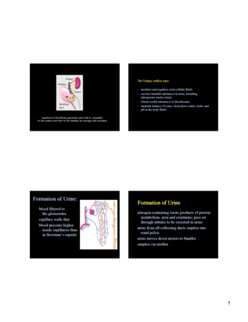
The Urinary outflow tract: monitors and regulates extra-cellular fluidsexcretes harmful substances in urine, includingnitrogenous wastes (urea)returns useful substances to bloodstreammaintain balance of water, electrolytes (salts), acids, andpH in the body fluidsnephrons in the kidney generate urine that is propelledto the ureters and then to the bladder for storage and excretionFormation of Urine:blood filtered tothe glomeruluscapillary walls thinblood pressure higherinside capillaries thanin Bowman’Bowman’s capsuleFormation of Urinenitrogen-containing waste products of proteinmetabolism, urea and creatinine,creatinine, pass onthrough tubules to be excreted in urineurine from all collecting ducts empties intorenal pelvisurine moves down ureters to bladderempties via urethra1
Formation of UrineThe urogenital system derives predominantly fromintermediate mesodermin healthy nephron, neither protein nor RBCsfilter into capsulein proximal tubule, most of nutrients and largeamount of water reabsorbed back tocapillariessalts selectively reabsorbed according tobody’body’s needswater reabsorbed with saltsDuring development, 3 successive kidneys form:pronephros in an early embryo2
Mesonephros in intermediate embryoA metanephros is always drained exclusively by one duct, theureter.In birds in reptiles the ureter separates from the nephric ductand enters the cloaca. In mammals, the ureter separates fromthe nephric duct and enters the bladderAs the embryo grows, the ureters lengthen, and thekidneys rotate and ascend along the dorsal body wallWolffian ducturogenital sinus uretercommonnephric ductbladderkidneytrigoneurethrarenal development begins when the ureteric bud invadeskidney mesenchyme (the metanephric blastema)3
the different compartments of the urinary outflow tract are lined withdistinct cell types that perform diverse functionsmaking a kidneythe kidneythe uretersthe bladderHow are the diverse cell types in the kidney, ureter and bladder formed?the kidney is radially patterned4
local proliferation at ureteric bud tips forms an ampullaThe kidney forms via interactions between 3 main celltypescollectingductsUreteric CKIDNEYThe ampulla splits to form two new tipsStroma4thGubWolffian duct2ndG3rdGThe collecting duct system grows by dichotomousbranching5
NEPHRONS FORM EXCLUSIVELY AT URETERIC BUD TIPS IN RESPONSE TO LOCALSIGNALSNephronprogenitors condense at ub tips, aggregateand trans-differentiate into epithelial cellsthat make up Comma and S-shaped bodiesnephrons differentiate from mesenchymal progenitorsDiverse cell types lining the nephron perform distinct functions6
Reciprocal signaling between epithelial andmesenchymal cell types is crucial for organ formationReciprocal Signaling is required for branching morphogenesis andfor nephron differentiation during renal developmentco-culture experiments demonstrate reciprocalsignaling between ureteric bud epithelial and nephron progenitorsbranching morphogenesis no ureteric bud, nephron progenitors undergo apoptosisXnephron induction7
Ret/Gdnf signaling exemplifies a reciprocal loop no nephron progenitors, no branching morphogenesisubThe Ret gene is expressed in ureteric bud tips where it controlsbranching morphogenesissignals from the ureteric bud control nephroninductionsignals from nephron progenitors control branchingmorphogenesisGdnf secreted by nephron progenitors binds to Ret viathe Ret receptor (Gfra1) inducing branchingmorphogenesisnephronprogenitorsureteric budRet/Gfra1Gdnf8
deletion of Ret, Gdnf or the Ret receptor Gfra1 resultsin renal agenesis or hypoplasiaConnecting the upper and lower urinary tractphysical or functional blockage that impedes urine flow cancause renal scarring, hydronephrosis or end state renal diseaserenal filtrate must be efficiently propelled to the bladder forstorage and excretionhydronephrosis in utero9
urorectalseptumHow does the lower urinary tract form?urachushindgutcloacathe cloaca is partitioned into the hindgut and urogenital sinusby the urorectal septumAs the embryo grows, the ureters lengthen, and thekidneys rotate and ascend along the dorsal body wallWolffian ducturogenital sinus uretercommonnephric ductThe urogenital sinus forms the bladder and the urethraWolffian ductbladderurogenital sinusuretercommonnephric a10
Urine transport depends on peristalsisThe renal pelvis, ureters and bladder are lined with a atransitional epithelium (the urothelium)ureters are surrounded by 2-3 coats of longitudinaland circular muscle that mediate myogenic peristalsismyogenic peristalsis is initiated in the renal pelvis moving abolus of urine to the ureter then to the bladder.TransitionalEpitheliummuscleThe ureter is initially joined to the Wolffian duct (futurevas-deferens) not to the bladderThe BladderEpithelium lined with uroplakinplaques that form a water-proof barrierWolffian ducturogenital sinus ureterrugae (folds)allow expansion of the bladderas it fillscommonnephric e connections are established when the ureter orifice is transposedfrom the posterior Wolffian duct (the common nephric duct) to the bladder11
Wolffian ductWolffianductAccepted model of ureter transpositionureteruretercommonnephric ducturogenital sinusurogenital sinusureter transposition in the mouseUrine transport depends on proper connections between theureters and the bladder trigoneformation of the trigone from the common nephricduct repositions the ureters in the bladderAccording to the accepted model, trigone formation isconsidered to be crucial for repositioning the ureter orificecommon nephric ductThe trigone is defined as the portion of the urogenital sinusthat lies between the ureters and sex ductsTrigoneduring ureter transposition, the cnd is incorporatedinto the bladder where it expands to form the trigoneeffectively separating the ureter orifice from the Wolffian duct12
the flap valve is an anti-reflux mechanism that prevents urineback flowuretermuscleureterproper positioning of the ureter orifice is necessary for: formation of patent connections along the outflow tract preventing refluxdefects in position, can cause obstruction or reflux,inducing severe renal damageintra-mural uretermusclefailure in urinary tract function can cause refluxnephropathy (kidney damage)its function depends on proper insertion of the ureter orificein the bladderusing mouse models to re-assess the mechanism ofureter transposition:How are these connections established?Ureter transposition depends on apoptosis of the commonnephric duct, which does not form the trigonekidneyWolffianducturetercommon nephric ductexpression of Jelly Fish green fluorescent protein in the mousecommon nephric duct of this transgenic mouse enables us tofollow its fate during ureter insertion13
A revised model of ureter transpositionthe common nephric duct is absorbed into the expandingurogenital sinus. The ureter makes directcontact with and inserts into the urogenital sinus**forget this revised model of ureter transposition when you take yourboards; the new model is published but not in the text books yet. Remember ithowever as an example of how modern tools will allow us to directly examineother embryological models of organogenesis!!apoptosis of the common nephric duct enables the ureterorifice to detach from the Wolffian ductcontinued growth and expansion of the urogenital sinusmoves the ureter orifice further anterior to the bladder neck14
Formation of Urine: blood filtered to the glomerulus capillary walls thin blood pressure higher inside capillaries than in Bowman’s capsule Formation of Urine nitrogen-containing waste products of protein metabolism, urea and creatinine, pass on through tubules to be excreted in urine