Transcription
Cat Anatomy and Physiology4 - H C AT P R O J E C T U N I T 3 E M 4 2 8 9 E
ACKNOWLEDGEMENTS:Our thanks to the 4-H leaders who contributed to the first edition: John and Sandy Edminster,Thurston County, and Mary Smith and Sue Hall, King County. Also to Roy J. Hostetler, DVM, formerExtension Veterinarian.Revised 2008 by Michael A. Foss, DVM, Skamania County; Nancy Stewart, King County; and Jean Swift,Skagit County.RESOURCES:The Anatomy of Domestic Animals, 5th Edition, Sisson and Grossman, WB Saunders Co., Philadelphia,1975.The Cornell Book of Cats, 2nd Edition, edited by Mordecai Siegal, Villard, New York, revised 1997.Reader’s Digest Book of the Cat, edited by Alice Philomena Rutherford, Readers Digest Association Ltd.,Montreal, 1992.The Royal Canin Cat Encyclopedia, Aniwa Publishing, Paris, 2001.Dear Leaders and Parents:This manual is primarily for 4-H members who have learned all the material in the previous two unitsand are at the senior level of 4-H (Grades 9-12). The focus of this unit is anatomy and physiology.Knowledge of the cat’s body and how it functions is important to every cat owner and will prove usefulin the pet’s daily care. It will also be helpful when giving the veterinarian needed information and inthe treatment of cat ailments and injuries.
CONTENTSChapter 1The Evolution of the Cat . 4Chapter 2The Five Senses. 9Chapter 3Musculo-Skeletal System. 12Chapter 4Respiratory and Circulatory Systems. 16Chapter 5Digestive and Urinary Systems. 18Chapter 6Reproductive System. 20Chapter 7Care of the Pregnant Cat and Kittens. 22Chapter 8Behavior. 25Chapter 9Vocabulary. 26Worksheets. 30Word Search and Crossword Puzzles. 363
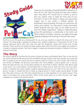
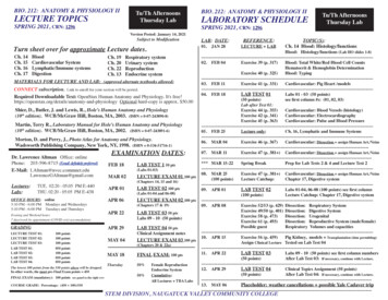
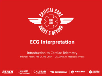
CHAPTER 1The Evolution of the CatCat-like animals first came into existence about65 million years ago. The oldest fossils showeda similarity to modern cats. These date back to20 million years ago. Cats were the last in lineto be domesticated. The oldest evidence of thedomestication of cats is 4500 B.C. in Egypt.Canoidea). The Aeluroidea evolved in threegroups, the Dinictis which gave rise to the felines,Hyaenids (hyenas), and Viverrids which becamecivets and mongooses. The Arctoidea evolvedinto four groups: Canids (dogs, wolves, foxes),Mustelids (weasels, mink, skunks, badgers),Procyonids (raccoons), and Ursinids (bears). Inthis chapter, we will only continue with theDinictis line which eventually led to the domesticcat of today.Cats are an evolutionary marvel! Though humansare at the top of the evolutionary pyramid, beingthe most successful of all animals, the cat is notthat far below. Cats represent the most supremelyefficient muscular machines in their ability tojump, twist, and turn. The ratio of their strengthto their size is far superior to humans.III. DinictisDinictis came into being about 53 million yearsago in the Eocene Age. With longer legs and tailand cat-like teeth for stabbing prey, Dinictis hadbecome a better hunter. Dinictis developed in twodifferent directions, Nimravidae (Paleofelids) andFelidae (Neofelids).As animals evolved, cats developed into the mostefficient hunter of all. Their keen sense of sight,hearing, and smell make it easier to locate theirprey. Strong legs to pursue and/or spring on theirprey along with sharp claws and teeth to kill andtear apart the prey make cats very formidable. Ithas been documented that cats have a degree ofreasoning ability. This moves them higher on theevolutionary ladder. This also makes cats moreefficient hunters, especially since all but lions aregenerally lone hunters.IV. Nimravidae and FelidaeNimravidae was the first saber toothed cat thatexisted for approximately 30 million years. Thislarge saber toothed cat (known as Eusmilus) wasthe size of a small puma, had claws that were notcompletely retractable, and walked on its pads.They were not as efficient as the Felidae and weremore limited in intelligence, which eventually ledto their dying out.The Evolutionary TreeI. Miacids:Felidae developed in two stages. The first stage wasthe Proailurus which developed in the OligoceneAge. This stage evolved into Pseudoailurus, whicharose in Europe and North America during theMiocene Age 23 million years ago.Miacids existed in the Paleocene Age about 65million years ago. They were the first ancestorof cats. Miacids fed primarily on flesh and bloodof other vertebrates. They had short legs, longbodies, and were weasel- to wolf-sized. They livedin the forests. Like all carnivores (meat eatinganimals), they had carnassial teeth—large sharpmolars and premolars in the upper and lower jawsthat cut food in a scissoring action. They also hadlarge sharp canine teeth for tearing flesh.V. PseudoailurusII. Aeluroidea and Arctoidea:Pseudoailurus evolved into four groups. Theearliest was the famed saber-toothed tiger—Smilodon. These creatures appeared 12 millionyears ago and survived for about 2 million years—into the Epoch of Man. It then died out.Miacids began to evolve into different familiesin the Eocene to Oligocene Ages, 53 to 34million years ago. The two branches were knownas Aeluroidea (or Feloidea) and Arctoidea (orPseudoailurus is the direct ancestor of the moderncat. It had a flattish skull, acute hearing, andwalked almost flat-footed. The oldest fossils showa close similarity with the modern cat.4
Starting in the Pliocene Age, 10 million yearsago, the three modern branches began to evolve.Those branches are Acynonyx, Panthera, and Felis.Acynonyx are the cheetahs. They have an ossifiedhyoid bone that makes them unable to roar. Theyalso have unretractable claws. Cheetahs are foundonly in the Old World.members of Felis catus are cougar, lynx, ocelot,bobcat, margay, serval, and caracal. Felis sylvestrisincludes the Scottish wildcat, Spanish wildcat,and Indian desert wildcat. It is unlikely thatthese wildcats played a role in the developmentof the domestic cat. They are very reclusive andextremely fearful of humans. However, they dohave the ability to crossbreed with domestic cats.Panthera are the large cats found in both theOld and New Worlds. These are the lions, tigers,leopards, and jaguars. They have a hyoid boneat the base of the tongue. It is partly cartilage,allowing it to move freely, enabling big cats to roar.The first domestication of cats appears to be8,000 years ago—compared to dogs which weredomesticated 50,000 years ago. Overwhelmingevidence points to ancient Egypt and the Africanwildcat (Felis sylvestris libyca) as being the originof the domesticated cat.Felis is the largest of the cat families. This familyof cats has an ossified hyoid bone and thusis unable to roar. Some of the better-known5
WORKSHEET—CHAPTER 11. List the ages in order.2. What two groups branched out from Miacids?3. Dinictis had two branches. Which one became extinct?4. From which sylvestris genus did the domestic cat originate? What is its Latin name?5. What is the common ancestor for the cat and the dog?6. Why did the Nimravidae become extinct?7. Catlike fossils were found that date back to 20 million years ago, but the oldest fossils most closelyresembling the modern cat date back to million years ago.8. What is a hyoid bone?9. What has happened to the hyoid bone in the cheetahs and the small cats? What are they unable todo because of this?10. What made the feline the most efficient hunter of all?6
PaleoceneAeluroideaFrom 65 million years mravusMioceneFrom 23 million years agoEusmilus(saber-toothedcat)From 34 million years agoOligoceneFAMILY TREE OF THE CATFrom 53 million years cougars, lynx,bobcats, ocelots,margay, servals,caracal)From 10 million years nthera(lions, tigers,leopards, jaguars)PseudoailurusFelisProailurusCanids(dogs, wolves,foxes)Felidae(Neofelids)Mustelids(weasels, mink,skunks, ris(wildcats—Scottish, Spanish,Indian desert,African)domestic cat7
FELINE EVOLUTION WORD CTAABSLINCW D otsRaccoonsAeluriodeaDinictisLynxOligoceneSaber Toothed tic oailurusTreeWild8
CHAPTER 2The Five SensesVisionnot feel well. It is not specific to any one area andmeans you should examine your cat closely for aproblem.Cats have exceptional eyesight. Particularly inthe early evening and at night, they see betterthan humans. Most cats can see kinds of light,such as ultraviolet, that are invisible to humans.In the course of their development, the abilityto see at night while hunting was favored overthe ability to see colors during the day. As far asscientists have been able to tell, cats are more orless colorblind.In the center of the eye is the pupil. It is actuallya hole, formed by the iris, which expands orcontracts to let in the correct amount of light.When exposed to bright light, the pupils contractinto linear slits. In darkness they open very wideto allow in as much light as possible.Behind the pupil is the lens, which focusesimages on the back of the eyeball. It is composedof strong, crystal-like fibrous tissue. Light rayspassing through the lens are bent to rest on avery sensitive area on the back of the eye, theretina. It is richly lined with nerves. The nerves inthe retina receive light and change it into nerveimpulses or signals. Impulses are transmitted tothe brain by way of the optic nerve. The braininterprets these impulses and lets the cat “see.”One phenomenon of cat vision is the tapetumlucidum, or “eyeshine”, the glowing of cat’s eyesin the dark. A layer of iridescent cells at the backof the eyeball reflects the light, which adds to thecat’s ability to see at night.A cat’s eyes are complicated and delicate organs.Even small injuries can render a cat blind. Theeyes appear as large, round globes or orbs, witha transparent covering known as the cornea.Around the cornea is a ring of white, shiny tissuecalled the sclera. The nictitating membrane,or third eyelid, is located in the lower part ofthe cat’s eye. This eyelid serves as protectionfor the eye. Cats have partial vision throughthe third eyelid, and often use it as protectionwhen fighting or when traveling through denseunderbrush. When the nictitating membrane isclearly visible, it is usually a sign that the cat doesUpper eyelidLensPupilCorneaIrisOpticnerveRetinaLower eyelidScleraCross Section of EyeHearingThe cat’s pupil narrowsin bright light and openswide in dim light.Cats are exceedingly sensitive to sound; theirrange of hearing extends well above and belowthe range of human hearing. It is probable thatthe cat has a keener sense of hearing than mostdogs, since it depends more upon sight andhearing than smell when hunting.Cats, like all four-legged animals, have cupped9
ears which serve as receptors and conductors ofsound. When listening, the cat will move its headthis way and that, turning its ears in the directionof the sound. The sound travels down throughthe outer ear canal, which is fitted with smallknobs or protuberances. The outer ear tapers andnarrows near the cat’s skull, then turns downwardand inward, ending in a delicate membraneknown as the eardrum. All of the hearing facultiesare protected within the skull.do. Fastidious about odors, they dislike many ofthe same smells that humans do. They will try tocover up disagreeable smells.Most cats have a particular fondness for catnip,a member of the mint family. It is believed theyare excited by the smell. Some authorities believethat the odor of catnip stimulates the cat sexually.Catnip is harmless and you can give your cat asmuch of the “feline snuff” as it wants. It mayenjoy nibbling on the leaves of a catnip plant,or playing with a catnip-stuffed toy. Some catsare not affected by catnip, some become justquietly excited, while others roll, purr, and growlin ecstasies of delight. The occasional cat maybecome disagreeable.Beyond the eardrum are three small, delicatebones: the hammer, anvil, and stirrup. The namesdescribe the shapes of the bones that function totransmit sound waves into a section of the innerear known as the cochlea. The cochlea, a snailshaped canal, contains the auditory nerve whichchanges sound waves into nerve impulses (nervesignals) and relays sound messages to the brain.Near the cochlea are three horseshoe-shaped tubesknown as the semicircular canals. They containfluid and fine hairs called cilia that function tomaintain the cat’s excellent sense of balance.Sense of TasteThe cat’s tongue is long and flat, with almostparallel sides. It tapers slightly in front andsomewhat more in the back of the mouth. Theupper surface of the tongue is covered with rasplike papillae which enable the cat to scrape everypiece of meat off a bone or to lick its coat clean.The tongue is covered with taste buds, particularlyat the tip and at the back of the throat. Thesetaste buds react to chemical stimuli to producesensations of acidity, sweetness, bitterness, andsaltiness.Connecting the middle ear to the throat is theEustachian tube. Its main purpose is to equalizepressures. Without this safety device, the eardrumwould be ruptured when subjected to greatpressure. When pressure is exerted on its ears, acat swallows and sticks out its tongue.Semicircular canalsStirrupSense of Touch (Hair and Skin)Auditory nerveThe least important sense in a cat is the sense oftouch. Both the skin and the hair play a role inthat sense.AnvilHammerCochleaSense of SmellFirst of all, the hair or fur serves as insulationagainst heat and cold. Hair also protects the catagainst insect bites, stings, thorns, and otherdangers and annoyances. The cat raises its hair,particularly the hair along its neck and spine, as aprotective device when frightened or threatened.With its hackles raised, the cat assumes a waryand defiant position. Back arched, tail hairsbristling, muscles tensed, it turns itself broadside.In this attitude, the cat appears larger and moreferocious to its attacker.The cat’s olfactory nerves, those concerned withits sense of smell, are not as sensitive as those ofsome other animals, but are quite adequate. Catscan scent people, animals, and other objects ata considerable distance, but they do not rely onthis sense while hunting as much as other animalsCats shed their hair according to climaticconditions and their state of health. Hair isshed naturally year round, especially in thespring and fall. Excessive shedding is a warningsignal of possible disease, poor diet, parasites,or overheating. Therefore, it is important to payEardrumEustachian TubeInside of Cat’s Ear10
attention to the condition of your cat’s hair.This should be kept in mind when you use anyinsecticides or medications on a cat’s skin. A toxicsubstance may prove fatal if absorbed through theskin.The cat’s skin is made up of an outer layer, theepidermis, and an inner layer, the dermis. Theepidermis consists of four sub-layers, with theinnermost providing for the regeneration of skincells.The skin has many glands. Sweat glands arelocated only on the foot pads. Sweating is only asmall portion of thermoregulation (keeping thebody at a correct operating temperature). The catcools itself primarily by panting. Cats also haveglands in their skin that are connected with thehair follicles, known as sebaceous glands. Theysecrete an oily substance known as sebum thatsolidifies when exposed to the air. It coats thehairs, thus protecting the fur and making it glossy.In a healthy state, the cat’s skin is always elasticand pliable, with the ability to regenerate at arapid pace.Skin functions to protect the body from theexternal environment. Skin protects againstinjuries and prevents excess loss of water,electrolytes, and large molecules. Skin is thefirst layer of defense in the immune system as itphysically blocks bacteria, viruses, fungus, andother pathogenic (disease causing) organisms.While the cat’s skin is somewhat waterproof,it is not impermeable. That is, certain oils andmedicines can be absorbed through the skin.11
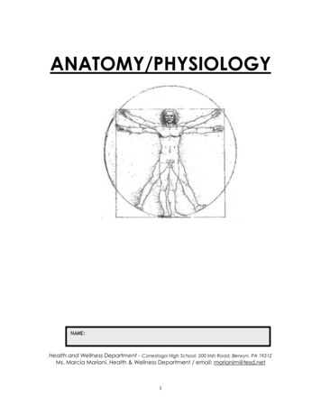
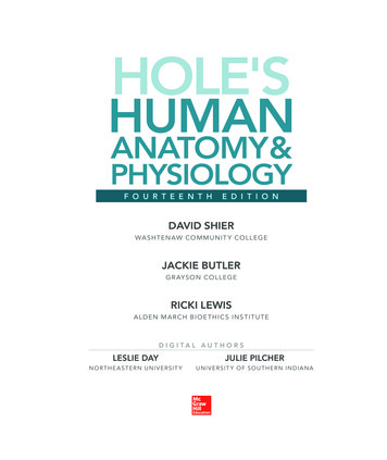
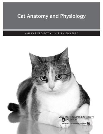
CHAPTER 3Musculo-Skeletal SystemThe cat’s skeleton is not so different from thehuman skeleton. The cat has more bones—230 asopposed to 206—but many are identical to thosein the human being. Cats have 13 ribs; humanshave 12. Cats do have clavicles (collar bones)but unlike humans, they are not attached toother bones. The outside of a bone, the cortex, iscomposed of minerals and protein and gives thebone its rigidity. Inside the bone is the marrowcavity that produces red blood cells.all fused into one functional unit. Inside the skullare air filled pockets called sinuses. The cat has afrontal sinus and a maxillary sinus. A major partof the skull, which holds the upper teeth, is calledthe maxilla, and the lower teeth are held by themandible. The small bones that provide structureto the nose are called nasal bones.As mentioned above, the skull holds the teeth. Inthe adult cat there are a total of 30 teeth. Thereare 12 incisors, four canines, 10 premolars (six ontop, four on the bottom) and four molars. In thekitten there are 26 deciduous teeth. The numbersof incisors, canines, and premolars are the samebut there are no molars in kittens.The cat’s muscles are tough and well-coordinatedand help to make the cat an agile hunter. Basically,the cat’s muscles are designed for walking, running,leaping, and twisting. Because of their unusualmusculature, cats are extremely nimble. The cat’smuscular control and skeletal flexibility enable it toright its body during a fall with incredible speed—atrick that is unique to cats.The front or foreleg bones consist of the scapulaor shoulder blade, humerus, radius, ulna, carpusor wrist bones, metacarpus, and phalanges ordigits. The hind leg bones are the pelvis, femur,patella or kneecap, tibia, fibula, tarsus or hock,metatarsus, and phalanges or digits.SkeletonThe body of the skeleton consists of five majorareas; the spinal column, skull, ribs, forelegs, andhind legs. The spinal column is composed of fiveregions, the cervical, thoracic, lumbar, sacral, andcaudal regions. The first region holds the cervicalor neck vertebrae. The first two cervical vertebraehave special names, the atlas and the axis. Thereare a total of seven cervical vertebrae. The thoracic(chest) region has 13 vertebrae and each vertebrais associated with a pair of ribs. The lumbar areahas seven vertebrae and ends at the pelvis. Thepelvis contains three sacral vertebrae that are allfused together. It usually takes 18 to 23 caudalvertebrae to make the tail. The cat’s spine is veryflexible. It is probably the most flexible of allmammals. A cat can arch its back in a ”U” shape.JointsJoints are formed by the union of two or morebones. There are many joints within the cat’s body.Joints are usually held together by connectivetissue called ligaments and tendons. Ligaments areconnective tissue that is attached to bone on bothends. Tendons are connective tissue that is attachedto bone on one end and muscle on the other. Thejoints are cushioned by cartilage—a cushioninglayer over the ends of the bones meeting at a joint.There are two types of joints. The hip and shoulderjoints are ball and socket. They are able to movein a forward, backward, and sideways motion. Theother joints are hinge type and are only able to flexand move forward and back. The major joints ofthe foreleg are the shoulder, the elbow, and carpusor wrist. The major joints of the hind leg are thehip, stifle or knee, and hock or ankle.There are 13 pairs of ribs. All are attached at thetop to a thoracic vertebra. Most are attached at thebottom to the sternum or breastbone. Collectively,the spine, ribs, and sternum form the thorax thathouses the heart and lungs. This rigid structure isneeded to allow breathing to occur.LocomotionThe cat’s skeleton serves as a strong frameworkfor the muscular system. This structural systemallows for some very important actions such asThe skull is attached to the spinal column at theatlas. The skull actually consists of many bones12
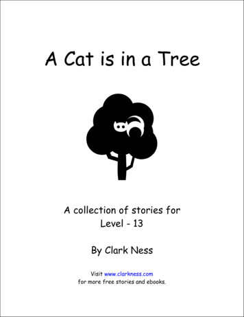
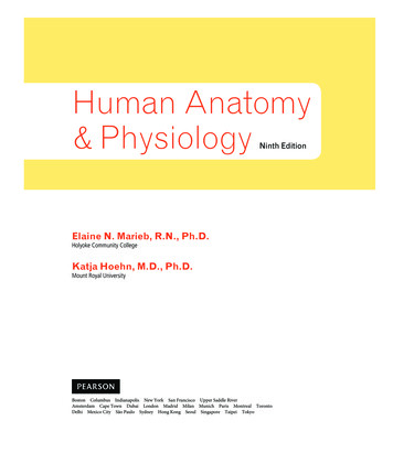
protection for vital organ and locomotion ofthe cat. The cat has several gaits, or patterns oflocomotion. The most common are: the walk,a 4-beat gait (meaning each foot lands on theground at a separate time); the trot, a 2-beat gaitwith contra-lateral (opposite side of body) frontlimb and rear leg striking the ground at the sametime; and the run, a 3-beat gait. Some cats willpace instead of trot. This is a 2-beat gait where theforeleg and the rear leg on the same side of thebody more forward at the same time.muscles. Muscles are usually named for the areas ofattachment. Often both origin (where the musclebegan) and insertion (where the muscle ends) areincluded in the name and sometimes its function.Dorsal refers to the top part of the body andventral refers to the bottom, or toward the ground.Muscles of the Head and NeckTemporal and Zygomaticoauriclaris musclespull the ears forward; Caudal Auricular musclespull the ear back. Levator nasolabialis raisesthe upper lip and dilates the nostrils. Massetercloses the jaw. It is the muscle of masticationor chewing. Buccinator forms part of the cheekand aids in movement of food in the mouth.Brachiocephalicus originates on the head andneck and extends to the shoulder.Unfortunately, motor vehicle accidents kill orinjure many cats each year. Most frequentlybroken bones are the bones of the forelegs andhind legs and the pelvis.Muscular SystemMuscles of the Dorsal BodyThe cat, like all mammals, has three kinds ofmuscles. There are striated muscles, cardiacmuscles, and smooth muscles. Striated musclesmake up the majority of muscles. Striated musclesare the ones involved in voluntary movement andare usually attached to bone. They allow the catto walk, breathe, and look around. These musclesare the flesh of the body—the legs, back, chest,and head. Striated muscle gets its name from itsappearance under the microscope as it has smallstripes or striations.The Trapezius muscle raises the head andshoulder. Latissimus dorsi flexes the shoulder.Thoracolumbar fascia serves as an anchor to anumber of back and abdominal muscles.Muscles of the Thorax, Abdomen, and TailIntercostal muscles connect the ribs to oneanother. They aid in breathing. Externalabdominal oblique is a large superficial musclethat forms part of the abdominal wall. Rectusabdominis originates from the stenum and insertson the pelvis. It makes a major ventral supportfor the abdomen. The Sarcocaudalis musclemanipulates the tail.Cardiac muscle is a specialized striated muscle.It is found only in the heart and contractsautomatically. There is no conscious control ofthe heart.Muscles of the ForelimbSmooth muscle gets its name because under themicroscope it does not have striations; it lookssmooth! It is the muscle of organs. It is notunder voluntary control. It is found in the wallof the digestive tract where it moves food fromthe stomach into the intestines and through theintestines without any conscious control. Smoothmuscle is also in the walls of the urinary tract andreproductive tract. It is in the walls of arteries,bronchioles, and in the eye.Brachiocephalicus helps to extend the shoulder.Infraspinatus supports the shoulder joint.Deltoidus flexes the shoulder. Brachialis flexesthe elbow. Triceps extends the elbow. Flexor carpiradialis, Extensor carpi ulnaris, and Flexor carpiulnaris together control the carpus. Digital flexorsand digital extensors control the phalanges (toes).Muscles of the Pelvic LimbSkeletal Muscles of the CatSartorius muscles flex the hip and extend thestifle. Medial gluteal is the major muscle of therump. It flexes the hip. Biceps femoris flexesthe stifle. Tensor fascia lata helps flex the hip.Semitendinosus is the rear most muscle of thethigh. It extends the hip. Gastrocnemius flexes thestifle and extends the hock. Common and lateraldigital extensors extend the toes.Skeletal muscles function by crossing a joint andmoving bone or cartilage. Each muscle has aspecific action or job. The muscle either extendsthe joint (makes the angle of the joint larger)or flexes the joint (makes the angle of the jointsmaller). There are hundreds of muscles within thecat. The following is a list of some of the major13
NasalFrontalCanine ar VertebraeRibsSKELETON OF THE CATCervical vertebraeSternumThoracic vertebraeUlnaPhalangesTarsusIliumSacral vertebraeIschiumFemurPatellaTibiaFibulaCaudal vertebrae14
Levator dobrachialisDeltoidusTricepsBrachialisMAJOR MUSCLES OF THE CATPeronealGastrocnemiusSemitendinosusBiceps femorisGluteus maximusSacrocaudalisGluteus mediusSarcocaudalisLatissimus dorsiThorocolumbar fasciaExternal abdominal obliqueSartoriusIntercostalExtensor carpi ulnarisTibialis carnialisRectus abdominisFlexor carpi asseterCleidocephalicusExtensor carpi radialisDigital extensorTensor fascia lata15
CHAPTER 4Respiratory and Circulatory SystemsRespiratory SystemHeartThe respiratory system functions primarily totransfer oxygen from the air to the blood and toremove carbon dioxide from the blood and carryit out of the body into the air. Oxygen is vitalto the body’s maintenance. Carbon dioxide is awaste product of the maintenance process. Thecat breathes in air through its nose and mouth.The nose and mouth join to form the pharynx.The trachea is a tube that extends from thepharynx to the lungs. The epiglottis covers thetrachea when swallowing occurs to prevent foodand water from entering the lungs. At the lungsthe trachea branches into smaller tubes calledbronchi or bronchial tubes. The bronchi divideinto smaller and smaller bronchioles until theybecome dead end sacs called alveoli. It is in thealveoli where the actual exchange of oxygen andcarbon dioxide occur.The heart is a marvelously specialized muscle.The heart has four chambers with four valves andcontracts in a specific squeezing pattern. Thissqueezing action pumps blood through the heartout to the lungs and body. At a resting pulse of120 beats per minute, a cat’s heart will beat 63million beats in a year! It is the closure of theheart valves that gives the “lub dub” heart sounds.Blood VesselsThe blood vessels include arteries, veins, andcapillaries. Arteries are the vessels that carry bloodaway from the heart, and veins carry blood backto the heart. The largest artery is called the aorta.It carries freshly oxygenated blood to the body.Capillaries are tiny vessels where the arteries endand the veins begin. Capillaries are only largeenough for one blood cell to pass through ata time. It is in the capillaries where nutrients,oxygen, and waste products are exchanged withindividual body cells. Most live healthy cellsin the body are in contact with a capillary. Thelargest veins are called the cranial and caudal venacava. They carry the oxygen-depleted blood backto the heart. The heart then pumps that bloodto the lungs via the pulmonary artery. Followingoxygenation in the lungs, the blood is carriedback to the heart through the pulmonary vein,and the cycle begins again.All mammals have a muscle that separatesthe respiratory system (lungs) from the rest ofthe internal organs. This muscle is called thediaphragm. The diaphragm works with themuscles between the ribs called “intercostals”to produce inspiration (or breathing in).Exhalation (or breathing out) is musclerelaxation, causing the air to move out of thelungs and body.When the diaphragm is torn or ruptured,usually as a result of being hit by an automobile,a diaphragmatic hernia is created. This allowsabdominal organs into the chest, collapses thelungs, and creates a life threatening condition.BloodCirculatory SystemBlood is composed of cells suspended in a specialfluid called plasma. The majority of the cells inthe blood are red blood cells. Red blood cellscontain hemoglobin, which functions by carryingoxygen and carbon dioxide. It is hemoglobin thatgives blood its characteristic red color.The circulatory system consists of the heart,blood, blood vessels, lymphatic system, andspleen. The circulatory system functions are todeliver oxygen and nutrients to the cells of thebody and to remove waste products.Blood also carries white blood cells. The job of thewhite blood cells is to fight infection or invasionby anything foreign. The blood also includesplatelets. Platelets are small cells that help inblood clotting.The respirat
Panthera are the large cats found in both the Old and New Worlds. These are the lions, tigers, leopards, and jaguars. They have a hyoid bone at the base of the tongue. It is partly cartilage, allowing it to move freely, enabling big cats to roar. Felis is the largest of the cat families. This family of