Transcription
Human Anatomy& PhysiologyNinth EditionElaine N. Marieb, R.N., Ph.D.Holyoke Community CollegeKatja Hoehn, M.D., Ph.D.Mount Royal UniversityBoston Columbus Indianapolis New York San Francisco Upper Saddle RiverAmsterdam Cape Town Dubai London Madrid Milan Munich Paris Montreal TorontoDelhi Mexico City São Paulo Sydney Hong Kong Seoul Singapore Taipei Tokyo# 105016 Cust: Benjamin Cummings/CA Au: Marieb Pg. No. iTitle: Anatomy & Physiology Server: S4CA03 MARI3268 09 SE FM NASTA.indd 1C/M/Y/KShort / NormalDESIGN SERVICES OFcarlislePublishing Services11/8/11 8:59 AM
Editor-in-Chief: Serina BeauparlantAcquisitions Editor: Gretchen PuttkamerAssociate Project Editor: Shannon CuttDirector of Development: Barbara YienDevelopment Editor: Alice FugateArt Development Manager: Laura SouthworthSenior Managing Editor: Debbie CoganProduction and Design Manager: Michele MangelliProduction Supervisor: David NovakMedia Producer: Aimee PavyEditorial Assistant: Lisa DamerelText Designer: tani hasegawaCover Designer: Riezebos Holzbaur Design GroupArt Houses: Imagineering STA Media Services Inc. and ElectronicPublishing Services Inc., NYCArt Coordinator: Jean LakePhoto Image Lead: Donna KalalPhoto Researcher: Kristin PiljayCopyeditor: Anita WagnerProofreader: Martha GhentCompositor: S4Carlisle Publishing ServicesSenior Manufacturing Buyer: Stacey WeinbergerMarketing Manager: Derek Perrigo and Susan BlumenthalCover photo of Olympic Gold Medalist Hope Solo Ina Fassbender/Reuters.Credits and acknowledgments borrowed from other sources and reproduced, with permission, in thistextbook appear on the appropriate page within the text or on p. C-1.Photo and illustration credits follow the Glossary.Copyright 2013, 2010, 2007 Pearson Education, Inc. All rights reserved. Manufactured in the UnitedStates of America. This publication is protected by Copyright, and permission should be obtainedfrom the publisher prior to any prohibited reproduction, storage in a retrieval system, or transmissionin any form or by any means, electronic, mechanical, photocopying, recording, or likewise. To obtainpermission(s) to use material from this work, please submit a written request to Pearson Education,Inc., Permissions Department, 1900 E. Lake Ave., Glenview, IL 60025. For information regardingpermissions, call (847) 486-2635.Many of the designations used by manufacturers and sellers to distinguish their products are claimedas trademarks. Where those designations appear in this book, and the publisher was aware of atrademark claim, the designations have been printed in initial caps or all caps.Library of Congress Cataloging-in-Publication DataMarieb, Elaine NicponHuman anatomy & physiology / Elaine N. Marieb, Katja Hoehn.—9th ed.p. ; cm.ISBN-13: 978-0-321-74326-8 (student ed.)ISBN-10: 0-321-74326-1 (student ed.)I. Hoehn, Katja. II. Title.[DNLM: 1. Anatomy. 2. Physiological Phenomena. QS 4]LC classification not assigned612—dc232011038702ISBN 10: 0-13-282874-X (High School Binding)ISBN 13: 978-0-13-282874-1 (High School Binding)1 2 3 4 5 6 7 8 9 10—RRD—15 14 13 12 11www.PearsonSchool.com/Advanced# 105016 Cust: Benjamin Cummings/CA Au: Marieb Pg. No. iiTitle: Anatomy & Physiology Server: S4CA03 MARI3268 09 SE FM NASTA.indd 2C/M/Y/KShort / NormalDESIGN SERVICES OFcarlislePublishing Services11/8/11 8:59 AM
About the AuthorsWe dedicate this work to our students both present and past,who always inspire us to “push the envelope.”Elaine N. MariebFor Elaine N. Marieb, taking the student’s perspective into account has always been an integral part of her teaching style. Dr.Marieb began her teaching career at Springfield College, whereshe taught anatomy and physiology to physical education majors. She then joined the faculty of the Biological Science Division of Holyoke Community College in 1969 after receivingher Ph.D. in zoology from the University of Massachusettsat Amherst. While teaching at Holyoke Community College,where many of her students were pursuing nursing degrees,she developed a desire to better understand the relationshipbetween the scientific study of the human body and the clinicalaspects of the nursing practice. To that end, while continuingto teach full time, Dr. Marieb pursued her nursing education,which culminated in a Master of Science degree with a clinicalspecialization in gerontology from the University of Massachusetts. It is this experience that has informed the development ofthe unique perspective and accessibility for which her publications are known.Dr. Marieb has partnered with Benjamin Cummings forover 30 years. Her first work was Human Anatomy & Physiology Laboratory Manual (Cat Version), which came out in 1981.In the years since, several other lab manual versions and studyguides, as well as the softcover Essentials of Human Anatomy& Physiology textbook, have hit the campus bookstores. Thistextbook, now in its 9th edition, made its appearance in 1989and is the latest expression of her commitment to the needs ofstudents studying human anatomy and physiology.Dr. Marieb has given generously to provide opportunitiesfor students to further their education. She contributes to theNew Directions, New Careers Program at Holyoke Community College by funding a staffed drop-in center and by providing several full-tuition scholarships each year for women whoare returning to college after a hiatus or attending college forthe first time and who would be unable to continue their studieswithout financial support. She funds the E. N. Marieb ScienceResearch Awards at Mount Holyoke College, which promotesresearch by undergraduate science majors, and has underwritten renovation and updating of one of the biology labs in ClappLaboratory at that college. Dr. Marieb also contributes to theUniversity of Massachusetts at Amherst where she generouslyprovided funding for reconstruction and instrumentation ofa cutting-edge cytology research laboratory. Recognizing thesevere national shortage of nursing faculty, she underwrites theNursing Scholars of the Future Grant Program at the university.In 1994, Dr. Marieb received the Benefactor Award fromthe National Council for Resource Development, AmericanAssociation of Community Colleges, which recognizes herongoing sponsorship of student scholarships, faculty teachingawards, and other academic contributions to Holyoke Community College. In May 2000, the science building at HolyokeCommunity College was named in her honor.Dr. Marieb is an active member of the Human Anatomyand Physiology Society (HAPS) and the American Associationfor the Advancement of Science (AAAS). Additionally, whileactively engaged as an author, Dr. Marieb serves as a consultantfor the Benjamin Cummings Interactive Physiology CD-ROMseries.When not involved in academic pursuits, Dr. Marieb isa world traveler and has vowed to visit every country on thisplanet. Shorter term, she serves on the scholarship committeeof the Women’s Resources Center and on the board of directorsof several charitable institutions in Sarasota County. She is anenthusiastic supporter of the local arts and enjoys a competitivematch of doubles tennis.iii# 105016 Cust: Benjamin Cummings/CA Au: Marieb Pg. No. iiiTitle: Anatomy & Physiology Server: S4CA03 MARI3268 09 SE FM NASTA.indd 3C/M/Y/KShort / NormalDESIGN SERVICES OFcarlislePublishing Services11/8/11 8:59 AM
ivAbout the AuthorsKatja HoehnDr. Katja Hoehn is an associate professor in the Departmentof Chemical and Biological Sciences at Mount Royal University in Calgary, Canada. Dr. Hoehn’s first love is teaching. Herteaching excellence has been recognized by several awards during her 17 years at Mount Royal University. These include aPanCanadian Educational Technology Faculty Award (1999),a Teaching Excellence Award from the Students’ Associationof Mount Royal (2001), and the Mount Royal DistinguishedFaculty Teaching Award (2004).Dr. Hoehn received her M.D. (with Distinction) fromthe University of Saskatchewan, and her Ph.D. in Pharmacology from Dalhousie University. In 1991, the DalhousieMedical Research Foundation presented her with the MaxForman (Jr.) Prize for excellence in medical research. During her Ph.D. and postdoctoral studies, she also pursued herpassion for teaching by presenting guest lectures to first- and# 105016 Cust: Benjamin Cummings/CA Au: Marieb Pg. No. ivTitle: Anatomy & Physiology Server: S4CA03 MARI3268 09 SE FM NASTA.indd 4second-year medical students at Dalhousie University and atthe University of Calgary.Dr. Hoehn has been a contributor to several books and haswritten numerous research papers in Neuroscience and Pharmacology. She oversaw a recent revision of the Benjamin Cummings Interactive Physiology CD-ROM series modules, andcoauthored the newest module, The Immune System.Following Dr. Marieb’s example, Dr. Hoehn provides financial support for students in the form of a scholarship thatshe established in 2006 for nursing students at Mount RoyalUniversity.Dr. Hoehn is also actively involved in the Human Anatomy and Physiology Society (HAPS) and is a member of theAmerican Association of Anatomists. When not teaching, shelikes to spend time outdoors with her husband and two sons,compete in triathlons, and play Irish flute.C/M/Y/KShort / NormalDESIGN SERVICES OFcarlislePublishing Services11/8/11 8:59 AM
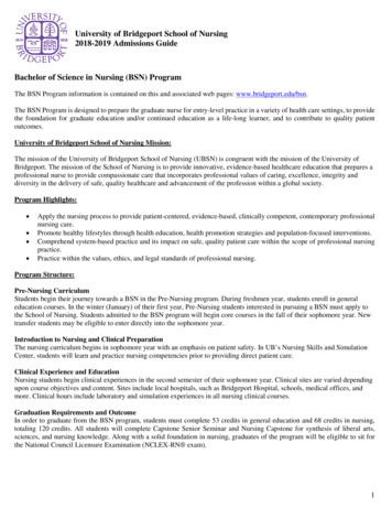
Introduce yourself to the chapterImproved readability and navigability makes thetext more accessible and easier to study.527Chapter 14 The Autonomic Nervous System Chapter OutlinesChapter outlines providea preview of the chapterand help you locateinformation easily.Learning ObjectivesLearning objectivesare integrated into thechapter and give you a previewof what content is to comeand what you are expectedto learn.Bulleted NarrativeThe narrative has beenbulleted wherever possibleto make the text easier toread and navigate.Check YourUnderstandingConcept checkquestions are tied tothe sections' LearningObjectives and ask youto stop, think, and checkyour understandingbefore moving on.Dilates the bronchioles in the lungs, increasing ventilation(and thus increasing oxygen delivery to body cells)Causes the liver to release more glucose into the blood to accommodate the increased energy needs of body cells14SympatheticEyeHeartEyeBrain stemSalivaryglandsAt the same time, the sympathetic division temporarilydamps nonessential activities, such as gastrointestinal tract motility. If you are running from a mugger, digesting lunch canwait! It is far more important to give your muscles everythingthey need to get you out of danger. In such active situations,the sympathetic division generates a head of steam that enablesthe body to cope with situations that threaten homeostasis. Itprovides the optimal conditions for an appropriate response tosome threat, whether that response is to run, see distant objectsbetter, or think more clearly.We have just looked at two extreme situations in which oneor the other branch of the ANS dominates. Think of the parasympathetic division as the D division [digestion, defecation,and diuresis (urination)], and the sympathetic division as theE division (exercise, excitement, emergency, embarrassment).Table 14.4 (p. 536) presents a more detailed summary of howeach division affects various organs.Remember, however, that the two ANS divisions rarely workin an all-or-none fashion as described above. A dynamic antagonism exists between the divisions, and both make continuousfine adjustments to maintain reasStomachLiverand gallbladderPancreasL1Liver andgallbladderAdrenalglandLumbarBladderBladderThe Autonomic Nervous SystemGenitalsSacralCheck Your Understanding 1. Name the three types of effectors of the autonomic nervoussystem.2. Which relays instructions from the CNS to muscles morequickly, the somatic nervous system or the ANS? Explain why.3. WhichofOverviewthe ANSwouldpredominateif youwere(pp.524–527)Dilatesthebranchbronchiolesin thelungs,increasingventilationthe beachComparisonenjoyingdeliverytheofsunandthe )theSomaticandofAutonomicwaves? Which branchwould predominateyou were on aNervousSystems (pp.if525–526)Causes the liver to release more glucose into the blood to acsurfboard and a sharkappearedwithinafewfeetofyou?ANS Divisions (pp. 526–527)commodate the increased energy needs of body cellsFor answers, see Appendix H.(pp.division527–533)temporarilyAt the same osacral)tract modamps nonessential activities,such as gastrointestinalDivision(pp. 527–529)tility.If youare running froma mugger,digesting lunch t! It is far more important to give your muscles everything(pp. 529–533)the toparasympatheticsympatheticdescribetheyForneedget you out ofanddanger.In such divisions,active situations,VisceralReflexes(p.533)site of CNSorigin,generateslocations aofheadganglia,and thatgeneralthe thesympatheticdivisionof steamenablesfiberpathways.the body to cope withsituationsthat threatenhomeostasis. ItANSPhysiology(pp. 533–539)provides the optimal ers and ReceptorsAnatomically,the sympatheticandisparasympatheticsomethreat, whetherthat responseto run, see distantdivisionsobjects(pp. 533–535)differinbetter, or think more clearly.The Effects of Drugs (p. 535)havelookedat two extremesituationsin which one WeSitesof justorigin.Parasympatheticare craniosacral—InteractionsoffiberstheAutonomicor thebranchof theANSdominates.the paraDivisions(pp. 535–537)theyotheroriginatein heticdivisiontheD division[digestion,defecation,Controlof AutonomicFunctionSympatheticfibers asarethoracolumbar—theyoriginatein ],andthesympatheticdivisionasthelumbar regions of the spinal cord.E ve(exercise,lengths oftheir fibers.The parasympatheticdiviHomeostaticImbalancesof the ANSTable14.4536)presents a moredetailed summary of howsionhas(p.longpreganglionic(p. 539) and short postganglionic fibers.eachdivisionaffects variousThesympatheticdivisionorgans.has the opposite srarelyworkDevelopmentalAspectsof theANSpreganglionic fibers are short andANSthepostganglionicfibersin anfashionas described above. A dynamic antag(p. 539)areall-or-nonelong.onism exists between the divisions, and both make continuousfine adjustments to maintain homeostasis.1. Name the three types of effectors of the autonomic nervoussystem.524quickly, the somatic nervous system or the ANS? Explain why.3. Which branch of the ANS would predominate if you werelying on the beach enjoying the sun and the sound of thewaves? Which branch would predominate if you were on asurfboard and a shark appeared within a few feet of you?For answers, see Appendix H.ANS AnatomyFor the parasympathetic and sympathetic divisions, describethe site of CNS origin, locations of ganglia, and generalfiber pathways.Sites of origin. Parasympathetic fibers are craniosacral—they originate in the brain (cranium) and sacral spinal cord.Sympathetic fibers are thoracolumbar—they originate in thethoracic and lumbar regions of the spinal cord.Relative lengths of their fibers. The parasympathetic division has long preganglionic and short postganglionic fibers.The sympathetic division has the opposite condition—thepreganglionic fibers are short and the postganglionic fibersare long.MasteringA&P # 105016 Cust: Benjamin Cummings/CA Au: MariebTitle: Anatomy & PhysiologyServer: S4C# 105016 Cust: Benjamin Cummings/CA Au: Marieb Pg. No. vTitle: Anatomy & Physiology Server: S4CA03 MARI3268 09 SE FM NASTA.indd 514The human body is exquisitely sensitive to changes in its hough sympathetic innervation to the skin is mapped to thecervicalregion here, all nerves to the peripherycarry postganglionicsympatheticand engagesin a lifelongstruggleEye to balance competing demandsEyefibers.Brain stemfor resources under ever-changing conditions. Although all body systems contribSalivarySkin*ute,the stability of our internal environment dependslargely on the autonomic nervousglandsCranial thatgangliaLocationof theirMostparasympatheticare smooth and cardiac musclesystem(ANS),the ganglia.system ofmotorneuronsinnervatesthe visceraleffectororgans. every moment, signals streamfrom visceral organs into the CNS, and autonomic nervesgangliaHeartCervicalmake adjustmentsas necessaryensureoptimalsupportfor body activities. In response toFigure14.3 illustratesthese heANSshuntsblood to “needy”areas, speeds or slows heart rate,are summarized in Table 14.1.LungsLungsWe begindetailedof the ANSthe anaadjustsbloodourpressureandexplorationbody temperature,andwithincreasesor decreases stomach secretions.T1tomicallysimplerparasympatheticMost ofthis fine-tuningoccursdivision.without our awarenessHeart or attention. Can you tell whenyour arteries are constricting or your pupils are dilating? Probably not—but if you’veStomachever been stuckThoracicin a checkoutline, and yourDivisionfull bladder was contracting as if it had aParasympathetic(Craniosacral)mindof its own, you’ve been very aware of visceralactivity. The ANS controls all thesePancreasStomachThe parasympathetic division is also called the craniosacralfunctions, both those we’re aware of and those we’re not. Indeed, as the term autonomicdivision because its preganglionic fibers spring fromLiveropposite(auto5 self; nom 5 govern) implies, this motor subdivisionof the peripheral nervousPancreasand gallendsof the CNS—the brain stem and the sacral regionof thesystemcordhas (Figurea certainamountfunctional independence.bladderL114.4). Theofpreganglionicaxonsextend The ANS is also called subconsciouscontrol, or the generalfrom the CNS nearly all the way to the structures theyinnerAdrenalLiver and motor system, which indicates the locationvisceralof most of its effectors.vate.There the axonssynapse with postganglionic neuronsLumbargland lo gall-catedin terminal ganglia that lie close to or within the targetbladderorgans. Very short postganglionic axons issue from the terminalganglia and synapse with effector cells in their immediate area.OverviewBladderBladderDefine autonomic nervous system and explain its relationship to the are the somatic and autonomic nervous systems relative to effectors, efferent# 105016 Cust: Benjamin Cummings/CA Au: Marieb Pg. No. 5272. Which relays instructionsServer:from theCNS to muscles moreTitle: Anatomy & PhysiologyS4C GenitalsFigure 14.3 The subdivisions of the ANS. The parasympatheticand sympathetic divisions differ anatomically in the (1) sites wheretheir nerves originate, (2) relative lengths of their preganglionic andChapter14 nglionicfibers,locations oftheir gangliahere by synapse sites).Check Your UnderstandingAnatomically, the sympathetic and parasympathetic divisionsdiffer inReading Questionskeep you on track.ParasympatheticPg. No. 4.3 Thesubdivisionsof the ANS.The parasympatheticand Comparesympatheticdivisionsdiffer anatomicallyinsites cand sympathetic divisions.DESIGN theSERVICESOFC/M/Y/Ktheirnerves originate, (2) relative lengths of their preganglionic andcarlisleShort/ Normal fibers, andPublishingServices of their ganglia (indicatedpostganglionic(3) locationshere by synapse sites).*Although sympathetic innervation to the skin is mapped to the cervicalregion here, all nerves to the periphery carry postganglionic sympatheticfibers. Location of their ganglia. Most parasympathetic ganglia arelocated in the visceral effector organs. Sympathetic ganglialie close to the spinal cord.Figure 14.3 illustrates these and other key differences, whichare summarized in Table 14.1.We begin our detailed exploration of the ANS with the anatomically simpler parasympathetic division.Parasympathetic (Craniosacral) DivisionThe parasympathetic division is also called the craniosacraldivision because its preganglionic fibers spring from oppositeends of the CNS—the brain stem and the sacral region of thespinal cord (Figure 14.4). The preganglionic axons extendfrom the CNS nearly all the way to the structures they innervate. There the axons synapse with postganglionic neurons located in terminal ganglia that lie close to or within the targetorgans. Very short postganglionic axons issue from the terminalganglia and synapse with effector cells in their immediate area.C/M/Y/KShort / NormalC/M/Y/KShort / NormalDESIGN SERVICES OFcarlislePublishing ServicesDESIGN SERVICES OFcarlislePublishing Services11/8/11 8:59 AM
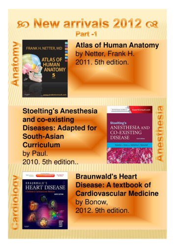
Follow complex processes step by stepFocus Figures help you grasp tough topics in A&P by walking you through carefullydeveloped step-by-step illustrations that use a big-picture layout and dramatic artto provide a context for understanding the process.FOCUSOverviewEach Overview quickysummarizes the keyidea of the figure.Big PictureOrientationThe big picture providesyou with a concretestarting point for theprocess.Bulk Flow Across Capillary WallsFigure 19.17 Bulk fluid flow across capillary walls causescontinuous mixing of fluid between the plasma and theinterstitial fluid compartments, and maintains theinterstitial environment.The big pictureFluid filters from capillaries at their arteriolarend and flows through the interstitial space.Most is reabsorbed at the venous end.ArterioleFluid moves throughthe interstitial space.For all capillary beds,20 L of fluid is filteredout per day—almost 7times the total plasmavolume!Blue TextThis text acts as theteacher's voice andexplains difficultconcepts. In somefigures the text isbroken into numberedsteps to help youmore easily understanddifficult processes.Net filtration pressure (NFP) determines the direction offluid movement. Two kinds of pressure drive fluid flow:Hydrostatic pressure (HP)Osmotic pressure (OP) Due to fluid pressing against aboundary HP “pushes” fluid across theboundary In blood vessels, is due to bloodpressure Due to nondiffusible solutes thatcannot cross the boundary OP “pulls” fluid across theboundary In blood vessels, is due toplasma proteins)17 L of fluid perday is reabsorbedinto the capillariesat the venous end.Boundary“Pulls”Venule# 105016 Cust: Benjamin Cummings/CA Au: Marieb Pg. No. viTitle: Anatomy & Physiology Server: S4CA03 MARI3268 09 SE FM NASTA.indd 6C/M/Y/KShort / NormalAbout 3 L per dayof fluid (and anyleaked proteins) areremoved by thelymphatic system(see Chapter 20).LymphaticcapillaryDESIGN SERVICES OFcarlislePublishing Services11/8/11 8:59 AM
ughace.mphaticpillaryMasteringA&P Focus FigureTutorialsAll Focus Figures have relatedtutorials in MasteringA&Pthat your teacher canassign and that will guideyou through the figuresstep by step.How do the pressures drive fluid flow across a capillary?Net filtration occurs at the arteriolar end of a capillary.CapillaryBoundary(capillary wall)Hydrostatic pressure in capillary“pushes” fluid out of capillary.HPc 35 mm HgOsmotic pressure in capillary“pulls” fluid into capillary.OPc 26 mm HgInterstitial fluidHydrostatic pressure ininterstitial fluid“pushes” fluid intocapillary.HPif 0 mm HgOPif 1 mm HgOsmotic pressure ininterstitial fluid “pulls”fluid out of capillary.To determine the pressure driving thefluid out of the capillary at any givenpoint, we calculate the net filtrationpressure (NFP)––the outward pressures(HPc and OPif) minus the inwardpressures (HPif and OPc). So,NFP (HPc OPif) – (HPif OPc) (35 1) – (0 26) 10 mm Hg (net outward pressure)As a result, fluid moves from the capillaryinto the interstitial space.NFP 10 mm HgNet reabsorption occurs at the venous end of a capillary.CapillaryBoundary(capillary wall)Hydrostatic pressure in capillary“pushes” fluid out of capillary. Thepressure has dropped because ofresistance encountered along thecapillaries.Osmotic pressure in capillary“pulls” fluid into capillary.Interstitial fluidHPc 17 mm HgOPc 26 mm HgHPif 0 mm HgHydrostatic pressure ininterstitial fluid “pushes”fluid into capillary.OPif 1 mm HgOsmotic pressure ininterstitial fluid “pulls” fluidout of capillary.NFP –8 mm Hg# 105016 Cust: Benjamin Cummings/CA Au: Marieb Pg. No. viiTitle: Anatomy & Physiology Server: S4CA03 MARI3268 09 SE FM NASTA.indd 7Again, we calculate the NFP:NFP (HPc OPif) – (HPif OPc) (17 1) – (0 26) –8 mm Hg (net inward pressure)Notice that the NFP at the venous end isa negative number. This means thatreabsorption, not filtration, is occurringand so fluid moves from the interstitialspace into the capillary.C/M/Y/KShort / NormalDESIGN SERVICES OFcarlislePublishing Services11/8/11 8:59 AM
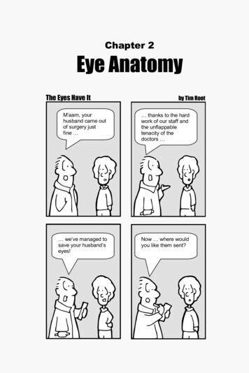
ing an elongated tube called the T tubule (T for “transverse”).Figure 9.4 Myosin heads forming cross bridges that generate muscular contractile force. Part of a sarcomere is seen in atransmission electron micrograph (277,000 ).fusing tubelike caveolae (inpocketings of the sarcolemma),the lumen (cavity) of the T tubule is continuous with theextracellular space.Along its length, each T tubule runs between the paired terminal cisterns of the SR, forming triads, successive groupingsof the three membranous structures (terminal cistern, T tubule,Study figures as you read the text9Sarcoplasmic Reticulum Shown in blue in Figure 9.5, the sar-the T tubules also encircle each sarcomere.coplasmicreticulum(SR)is an elaboratesmoothendoplasmicSelectpiecesof artprovidemorevisualcontent andnext,oftenhaveMusclecontraction is ultimately controlled by nervereticulum (see ndstep-by-steptextthatItshelpsyou betterstructure,initiated electrical impulses that travel along the sarcolemma.Because T tubules are continuations of the sarcolemma, theyfunctions,andarm.processes.surrounds your 3-D anatomycommunicatingwith arteach other at the H zone. Others calledterminalcisterns(“endsacs”)form larger,perpendicular crossStunning 3-D anatomy artis renderedin a always occurmore dynamic, realistic style that uses atedwiththeSRarelargenumbers ofhelp you visualize key anatomical structures.conduct impulses to the deepest regions of the muscle cell and-I bandPart of a skeletalmuscle fiber (cell)Z discA bandI bandH zoneZ discMlineSarcolemmaMyofibrilTriad: T tubule Terminalcisternsof the SR (2)Sarcolemma9Tubules ofthe SRMyofibrilsMitochondriaFigure 9.5 Relationship of thesarcoplasmic reticulum and T tubules tomyofibrils of skeletal muscle. The tubulesof the SR (blue) encircle each myofibril like a“holey” sleeve. These tubules fuse to form anet of communicating channels at the levelof the H zone and saclike elements calledterminal cisterns abutting the A-I junctions.The T tubules (gray) are inward invaginationsof the sarcolemma that run deep into the cellMasteringA&Pbetween the terminal cisterns. (See detailedview in Figure 9.11, pp.290-291) Sites of closecontact of these three elements (terminalcistern, T tubule, and terminal cistern) arecalled triads. # 105016 Cust: BenjaminM09 MARI3268 09 SE CH09.indd284Cummings/CA Au: Marieb Pg. No. 284Title: Anatomy & NEW!PhysiologyServer:andS4C Ranking/Art LabelingC/M/Y/KShort / NormalDESIGN SERVICES OFCARLISLE4/20/11 9:16 AMPublishing ServicesSorting Questions are drag anddrop activities that allow you toassess your knowledge of termsand structures as well as the orderof steps and elements involved inphysiological processes.# 105016 Cust: Benjamin Cummings/CA Au: Marieb Pg. No. viiiTitle: Anatomy & Physiology Server: S4CA03 MARI3268 09 SE FM NASTA.indd 8M09C/M/Y/KShort / NormalDESIGN SERVICES OFcarlislePublishing Services11/8/11 8:59 AM
old and new matrix. As blood concentrations of calcium rise,It is also evident that the brain, intestine, and skeleton havethe stimulus for PTH release ends. The decline of PTH reversesongoing conversations that help regulate the balance betweenits effects and causes blood Ca21 levels to fall.bone formation and destruction, with serotonin serving as aIn humans, calcitonin appears to be a hormone in search of ahormonal go-between. Serotonin is better known as a neufunction because its effects on calcium homeostasis are negligirotransmitter that regulates mood and sleep, but most of theble. When administered at pharmacological (abnormally high)body’s serotonin is made in the gut (intestine) and the blooddoses, it does lower blood calcium levels temporarily.brain barrier (see Chapter 12) bars it from entering the brain.These hormonal controls act to preserve blood calciumThe role of gut serotonin is still poorly understood. What ishomeostasis, not the skeleton’s strength or well-being. In fact,known is that when we eat, serotonin is secreted and circulatedif blood calcium levels are low for an extended time, the bonesvia the blood to the bones where it interferes with osteoblast acbecome so demineralizedClinicalthat theycoveragedevelop large,punched-outof bone turnoverafter eating may lock calciumand case studies tivity.haveReductionbeen expandedthroughout.looking holes. Thus, the bones serve as a storehouse from whichin bone when new calcium is flooding into the bloodstream.ionic calcium is drawn as needed.This is a troubling finding for those taking Prozac and otherantidepressant drugs that inhibit serot
Her first work was Human Anatomy & Physiol-ogy Laboratory Manual (Cat Version), which came out in 1981. In the years since, several other lab manual versions and study guides, as well as the softcover Essentials of Human Anatomy & Physiology textbook, have hit the campus bookstores. This textbook