Transcription
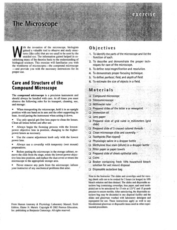
ith the invention of the microscope, biologistsgained a valuable tool to observe and study structures (like cells) that are too small to be seen by theunaided eye. The information gained helped in establishing many of the theories basic to the understanding ofbiological sciences. This exercise will familiarize you withthe workhorse of microscopes-the compound microscope-and provide you with the necessary instructions for itsproper use.WCare and Structure of theCompound MicroscopeObjectives1. To identify the parts ofthe microscope and list the.function of each.2. To describe and demonstrate the proper techniques for care of the microscope.3. To define tota/magnification and resolution.:4. To demonstrate proper focusing technique.'·'5. To definfparfoca/, field, and depth of fie/d.·G.'To estimate the size of objects in afield.,-.,,-.The compound microscope is a precision instrument andshould always be handled with care. At all times you mustobserve the following rules for its transport, cleaning, use,and storage:. When transporting the microscope, hold it in an uprightposition with one hand on its arm and the other supporting itsbase. Avoid jarring the instrument when setting it down. Use only special grit-free lens paper to clean the lenses.Clean all lenses before and after use. Always begin the focusing process with the lowestpower objective lens in position, changing to the higherpower lenses as necessary. Use the coarse adjustment knob only with the lowestpower lens. Always use a coverslip with temporary (wet mount)preparations. Before putting the microscope in the storage cabinet, remove the slide from the stage, rotate the lowest-power objective lens into position, and replace the dust cover or return themicroscope to the appropriate storage area. Never remove any parts from the microscope; informyour instructor of any mechanical problems that arise.','. pound microscope,'Siereomicroscope. ''I.-"m:e'.t'e''r'ru'·Ier'.,".', ,- :;. : .repar d slides of the letter eor riewspri nt ./ ',.':.:I ·mersionoil."'o e spaper : ':'".".'o Prepared slide or grid ruled inJnillimeters (grid.slide)';,.b··.f'rep re l slide of icrossedcoiored threads ', " d'Cleanmicroscopeslide and coverslip ''0 Toothpicks(flat tipped}' .'.". ". 0 Physiologic saline in a dropper bottle . . . '.;.0 ,Methylene blue stain (dilute) in a dropp r bottle::" .:0, :,Filter paper or paper towels ' ,. . . '. . . ' ; ". o·Prepared slideof cheek epithelial cells:·" '.::OCdins .''0 '. Beaker containing fresh '100/0 household bleach. . solution fOLwetmount disposal.o Disposable autoclave bag '.E t '" '.'From Human Anatomy & Physiology Laboratory Manual, SixthEdition, Elaine N. Marieb. Copyright 2002 Pearson Education,Inc. pUblishing a Benjamin Cummings. All rights reserved.-,',.Notet the Instructor: The slide's and coverslips used for view- ,ing cheek cells are to be soaked for 2 hours (or longer) in 10%. bleach solution and then drained. The slides and disposable autoclave bag (containing coverslips, lens paper, and used toothpicks) are to be autoclaved for 15 min at l2I oC and 15 pounds. pressure to ensure sterility. After autoclaving, the disposable autoclave bag may be discarded in any disposal facility and the.slides and glassware washed with laboratory detergent andreprepared for use. These instructions apply as well to anybloodstained glassware or disposable items used in other experimental procedures.
20The MicroscopeOcular lenses - -Ocular (eyepiece) ------:--H-----ArmRotating nosepiece - - - - - - - - - - - - - 'Objective lenses - - - - - - - - - -----Power switchStage------- ----Light controlMechanical s t a g e - - - - - - - - - . . . :7:--'777:-'1- ---- Mechanical stage controlsIris diaphragm l e v e r - - - - - - - - / - - - - Coarse adjustment knobCondenser---------./' - - - - Fine adjustment knobSubstage light - - - - - -···.··ce.c:.:.,.;L-------BaseFigure 1 Compound microscope and Its parts.Activity 1:Identifying the Parts of a Microscope1. Obtain a microscope and bring it to the laboratory bench.(Use the proper transport technique!) Record the number of your microscope in the summarychart.Compare your microscope with the illustration in Figure Iand identify he following microscope parts:Base: Supports the microscope. (Note: Some microscopesare provided with an inclination joint, which allows the instrument to be tilted backward for viewing dry preparations.)Substage light (or mirror): Located in the base. In microscopes with a substage light source, the light passes directlyupward through the microscope. If a mirror is used, lightmust be reflected from a separate free-standing lamp.Stage: The platform the slide rests on while being viewed.The stage has a hole in it to permit light to pass through bothit and the specimen. Some microscopes have a stageequipped with spring clips; others have a clamp-type mechanical stage as shown in Figure I. Both hold the slide inposition for viewing; in addition, the mechanical stage permits precise movement of the specimen.Condenser: Concentrates the light on the specimen. Thecondenser may have a height-adjustment knob that raises andlowers the condenser to vary light delivery. Generally, thebest position for the condenser is close to the inferior surfaceof the stage.Iris diaphragm lever: Ann attached to the condenser thatregulates the amount of light passing through the condenser.The iris diaphragm permits the best possible contrast whenviewing the specimen.Coarse adjustment knob: Used to focus on the specimen.
21The MicroscopeSummary Chart for Microscope#ScanningLow powerHigh powerOil immersionMagnification ofobjective lensxxxxTotal magnificationxxxxmmmmmmmmWorking distanceDetail observedLetter eField size (diameter)mmmmFine adjustment knob: Used for precise focusing oncecoarse focusing has been completed.Head or body tube: Supports the objective lens system(which is mounted on a movable nosepiece), and the ocularlens or lenses.Arm: Vertical portion of the microscope connecting the baseand head.Ocular (or eyepiece): Depending on the microscope, thereare one or two lenses at the superior end of the head or bodytube. Observations are made through the ocular(s). An ocularlens has a magnification of lOx. (It increases the apparentsize of the object by ten times or ten diameters). If your microscope has a pointer (used to indicate a specific area of theviewed specimen), it is attached to one ocular and can be positioned by rotating the ocular lens.Nosepiece: Generally carries three or four objective lensesand permits sequential positioning of these lenses oVer thelight beam passing through the hole in the stage. Use thenosepiece to change the objective lenses. Do not directly grabthe lenses.Objective lenses: Adjustable lens system that permits the useof a scanning lens, a low-power lens, a high-power lens, oran oil immersion lens. The objective lenses have differentmagnifying and resolving powers.2. Examine the objective lenses carefully; note their relative lengths and the numbers inscribed on their sides. Onmany microscopes, the scanning lens, with a magnificationbetween 4X and SX. is the shortest lens. If there is no scanning lens, the low-power objective lens is the shortest andtypically has a magnification of lOx. The high-power objective lens is of intermediate length and has a magnificationrange from 40X to SOX, depending on the microscope. Theoil immersion objective lens is usually the longest of the ob-mmmmJ.l.mjective lenses and has a magnifying power of 95.x to 100X.Some microscopes lack the oil immersion lens. Record the magnification of each objective lens of yourmicroscope in the first row of the chart above. Also, cross outthe column relating to a lens that your microscope does nothave.3. Rotate the lowest power objective lens until it clicks intoposition, and tum the coarse adjustment knob about 180 degrees. Notice how far the stage (or objective lens) travels during this adjustment. Move the fine adjustment knob 180 degrees, noting again the distance that the stage (or theobjective lens) moves. Magnification and ResolutionThe microscope is an instrument of magnification. In thecompound microscope, magnification is achieved throughthe interplay of two lenses-the ocular lens and the objectivelens. The objective lens magnifies the specimen to produce areal image that is projected to the ocular. This real image ismagnified by the ocular lens to produce the virtual imageseen by your eye (Figure 2).The total magnification (TM) of any specimen beingviewed is equal to the power of the ocular lens multiplied bythe power of the objective lens used. For example, if the ocular lens magnifies lOX and the objective lens being usedmagnifies 4SX, the total magnification is 4S0X (10 X 4S). Determine the total magnification you may achieve witheach of the objectives on your microscope and record the figures on the second row of the chart.The compound light microscope has certain limitations.Although the level of magnification is almost limitless, the
22The Microscope(c) RetinaG)Ocular lens . ,--------(a) Real imagepassing through the stage. If you are using a microscope withspring clips, make sure the slide is secured at both ends. Ifyour microscope has a mechanical stage. open the jaws of itsslide retainer (holder) by using the control lever (typically)located at the rear left corner of the mechanical stage. Insertthe slide squarely within the confines of the slide retainer.Check to see that the slide is resting on the stage (and not onthe mechanical stage frame) before releasing the controllever.3. With your lowest power (scanning or low-power) objective lens in position over the stage, use the coarse adjustmentknob to bring the objective lens and stage as close together aspossible.magnifiedlensObject- -4. Look through the ocular lens and adjust the light forcomfort using the iris diaphragm. Now use the coarse adjustment knob to focus slowly away from the e until it is asclearly focused as possible. Complete the focusing with thefine adjustment knob.5. Sketch the letter e in the circle on the summary chart justas it appears in the field (the area you see through the microscope).What is the total magnification?Figure 2 Image formation in lightmicroscopy. (a) Light passing through the objectivelens forms a real image. (b) The real image serves as theobject for the ocular lens, which remagnifies the imageand forms the virtual image. (c) The virtual image passesthrough the lens of the eye and is focused on the retina.resolution (or resolving power), that is, the ability to discriminate two close objects as separate, is not. The humaneye can resolve objects about 100 J-tm apart, but the compound microscope has a resolution of 0.2 J-tm under idealconditions. Objects closer than 0.2 J-tm are seen as a singlefused image.Resolving power is determined by the amount and physical propelties of the visible light that enters the microscope.In general, the more light delivered to the objective lens, thegreater the resolution. The size of the objective lens aperture(opening) decreases with increasing magnification, allowingless light to enter the objective. Thus, you will probably findit necessary to increase the light intensity at the higher magnifications.Activity 2:Viewing Objects Throughthe MicroscopeI. Obtain a millimeter ruler, a prepared slide of the letter eor newsprint, a dropper bottle of immersion oil, and somelens paper. Adjust the condenser to its highest position andswitch on the light source of your microscope. (If the lightsource is not built into the base, use the curved surface of themirror to reflect the light up into the microscope.)2. Secure the slide on the stage so that you can read theslide label and the letter e is centered over the light beam/XHow far is the bottom of the objective lens from the specimen? In other words, what is the working distance? Use amillimeter ruler to make this measurement summary. mmRecord the TM detail observed and the working distance inthe summary chart.How has the apparent orientation of the e changed top to bottom, right to left, and so on?6. Move the slide slowly away from you on the stage as youview it through the ocular lens. In what direction does the image move?Move the slide to the left. In what direction does the imagemove?At first this change in orientation may confuse you, but withpractice you will learn to move the slide in the desired direction with no problem.7. Today most good laboratory microscopes are parfocal;that is, the slide should be in focus (or nearly so) at the highermagnifications once you have properly focused. Withouttouching the focusing knobs. increase the magnification byrotating the next higher magnification lens (low-power or
The Microscopehigh-power) into position over the stage. Make sure it clicksinto position. Using the fine adjustment only, sharpen thefocus.* Note the decrease in working distance. As you cansee, focusing with the coarse adjustment knob could drive theobjective lens through the slide, breaking the slide and possibly damaging the lens. Sketch the letter e in the summarychart. What new details become clear?2310 xL . . )45 x100 x- What is the total magnification now?Record the TM, detail observed, and working distance in thesummary chart.As best you can, measure the distance between the objectiveand the slide (the working distance) and record it on the chart.Is the image larger or smaller?Approximately how much of the letter e is visible now?Is the field larger or smaller?StageXWhy is it necessary to center your object (or the portion of theslide you wish to view) before changing to a higher power?Figure 3 Relative working distances of thelOX, 45x, and lOOx objectives.opening in the stage is unobstructed. Place a drop of immersion oil over the e on the slide and rotate the oil immersionlens into position. Set the condenser at its highest point (closest to the stage), and open the
Base: Supports the microscope. (Note: Some microscopes are provided with an inclination joint, which allows the in strumentto betilted backward for viewing dry preparations.) Substage light (or mirror): Located in the base. In micro scopes with a substage light source, the light passes directly upward through the microscope. If a mirror is used, lightFile Size: 758KBPage Count: 9