Transcription
Pathogenesisand Pathology ofBovine PneumoniaRoger J. Panciera,DVM, PhDa, Anthony W. Confer,b,DVM, PhD *KEYWORDS Bronchopneumonia Fibrinous pneumonia PleuropneumoniaAspiration pneumonia Caseonecrotic pneumoniaInterstitial pneumonia Embolic pneumoniaVerminous pneumoniaeDespite availability and use of many bovine respiratory pathogen vaccines and newantimicrobial drugs as well as greater understanding of the pathogenesis of bovinerespiratory disease (BRD), pneumonia, ranging from subclinical to fatal, remainsa major cause of morbidity, mortality, and economic loss to the beef and dairy cattleindustries.1,2 When cattle are subjected to stresses, such as weaning, shipment, andcommingling with animals from other sources, transmission of various infectiousagents and proliferation of endogenous—yet potentially pathogenic—microbes occuroften, resulting in damage to the respiratory tract with subsequent upper or lowerrespiratory disease.3 Most fatal forms of BRD and often the outcome of this stress/infectious agent scenario are severe bacterial (including mycoplasmal) pneumonias.In addition, other forms of severe respiratory disease and pneumonia, such as acuteinterstitial pneumonia (AIP), exist whose pathogenesis are less well established.Finally, incidental and less frequent causes of bovine pneumonia include embolic,verminous, and aspiration pneumonias.This article focuses on pathogenesis and pathologic characteristics of selectedtypes of bovine pneumonia with emphasis on gross pathologic changes. Readersare referred to several recent articles and textbooks for more complete histopathologic descriptions.3–5 Emphasis is on bacterial and AIP, major causes of lossesprimarily in feedlot and stocker cattle. Bacterial pneumonia usually occurs within thefirst 6 to 10 days after stress, such as shipping or commingling, with interstitial pneumonias often occurring 70 or more days later.6 Bacterial pneumonia is second todiarrheal disease as a cause of illness and losses in dairy calves. In addition, severalaDepartment of Veterinary Pathobiology, Oklahoma State University, 212 McElroy Hall,Stillwater, OK 74078-2007, USAbDepartment of Veterinary Pathobiology, Oklahoma State University, 224 McElroy Hall,Stillwater, OK 74078-2007, USA* Corresponding author.E-mail address: anthony.confer@okstate.eduVet Clin Food Anim 26 (2010) clinics.com0749-0720/10/ – see front matter ª 2010 Elsevier Inc. All rights reserved.
192Panciera & Conferof the minor pneumonias of cattle are discussed on a lesion recognition and differentialdiagnosis basis.BOVINE BACTERIAL PNEUMONIAThe role of bovine respiratory viruses in precipitating severe BRD and bacterialpneumonias has long been known. Bovine herpesvirus-1 (BHV-1) (infectious bovinerhinotracheitis virus); parainfluenza virus-3 (PI-3); and bovine respiratory syncytialvirus (BRSV) are recognized as primary respiratory pathogens.5,7 During the past 25years, the roles of other viruses have been speculated on and investigated; several,including bovine rhinoviruses and adenoviruses, have been dismissed as minimalpathogens at best and bovine viral diarrhea virus (BVDV) has been recognized asa major pathogenic partner in BRD.8 A pathogenic role for bovine respiratorycoronavirus has been postulated, but if such a role exists, it is still under investigation.9BHV-1, PI-3, BRSV, and BVDV can cause some degree of acute respiratory disease.BHV-1 is well recognized as a cause of severe upper respiratory lesions, ranging fromhemorrhage to diphtheritic membranes.4 With the exception of certain instances ofBRSV, fatality is usually not associated with those infections alone. Instead, their rolesare primarily to assist in establishing a respiratory environment that is favorable tocolonization and replication by several pathogenic bacteria resulting in pneumonia.10–14 This is done through two major mechanisms. The first is by alteration inmucosal surfaces such that adhesion of bacteria to virus-infected cells is enhanced;further colonization occurs more readily in areas of virus-induced mucosal erosionthan in intact mucosa.15–17 The second is modification of the innate and adaptiveimmune systems through altered alveolar macrophage function, suppression oflymphocyte proliferation and induced apoptosis, and modified cytokine and otherinflammatory mediator release.17In an overview of the pathogenesis of pneumonia in feedlot cattle in a 1983 symposium on BRD, Thomson18 described only Mannheimia haemolytica (formerly Pasteurella haemolytica) and Pasteurella multocida as bacterial pathogens in the BRDcomplex. Since that time, Histophilus somni (formerly Haemophilus somnus), Arcanobacterium pyogenes, Mycoplasma bovis, and, most recently, Bibersteinia trehalosi(formerly Pasteurella trehalosi) have also been recognized as additional bacterialagents associated with severe bovine bacterial pneumonia (Table 1).19,20 Thesebacteria are ubiquitous in the cattle population as normal nasopharyngeal commensals and, after stress or viral infection, can proliferate and be inhaled into the lungs.Each has its own cadre of virulence factors, including biofilm, capsules, adhesins,toxins, and enzymes, that enhance its ability to colonize the lower airway, evade theimmune system, resist antimicrobial treatment, cause tissue destruction, and incitean intense inflammatory response.10Differences in various virulence factors possessed by each bacterial species areresponsible for the relative pathogenicity and lesions indicative of that particular infection (see Table 1). M haemolytica virulence factors include protein adhesins, capsularpolysaccharide, lipopolysaccharide (LPS), iron-binding proteins, secreted enzymes,and a ruminant-specific RTX toxin—leukotoxin (LKT).14 LPS and LKT are the twofactors responsible for most of the destructive lesions of M haemolytica infection.Specific adhesins include a glycoprotein, N-acetyl-D-glucosamine, that mediatesadherence to tracheal epithelial cells and activates the oxidative burst of bovineneutrophils. Heat-modifiable outer membrane protein A (OmpA) and the surface lipoprotein 1 mediate M haemolytica binding to bronchial epithelial cells. In addition, theM haemolytica capsule may function as an adhesin in addition to its antiphagocytic
Table 1Major BRD pathogenic bacteria and their virulence factorsCapsuleEndotoxinExotoxinsAdhesin ProteinsSecreted EnzymesOther FactorsMannheimiahaemolyticaYesLPSLKT Neuraminidase Sialoglycoprotease BiofilmPasteurellamultocidaYesLPSP multocidaToxin—rarely in BRD isolates OmpA Type IV fimbriae FHA Neuraminidase BiofilmHistophilus somniNoLOSNoneMycoplasma bovisNoNoPolysaccharide toxin (?) VSPsArcanobacteriumpyogenesNoNoPyolysin Collagen-binding protein Proteases DNAaseBibersteiniatrehalosiYesYesLKT (strain dependent) Two OmpA Fibrinogen-binding proteins Novel protease Superoxide dismutaseOmpALipoprotein IN-acetyl-D-glucosamineFibrinogen-binding proteins Biofilm IgBPs Histamine Biofilm Hydrogen peroxide BiofilmPathogenesis and Pathology of Bovine PneumoniaBacterium193
194Panciera & Conferproperties. Fibrinogen-binding proteins have been identified.21 Neuraminidase andsialoglycoprotease modify cell surfaces and may enhance bacterial adhesion. M haemolytica LPS has typical endotoxic and proinflammatory properties, causes vasculitis,and complexes with LKT enhancing LKT receptor production and augmenting LKTactivity. LKT induces dose-related changes in bovine leukocytes, ranging fromosmotic swelling, membrane pore formation, and necrosis to apoptosis or releaseof proinflammatory cytokines, oxygen-free radicals, and cellular protease.14,22–24Evidence of biofilm formation with reduced antimicrobial susceptibility has beendemonstrated.25P multocida virulence factors are less numerous than those identified in M haemolytica. Several adhesins, a thick polysaccharide capsule, and LPS are the majorfactors responsible for bacterial colonization, evasion of host defense, tissue destruction, and inflammation.13 Adhesins are responsible for bacterial adherence to andcolonization of cell surfaces. These include type IV fimbriae, OmpA, neuraminidase,and filamentous hemagglutinin (FHA). In addition, OmpA and various iron-bindingproteins, such as hemoglobin-binding protein A and transferrin-binding protein A,bind fibronectin; and other extracellular matrix proteins may aid in invasion. The importance of capsule as a virulence determinant in the pathogenesis of P multocida infection is due to its antiphagocytic properties. P multocida LPS is a potent stimulator ofinflammatory cytokines and a predominant inciter of pulmonary inflammation.13Evidence of biofilm formation with reduced antimicrobial susceptibility has beendemonstrated.25H somni are nonencapsulated, and the virulence factors include lipooligosaccharide(LOS) and various outer membrane proteins, especially transferrin-binding proteinsand immunoglobulin-binding proteins (IgBPs).12 LOS can mediate endothelial cellapoptosis and, through antigenic phase variation, can assist the bacterium to escapethe host immune response. LOS is the primary factor responsible for lesion formationby causing thrombosis, inflammation, and tissue destruction. IgBPs assist the bacterium to evade host defenses. They are surface-exposed fibrillar protein networks thatbind the Fc domain of bovine IgG2 and are responsible for virulent strains that areresistant to phagocytosis and complement-mediated serum killing.26 H somniproduces histamine, which in conjunction with anti–major outer membrane proteinIgE, may account for early respiratory lesions.12,27,28 Recently, biofilm production byH somni within the host was documented, which allows the bacterial colonies to evadehost defense and resist antimicrobial drugs. FHA proteins may be involved in thatprocess.29M bovis virulence factors include variable surface proteins (VSPs) that function asadhesins allowing the bacterium to colonize bronchioles. VSPs are responsible forphenotypic variation among M bovis strains and, through antigenic phase variations,allow for evasion of host immune responses.30,31 A polysaccharide toxin has beendescribed; however, the role or even existence of that toxin remains controversial.M bovis strains may produce hydrogen peroxide, which forms oxygen-free radicalsand causes host lipid peroxidation. The formation of biofilm has been associatedwith many M bovis strains, and that trait enhances immune and antimicrobial resistance and colonization.32A pyogenes produces a collagen-binding protein (CbpA) that allows it to bindcollagen and promotes adhesion to host cells.33 A cholesterol-dependent cytolysin(pyolysin) that is a pore-forming cytolysin/hemolysin has been characterized.34,35Adhesion may also be mediated by type II fimbriae and by two neuraminidases thatcleave sialic acids and expose cell receptors. Several extracellular matrix-bindingproteins that bind to collagen or fibronectin and exoenzymes (DNase and proteases)
Pathogenesis and Pathology of Bovine Pneumoniaassist in invasion of tissue and degradation of proteins and nucleic acids. In addition,A pyogenes can evade host defenses by invasion of epithelium by intracellular survivalin macrophages and by formation of biofilm.36Virulence factors of B trehalosi are not as well understood as those of many of theother BRD bacterial pathogens; however, there are many similarities with M haemolytica. Strains are encapsulated, and some strains are positive for LKT whereas othersare not.37 It is not clear whether or not strains that are LKT deficient are pathogenic.A novel protease has been demonstrated in B trehalosi isolated from bighorn sheep,and high antibodies to it were found in healthy sheep.38 Fibrinogen-binding proteinshave been identified.21 Adhesion to epithelial surfaces could be mediated by thetwo OmpA molecules expressed in B trehalosi.39Identification of pathogenic bacteria at necropsy depends on many factors,including type and number of antimicrobial treatments, extent of tissue decomposition, sample collection, holding, and shipping methods, transit time to laboratory,and method of bacterial detection. Traditional culture, immunohistochemistry, in situhybridization, and polymerase chain reaction (PCR) techniques are available in variouslaboratories across North America. Technique sensitivity and specificity differ amongthese tests, and results may vary depending on the test applied. For example, H somniwas cultured from only 10 of 65 cases of pneumonia, yet the bacterium was demonstrated by immunohistochemistry, in situ hybridization, and PCR in 17, 19, and 29 ofthe cases, respectively.40 In feedlots in particular, there is strong correlation betweenthe time of onset of pneumonia, acuteness of the lesion, and the etiologic agents thatcan be isolated from that lesion. For example, Booker and colleagues41 using immunohistochemistry demonstrated that in lungs from cattle dying of peracute to subacutepneumonia, M haemolytica, H somni, and M bovis were demonstrated in approximately more than 80%, less than 20%, and 50% to 60% of the lungs, respectively.In contrast, the incidences of M haemolytica, H somni, and M bovis were approximately 40%, 30%, and 90%, respectively, in chronic pneumonia; lungs were notexamined for P multocida. Using bacteriologic culture, Fulton and colleagues42demonstrated that when M haemolytica was isolated from lungs of cattle dying ofpneumonia the mean onset of fatal disease was approximately 19 days in the feedlot.In contrast, when M haemolytica was not isolated from the lungs, the mean day atonset was approximately 33 days on feed. The opposite was true for Mycoplasmasp isolation, whereas onset of disease was approximately 70 days for positive lungsand 29 days for negative lungs. To complicate matters further, multiple pathogenicbacteria are often isolated from individual cases of bovine pneumonia at necropsyresulting in an inability to identify the primary pathogen or pathogens. In severalstudies, more than 60% of the lungs from cattle dying of bacterial pneumonia contained more than one potentially pathogenic bacterial species.41–43 Therefore, relianceon microbiologic techniques applied to lungs at necropsy, using culture or anothermeans, such as PCR or immunohistochemistry, is likely not providing a clear representation of the role of specific pathogens in initiating the lesion. Finally, the characteristics and type of pneumonic lesion present at necropsy are determined by theinciting causative bacteria and concurrent or predisposing pathogens as well as theeffectiveness or ineffectiveness of host defense mechanisms. In addition, lesion characteristics are highly dependent on acuteness or chronicity of the lesion as well as thetypes, numbers, and length of treatments received.5Inhaled bacterial pathogens first colonize the bronchoalveolar junction, overcomehost defenses, incite inflammation at that site, and spread through contiguous airwaysor through adjacent components of lung tissue to produce three general types orpatterns of pneumonia. These are suppurative bronchopneumonia (also called lobular195
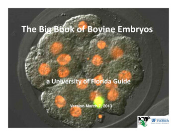
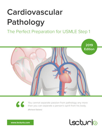
196Panciera & Conferbronchopneumonia), fibrinous pneumonia or pleuropneumonia (also called lobarpneumonia or fibrinous bronchopneumonia), and caseonecrotic pneumonia (alsocalled mycoplasmal pneumonia).4,44 These various types of bronchopneumonia areclassified based on the type of exudation present, initial site of bacterial localizationin the airway, influence of various bacterial virulence factors, host resistance, andrapidity and method of spread of the infection within the lung. In addition, specificbronchopneumonia subclassifications are more commonly associated with specificbacteria.Suppurative Bronchopneumonia (Lobular Bronchopneumonia)This form of pneumonia is the most common form seen in BRD of young dairy calvesand most often associated with P multocida infection, although other respiratorybacteria may also produce the lesion.3,4,13 Suppurative bronchopneumonia occurswhen bronchial colonization of moderately virulent bacteria initiates a suppurativebronchitis with progressive spread along airways resulting in an obviouslybronchiole-centered lesion within each lung lobule. The pneumonia is bilateral,cranioventrally distributed, and moderately firm (Fig. 1). In acute lesions, affectedlobes are fairly uniform in color varying from pink, pink-gray, dark red, red-gray, orgray with minimal to mild interlobular septal edema (Fig. 2). Pleuritis is usually notpresent; however, if present, it consists of small foci of pleural dullness to small clusters of fibrin strands. On cut surface within lobules, there are variable-sized, partiallydiscreet tan to gray foci indicative of a pattern of bronchiolar and peribronchiolarinflammation (Fig. 3). Remaining areas of lobules are pink to dark red representingvarious amounts of inflammation, congestion, and atelectasis. Intrabronchial purulentto mucopurulent exudate may be grossly obvious or in more subtle cases requirea gentle squeeze to express exudate.As bronchopneumonia becomes more chronic, palpation reveals a more lumpydistribution of consolidation with more obvious purulent bronchitis, bronchiectasis,and abscess formation. Inter- and intralobular fibrosis is present. Affected bronchiolesmay be prominent due to peribronchial fibrosis with dilated, mucoid to purulent,exudate-filled lumens. Shrunken, obscured lumens due to bronchiolitis obliteransbecome prominent histopathologically, but those changes are not readily grosslyrecognized. Focal fibrinous to fibrous adhesions may develop between parietal andvisceral pleura especially over underlying abscesses. In chronic cases, A pyogenesis usually cultured particularly from bronchiectatic airways and abscesses, whereasother parts of the lung may yield multiple bacterial species.Fibrinous Pneumonia or Fibrinous Pleuropneumonia (FibrinousBronchopneumonia, Lobar Pneumonia)This form of pneumonia is typical of that produced by M haemolytica and to a lesserextent H somni and is the most common form of acute pneumonia in weaned, stressedbeef cattle (shipping fever).3,4 Fibrinous pneumonia occurs when there is overwhelmingcentrifugal spread of the inflammatory process from the primary locus of colonization inthe bronchioles via extension from lobules into adjacent lobules within the cranioventrallobes. The rapid intra- and interlobular spread of M haemolytica is thought due to theeffects of LPS on vascular integrity, severe cytolytic effects of LKT on resident andresponding leukocytes, and the tissue destructive effects of released enzymes, oxygenradicals, and inflammatory mediators during that process.14,45Fibrinous pneumonia is a bilateral, cranioventrally distributed, very firm, minimallycompressible lung consolidation (Fig. 4). Fibrinous pneumonia is characterized bywide distension of interlobular septa with yellow gelatinous edema or coagulated
Pathogenesis and Pathology of Bovine PneumoniaFig. 1. Bronchopneumonia (suppurative bronchopneumonia, lobular bronchopneumonia).fibrin. Fibrin thrombi may be visible in distended interlobular lymphatics. Consolidatedlobes have a marbled appearance, a descriptive term indicating that each lobule isreasonably uniform in color with a multicolor patchwork or marbled pattern of lobulesranging from pink, pink-tan, dark red, to red-gray (Fig. 5). Large, irregular foci of pinktan coagulation necrosis are frequently within lobules and may involve entire lobules.These necrotic foci are usually outlined by a discrete pale line representing intenseaccumulations of inflammatory cells and result from intralesional vasculitis and thrombosis that develop. Bronchi contain fibrinous coagulum but not frank pus.Fibrinous pleuropneumonia (fibrinonecrotic pneumonia and pleuropneumonia) islikewise a bilateral, cranioventral, marbled pneumonia in which a fibrinous pleuritisFig. 2. Cranioventral, acute, suppurative bronchopneumonia (lobular pneumonia).197
198Panciera & ConferFig. 3. Acute suppurative bronchopneumonia demonstrating bronchiolar and peribronchiolar pattern of inflammation with pus-filled airways.of varying intensity is present (Fig. 6). The presence of pleuritis is an indication of theaggressiveness of the lung infection such that there is extension of infection andinflammation from alveoli to subpleural connective tissue and onto the visceral pleuralsurface. In early stages, pleural granularity, dullness, and fine fibrinous strands arepresent. Later, more intense broad sheets of yellow fibrin may obscure the appearance of the underlying lung. Fibrinous adhesions are likely present between parietaland visceral pleura. Fibrin-rich, yellow fluid within the pleural cavity is highly variablein quantity. In contrast to M haemolytica–associated pneumonia, H somni–associatedfibrinous pneumonia may have accompanying lesions of myocarditis, myocardialinfarction, and fibrinous synovitis.Chronic pneumonic changes similar to those seen in bronchopneumonia occur incattle that survive acute fibrinous pneumonia or pleuropneumonia.Fig. 4. Fibrinous pneumonia and fibrinous pleuropneumonia (lobar bronchopneumonia).
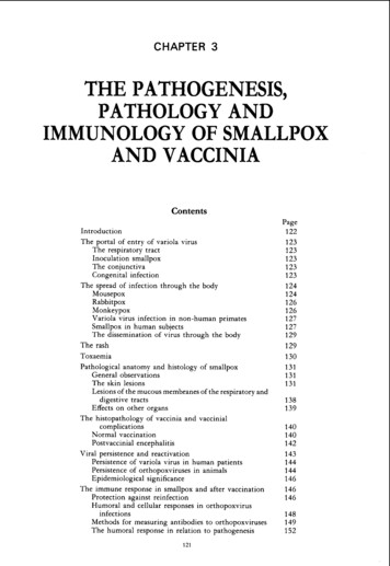
Pathogenesis and Pathology of Bovine PneumoniaFig. 5. Fibrinous pneumonia (lobar pneumonia) demonstrating marbled appearance due topale areas of coagulation necrosis and dark areas of acute pneumonia. Interlobular septaare distended with fibrin-rich exudate.Caseonecrotic Bronchopneumonia (Mycoplasmal Pneumonia)In recent years in North America, this type of pneumonia has been recognized as characteristic of chronic Mycoplasma infection, especially M bovis, which is more virulentthan other bovine Mycoplasma spp.32,46 In many studies of BRD, Mycoplasma sppcan be demonstrated in more than 70% of pneumonia cases usually in combinationwith other bacteria. Sole Mycoplasma spp isolation occurs in less than 20% of BRDcases.41,43Mycoplasma spp colonize the ciliated epithelium of the respiratory tract producinga mild mucopurulent bronchitis and bronchiolitis and, through persistent infection,pulmonary lesions may develop.31 In mild, subacute cases, the main lesion is largeperibronchial lymphocytic cuffs. Systemic spread of M bovis may lead to serofibrinoussynovitis and otitis media.47 Gross examination of the lung at this stage likely revealsno obvious changes or small patchy red to red-gray areas of cranioventral atelectasis.With M bovis infection, however, many calves develop chronic, multifocal, caseousnecrosis within the cranial lung lobes (caseonecrotic bronchopneumonia). Althoughthe pathogenesis of this lesion is still under investigation, bacterial antigen is demonstrated surrounding necrotic foci suggestive of a cause and effect relationship.48–50M bovis virulence factors responsible for this lesion are not currently understood. Atnecropsy, the cranioventral lobes are expanded with firm consolidation, which is oftenFig. 6. Fibrinous pleuropneumonia (lobar pneumonia) with extensive fibrinous exudate.199
200Panciera & ConferFig. 7. Caseonecrotic (Mycoplasma) pneumonia.visibly and palpably nodular (Fig. 7). Clusters of discrete yellow foci of caseousnecrosis are present within lobules, and these foci often range from 1 to 10 mm indiameter (Fig. 8). The surrounding lobular tissue is often mottled to uniformly grayto dark red. In more severe lesions, necrotic foci coalesce and can involve an entirelobule. When the lung is squeezed, the necrotic material falls out as a single massor multiple pieces. Sequestra may be seen. Expressed bronchial exudate may bemucoid to mucopurulent; the character of the exudate is likely determined by the presence or absence of other bacteria. A caseous to caseofibrinous synovitis or otitis mayaccompany the pneumonia.Fig. 8. Caseonecrotic bronchopneumonia typical of M bovis infection. Small (arrow) to large(asterisk) areas of coalescing foci of necrosis are present. (Courtesy of Dr Jeff Caswell,University of Guelph, Guelph, Ontario, Canada.)
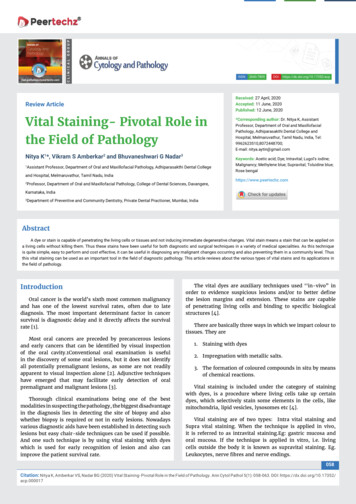
Pathogenesis and Pathology of Bovine PneumoniaINTERSTITIAL PNEUMONIAThe term, interstitial pneumonia, designates a lesion rather than a disease. The lesionmay be acquired by delivery of causative factors through pulmonary circulation or byairways. Whatever the route of exposure, primary damage occurs to structures in alveolar septa (ie, alveolar type I pneumocytes or vascular endothelial cells) and to bronchiolar Clara cells. Subsequently, the fibrin-rich plasma exudation phase occurscreating severe edema and intraalveolar hyaline membranes.3,5 This exudative phaseis followed by type II pneumocyte proliferation, and, with time, alveolar septal fibrosis.Type II pneumocyte hyperplasia can be extensive resulting in alveoli lined by cuboidalepithelium in an acinar or gland-like appearance (sometimes referred to as pulmonaryfetalization or adenomatosis).There are many established causes of interstitial pneumonia, which include, but arenot limited to, various chemical compounds, such as 3-methylindole (3MI); viruses,such as BRSV; migrating parasite larvae; immune mechanisms; toxic gases; andperhaps environmental conditions.3,51,52 In many instances, however, the causativefactor is not known. Interstitial pneumonia occurs in various patterns of lung involvement. The best characterized pathologically is diffuse AIP (Fig. 9), whereas less wellcharacterized is secondary interstitial pneumonia, which is distinguished by coexisting, probably preceding, cranioventral bronchopneumonia with extensive interstitialpneumonia in caudal areas of lungs (Fig. 10). A third, less well-characterized pattern,involves various areas of lung, predominantly caudal, dorsal with intervening areas ofmore normal, but prominently pale, overinflated lobules or groups of lobules, forminga so-called checkerboard pattern (Fig. 11).51Acute interstitial pneumoniaAIP, frequently referred to as acute bovine pulmonary edema and emphysema or, inthe United Kingdom, fog fever,5 occurs in pastured cattle moved to lush greenFig. 9. Acute interstitial pneumonia.201
202Panciera & ConferFig. 10. Secondary interstitial (so-called feedlot interstitial pneumonia).pastures or by exposure to chemicals, including plant toxins, such as Perilla mintketone and 4-ipomeanol from moldy sweet potatoes.53 Clinical signs usually developwithin 2 to 3 weeks of pasture changes or as short as 1 to 2 days after exposure toplant toxins. Classically, movement of cattle to lush green pastures results in excessingestion of L-tryptophan with conversion to 3MI by rumen flora.51,52 Metabolism ofFig. 11. AIP in a feedlot calf. Overinflated cranioventral lobules interspersed with darkerpneumonic lobules (checkerboard). Caudal dorsal lung contains diffuse gray areas of pneumonia. (Courtesy of Dr Amelia Woolums, University of Georgia, Athens, GA.)
Pathogenesis and Pathology of Bovine Pneumonia3MI in bronchiolar Clara cells and type I pneumocytes results in highly activated intermediates that cause pneumocyte necrosis and exfoliation setting in motion the pathologic processes resulting in interstitial pneumonia. This form of AIP is differentiablefrom AIP of feedlot cattle (discussed later) by epidemiology, known causative agents,and lesion characteristics.At necropsy, cattle often have abundant subcutaneous emphysema over the dorsalsubcutis, emphysema within the mediastinum, and frothy edema in the trachea. Lungsare diffusely wet, heavy, and fail to collapse. Lobules tend to be individualized(Fig. 12). Interlobular septa are distended with edema and gas bubbles. Palpablechanges may be subtle to obvious and range from palpably normal to somewhatrubbery in a diffuse distribution. Palpation of single lobules provides critical assessment of lung texture. Parenchyma ranges from pink to tan. Clear, watery, edema fluidreadily exudes from the cut surface.Interstitial pneumonia in feedlot cattleThis form of interstitial pneumonia is a sporadic cause of death in North Americanfeedlot cattle that usually occurs late within the feeding period.6,51,52 Most surveysrecognize the lesion in less than 10% of the total feedlot deaths with occasionallylarger outbreaks reported.6,41,42 Many causes have been investigated, and it ismost likely that interstitial pneumonia in feedlot cattle results from one or combinationsof several mechanisms. As in AIP of pastured cattle, a role for 3MI has been examined.In the feedlot disease, 3MI has been generally discounted because of the sporadicnature of the disease and lack of association with dietary change. In one feedlot study,however, blood levels of a 3MI metabolite were significantly higher in AIP than incontrol cattle suggesting a potential pathogenic role for 3MI.54 Interstitial pneumoniaalso was described with some frequency in feedlot heifers fed melengestrol acetate tosuppress estrus.55 Several studies have investigated the association of BRSV andinterstitial pneumonia with conflicting results; however, this may be because attemptsto culture or demonstrate viral antigen is done using postmortem-collected specimens, and negative results may not accurately account for the virus’ role in pathogenesis.51,54,56,57 Lesions of small airway disease, such as bronchiolar necrosis and, morecommonly, its healing stage, bronchiolitis obliterans, are frequently found histologically in feedlot cases of interstitial pneumonia.4,6,57 It was suggested that feedlot interstitial pneumonia might result from the effects of increased proinflammatory cytokineswithin lungs due to chronic bronchiolar disease, and endotoxin from gram-negativeFig. 12. AIP with lobules clearly separated by interlobular edema. Subtle differences areseen in various lobules. (Courtesy of Dr John King, Cornell University, Ithaca, NY.)203
204Panciera & Conferrespiratory infections may contribute alveolar and bronchiolar damage leading tointerstitial pneumonia.52Gross patterns of interstitial pneumonia i
and Pathology of Bovine Pneumonia Roger J. Panciera, DVM, PhDa, Anthony W. Confer, DVM, PhDb,* Despite availability and use of many bovine respiratory pathogen vaccines and new antimicrobial drugs as well as greater understanding of the pathogenesis of bovine respiratory disease (BR