Transcription
CLINICALGROUPvvISSN:2640-7809DOI: https://dx.doi.org/10.17352/acpReceived: 27 April, 2020Accepted: 11 June, 2020Published: 12 June, 2020Review ArticleVital Staining- Pivotal Role inthe Field of PathologyNitya K1*, Vikram S Amberkar2 and Bhuvaneshwari G Nadar3Assistant Professor, Department of Oral and Maxillofacial Pathology, Adhiparasakthi Dental College1and Hospital, Melmaruvathur, Tamil Nadu, IndiaProfessor, Department of Oral and Maxillofacial Pathology, College of Dental Sciences, Davangere,2*Corresponding author: Dr. Nitya K, AssistantProfessor, Department of Oral and MaxillofacialPathology, Adhiparasakthi Dental College andHospital, Melmaruvathur, Tamil Nadu, India, Tel:9962623510,8072448700;E-mail: nitya.aytin@gmail.comKeywords: Acetic acid; Dye; Intravital; Lugol’s iodine;Malignancy; Methylene blue; Supravital; Toluidine blue;Rose bengalhttps://www.peertechz.comKarnataka, IndiaDepartment of Preventive and Community Dentistry, Private Dental Practioner, Mumbai, India3AbstractA dye or stain is capable of penetrating the living cells or tissues and not inducing immediate degenerative changes. Vital stain means a stain that can be applied ona living cells without killing them. Thus these stains have been useful for both diagnostic and surgical techniques in a variety of medical specialities. As this techniqueis quite simple, easy to perform and cost effective, it can be useful in diagnosing any malignant changes occurring and also preventing them in a community level. Thusthis vital staining can be used as an important tool in the field of diagnostic pathology. This article reviews about the various types of vital stains and its applications inthe field of pathology.IntroductionOral cancer is the world’s sixth most common malignancyand has one of the lowest survival rates, often due to latediagnosis. The most important determinant factor in cancersurvival is diagnostic delay and it directly affects the survivalrate [1].Most oral cancers are preceded by precancerous lesionsand early cancers that can be identified by visual inspectionof the oral cavity.1Conventional oral examination is usefulin the discovery of some oral lesions, but it does not identifyall potentially premalignant lesions, as some are not readilyapparent to visual inspection alone [2]. Adjunctive techniqueshave emerged that may facilitate early detection of oralpremalignant and malignant lesions [3].Thorough clinical examinations being one of the bestmodalities in suspecting the pathology, the biggest disadvantagein the diagnosis lies in detecting the site of biopsy and alsowhether biopsy is required or not in early lesions. Nowadaysvarious diagnostic aids have been established in detecting suchlesions but easy chair-side techniques can be used if possible.And one such technique is by using vital staining with dyeswhich is used for early recognition of lesion and also canimprove the patient survival rate.The vital dyes are auxiliary techniques used ‘‘in-vivo” inorder to evidence suspicious lesions and/or to better definethe lesion margins and extension. These stains are capableof penetrating living cells and binding to specific biologicalstructures [4].There are basically three ways in which we impart colour totissues. They are1.Staining with dyes2. Impregnation with metallic salts.3. The formation of coloured compounds in situ by meansof chemical reactions.Vital staining is included under the category of stainingwith dyes, is a procedure where living cells take up certaindyes, which selectively stain some elements in the cells, likemitochondria, lipid vesicles, lysosomes etc [4].Vital staining are of two types: Intra vital staining andSupra vital staining. When the technique is applied in vivo,it is referred to as intravital staining.Eg: gastric mucosa andoral mucosa. If the technique is applied in vitro, i.e. livingcells outside the body it is known as supravital staining. Eg.Leukocytes, nerve fibres and nerve endings.058Citation: Nitya K, Amberkar VS, Nadar BG (2020) Vital Staining- Pivotal Role in the Field of Pathology. Ann Cytol Pathol 5(1): 058-063. DOI: https://dx.doi.org/10.17352/acp.000017
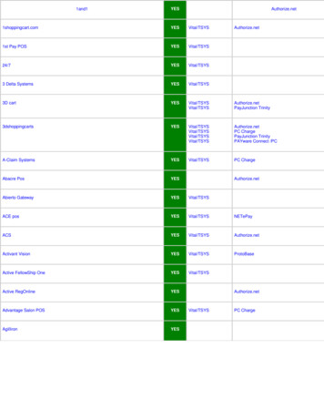
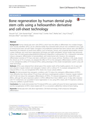
ogy-and-pathologyThus this article gives a brief review on various dyes used Used to stain various cell organelles – Janus Green stainsin detecting oral lesions and their applications in the field ofcell mitochondria, DIOC ( 3,3 Dihexyloxa carbocyaminepathology.Iodide) is used stain endoplasmic reticulum as afluroscent dye, Rhodamine 1,2,3 fluroscenceHistoryAn English anatomist named John Hunter in the eighteenthcentury first introduced vital staining. Most of the stains used Principle: The principle behind vital staining is that particlescells]. The uptake of the dye may be because of phagocytosisand the coloured particle is seen within the cytoplasm of thecell [4].Classification [4]Intravital Staining Toluidine Blue Lugol’s Iodine Methylene Blue Acetic Acid Rose BengalSupravital Staining DIOC for endoplasmic reticulum Janus green stain Rhoda mine 123 Tryphan blue Nile redApplications [5]To determine the cell viability: Evans and Schulman notedthat when leukocytes suspended in solution of tryphan blue,readily took up the dye upon mechanical injury. This is bysupravital staining.Chromoendoscopy: Is a procedure of using vital stains toidentify abnormal mucosa. In patients who are at increased riskToluidine blue (Tb)Discovered by William Henry Perkin in1856, after which itwas primarily used in dye industry. The earliest technique ofvital staining was developed by Paul Ehrlich in 1885, involvedthe immersion of freshly removed tissue in methylated blue.In 1960, Suggestion was that TB may stain malignant epitheliaof the mucous membrane in vivo, whereas normal tissue failedto retain the dye. TB was first applied for in vivo staining byReichart in 1963 for uterine cervical carcinoma in situ [6].Chemistry: Also known as Tolonium chloride, methylanalineaminotoluene which belongs to Thiazine group, which ispartially soluble in water and alcohol.TB is an acidophilic metachromatic dye, that selectivelystains acidic tissue components ( sulfates, carboxylates andphosphate radicals). TB has affinity for nucleic acids andtherefore binds to nuclear material of the tissues with highDNA and RNA content with molecular weight of 305.84Composition: 100 ml of 1% TB contains 1 gm of toluidineblue powder, 10 ml of 1% acetic acid, 4.19 ml of absolute alcoholand 86 ml of distilled water, pH maintained at 4.5.Principle of staining: As the TB has affinity for nucleicacids, the principle is based on the fact that dysplastic andneoplastic cells contain quantitatively more nucleic acids thannormal tissues. Also malignant epithelium may contain widerintracellular canals than that of normal epithelium, which mayfacilitate penetration of the dye .The other proposals about theuptake of TB in dysplastic and carcinomas include the highdensity of nuclear material, loss of cell cohesion and increasedmitosis [6,7].Procedure: detection, in such cases it is applied through spray catheter.Apply 1% of acetic acid for 20seconds to remove ropeysaliva and then apply 1% toluidine blue either withcotton swab or can be given as rinse.Here, the dye stains the glycogen in normal squamousepithelium a dark brown. Areas that are unstained particularlyRinse the mouth twice with water for 20s to removedebris.for squamous cell carcinoma, vital staining with lugol’s iodineis performed at the time of upper endoscopy to aid in cancerNerve fibres: Using Methylene blue by Coers and Woolfwhich is a supravital staining.on living tissue.of coloured matter are engulfed by macrophages [phagocyticUsed to stain various inclusions within the cell : Nile redfluroscence stains lipid vesicles.for vital staining are dyes which are used in textile industry.These dyes are diluted to a large extent before they can be usedformitochondria. Then, 2 rinses with 1% acetic acid are done to reducethose that are larger than 5 mm are likely to be dysplastic /the mechanically retained stain. Finally rinse the mouthmalignant and can be targeted for biopsy. This is an intravitalwith water. Then the color change is assessd [7].staining, which is quick and easy to perform. To obtain cytological details of Protozoa.Interpretation: Positive- dark royal blue; Doubtful- paleblue; Negative- no colour change Figure 1.059Citation: Nitya K, Amberkar VS, Nadar BG (2020) Vital Staining- Pivotal Role in the Field of Pathology. Ann Cytol Pathol 5(1): 058-063. DOI: https://dx.doi.org/10.17352/acp.000017
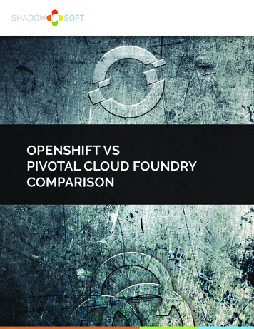
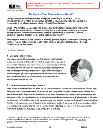
ogy-and-pathologyFigure 1: A. The lesion present over the lateral border of the tongue. B: lesion afterstaining with 1% toluidine blue [9].False positive results are seen with following lesions:Epithelial hyperplasia, hyperkeratotic lesions, inflammatoryand traumatic lesions, hyperplastic candidiasis can retain60% of stain. The decision making can also be attributed tothe experience of the clinician. Repeat the test after 10-14 daysto allow the inflammatory lesions to resolve. This reduces thefalse positivityFalse negative results are recognized in Low grade dysplasia,lichenoid reaction [5,8].Advantages It is inexpensive and simple procedure Can be used for screening high- risk patients who mayhave asymptomatic malignant lesions of oral cavity. Helpful for surgeon in operating room to evaluate freesurgical margins. Toluidine blue staining of oral epithelium will notinterfere with histologic staining or interpretation.Disadvantages Both false positive and false negative results are more. Filiform papillae retain the dye due to high proteinsynthesis rate. TB appears to stain only three to four cells deep and thusreflects changes in the epithelial layer alone. Invadedunderlying tissue is not penetrated by the dye. So theextent of submucosal spread is difficult to appreciate.As the dye reacts with nucleic acid, there is a possiblemutagenic effect of TB when vitally stained cells are exposedto high energy radiation [8,9].Lugol’s iodine(Li)Lugol’s iodine was first made in 1829, which is named afterthe French physician Lugol (1786–1851). Upto the end of 19thcentury, it was used as an antiseptic and disinfectant.Principle: Is based on glycogen content of the cytoplasmand the reaction is known as the iodine–starch reaction,visualized by a colour change. As there is enhanced glycolysisin cancer cells, do not promote the iodine–starch reaction.Hence it appears mustard yellow or saffron colour in dysplasticepithelium whereas normal epithelium appears mahoganybrown colour due to high glycogen content. The vital dye withLugol’s solution is also called Schiller’s test and during mucosalexamination, Lugol’s iodine is applied on the suspicious lesions[5,11].Procedure: Rinse with water/ carbocisteine syrup 250 mg/5ml and dry with a gauze to clear the mucin. Then apply Lugol’siodine until parakeratinized epithelium is stained a brown orblack. After one to two minutes, interpret the stain reaction.Most effective method is to stain the lesion with 3% Lugol’ssolution followed by 5% Lugol’s solution. A study done byNagaraju K (2010) [11] have used both toluidine blue and lugolsiodine, showed that the combination can be used as a pretherapeutic assessment of biologic aggressiveness of lesions.Interpretation:Normalmucosa-brown;mucosa- do not take up stain [11,12] Figure 2.DysplasticMethylene blue (Mb)In 1990, MB has been used to detect gastric, prostrate andbladder cancer. In the diagnosis, the accuracy of MB techniqueis used for identification of intestinal metaplasia, carcinoma ordysplasia.The exact mechanism for the uptake of methylene bluein epithelial tissue may resemble that of toluidine blue in theacidophilic characteristic of cells with abnormal concentrationof nucleic acid [14].Composition Solution A: 1 % methylene blue; 1% malachite ; 0.5%eosinGlycerol &dimethylsulfoxide. Solution B: Pre and post rinse solution had 1% lacticacid; Purified water.Procedure: The application of methylene blue was asfollows:A 5-minute teeth brushing procedure is required beforetesting. Rinse the mouth with Bottle B for 20 seconds toremove food debris and excess saliva.Other names for Lugol’s solution are I2KI (iodine–potassium iodide), markodine, strong solution (systemic),aqueous iodine solution. In 1929, Schiller W described Iodinetest to delineate areas of cervical pre cancers [10].Composition: Iodine – 2 grams; Potassium iodide – 4grams; Distilled water – 100 ccFigure 2: A: lesion present on the buccal mucosa.B: lesion is stained with lugol’s iodine solution. The margins appeared to be illdefined shows prominent after staining [13].060Citation: Nitya K, Amberkar VS, Nadar BG (2020) Vital Staining- Pivotal Role in the Field of Pathology. Ann Cytol Pathol 5(1): 058-063. DOI: https://dx.doi.org/10.17352/acp.000017
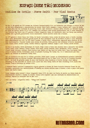
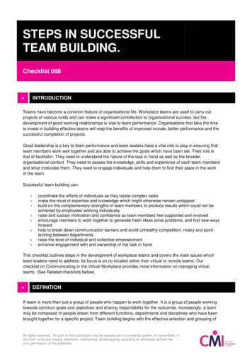
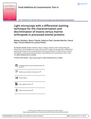
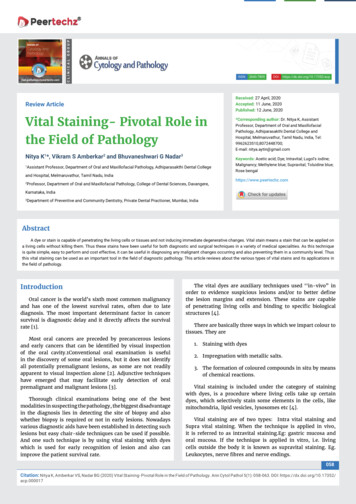
ogy-and-pathologyThe mucosa in the target area was gently dried withgauze and power air spray to ensure that the lesion is notcontaminated with saliva. Gargle and rinse with 1% methyleneblue dye (Bottle A) for 20 seconds, then expectorated. Thenrinse again with Bottle B for 20 seconds to wash out the excessdye Figure 3.Interpretation: The pattern of dye retention- assessed bythe intensity of stain retention on the lesion. Local, stippled,patchy and deep blue stains were marked as positive ( )reaction . Wide, shallow or faint blue stains were marked asnegative (–) reaction [13-15].It plays a major role in demarcating the margins of lesionand thereby enabling clinician to estimate the correct size andextent of the lesion for proper management [15].Acetic acid stainingIn the past, 3-5% acetic acid was used as a vital stainingfor the detection of oral cancers in developing countries.Sankarnarayan, et al. [16], used 3% acetic acid for the detectionof cervical cancer , further Bhalang, et al. [17], used 5% as aclinical marker for detection of oral cancer.Composition: Of 1% acetic acid rinse - 1 ml of glacial aceticacid and 99 ml distilled waterPrinciple: Application of acetic acid causes reversiblecoagulation / precipitation of cellular proteins and causesswelling of the epithelial tissue, particularly abnormalsquamous epithelial areas, dehydration of the cells and it helpsin coagulating and clearing the mucous secretions.The normal squamous epithelium appears pink and thecolumnar epithelium red, due to the reflection of light fromthe underlying stroma, which is rich in blood vessels. If theepithelium contains a lot of cellular proteins, acetic acidcoagulates these proteins, which may obliterate the colour ofthe stroma.The resulting aceto- whitening is seen distinctly ascompared with the normal pinkish colour of the surroundingnormal squamous epithelium [17].Procedure: A piece of gauze soaked with 5% acetic acid isapplied to a cleaned and dried lesion for 60 seconds.After that gauze is removed and characteristic color changecan be noted. Colorchange to opaque white is consideredpositive and transparent white as negative Figure 4.Figure 4: a: Patient with tobacco chewing habit. b: Application of acetic acid wasdone.c: Histopathology revealed hyperkeratosis [18].Advantages: Advantage is associated by false positivereaction with small aphthous-like ulcerated lesion thatmight not routinely be biopsied turned opaque white after theapplication of acetic acid and the histopathologic result wasmoderate epithelial dysplasia.Disadvantages: Burns the oral mucosaFalse positive results: The acetowhite appearance is notunique to early cancer. It is also seen in other conditionswhen increased nuclear protein is present, as in immaturesquamous metaplasia, in healing and regenerating epithelium,inflammation and hyperkeratosis [18].Rose bengal stain(Rb):It is 4, 5, 6, 7 tetrachloro-2, 4, 5, 7 tetraiododerivate offluorescein, that can stain the desquamated ocular epithelialcells. It was observed by Norn, that with an exposure for 1second, RB predominantly stains the cell membranes. Anincreasing the concentration or time of exposure, it producespredominant nuclear staining. RB staining has been evenused to delineate the extent of the corneal and conjunctivalneoplasms. RB is also used to detect oral epithelial dysplasiaand OSCC [19].Norn established the concept that RB stains the cells,wherever there is poor protection of the surface epithelium bythe preocular tear film. This concept has also been extendedto the interpretation of other lesions, such as herpes simplexand zoster, dysplasias or squamous metaplasia of conjunctivalsquamous neoplasms. Mucus/ mucous layer may block theRB uptake. A primary epithelial abnormality i.e, dysplasia,metaplasia, virus infected cells or other forms of epithelialkeratitis, can render the inability of epithelium to interact withthe mucous layer, thus allowing the RB staining [19,20].Procedure: Distilled water is used to rinse the mouth for 1minute in order to clean the lesions.Apply RB solution with cotton with 2 mins. Again thedistilled water is used to rinse the mouth for 1 min to removeexcess stainInterpretation: Pink- positive; no color change- negativeFigure 5.Figure 3: A: Lesion presenting clinically red and white homogenous patch.B: Vital staining with Methylene blue showing deep staining.C: Histopathology image revealed severe dysplasia [14].False negative results could be due to the late clinicalexpression of genetically induced changes in the cells061Citation: Nitya K, Amberkar VS, Nadar BG (2020) Vital Staining- Pivotal Role in the Field of Pathology. Ann Cytol Pathol 5(1): 058-063. DOI: https://dx.doi.org/10.17352/acp.000017
ogy-and-pathologyand flouroscent properties are similar to that of DAPI. This canbe preferentially used with living, unfixed cells [22].The absorption maximum is at 340nm and the emissionmaximum is 450nm, which emits blue colour.Advantages: It is quick and easy to use.Biopsy can be avoided/not required in intra vital staining. Itis a non-invasive procedure. It is inexpensive and can be usedBAfor screening oral cancers. Eg. Toluidine blue test and usingFigure 5: A. Intense positive staining with Rose Bengal stain in a malignant lesionB. Histopathology revealed Well differentiated squamous cell carcinoma [20].or inability of the stain to penetrate the deeper layers ofepithelium showing the dysplastic changes. Sensitivity-93.9%,specificity-73.7% and positive predictive value- 55.4%.lugol’s solution for screening esophageal carcinomas throughChromoendoscopy.It provides more cytological details of the cell, wherespecific organelles can be stained.Disadvantages: Most of these stains are dyes and areA study done by Mittal N stated that RB staining is morepromising in detection of dysplasia in precancerous orextremely toxic, which can result in death, so it has to bediluted to a larger extent.clinically benign lesions, when compared with toluidine blueSome of the dyes like flourochromes are carcinogenic andbecause even the cases of mild dysplasia can be detected by RBmutagenic. So one should avoid breathing any of the powderstaining [20].or allowing it to come in contact with the skin while preparingthe solutions.Supra vital stainsAcridine orange: Acridine orange can be used as vital stain.It stains green for DNA, and orange for RNA under fluorescentmicroscopy. It also stains lysosomes [21].DIOC stain for endoplasmic reticulum: It is a lipophiliccarbocyanine dye, preferentially staining certain membranewith the dye [24].ConclusionAs the oral cavity is easily accessible for the physicalexamination, various non-invasive diagnostic methods canstructures in living cells.It can be used to demonstrate the dynamics of endoplasmicreticulum. It works with living cells. Emission is at about600nm i.e. red colour [22].Rhodamine 123 Fluorescence For Mitochondria:Some vital dyes, like Tryphan blue stain clothes and skinfor a long time. So care should be taken to avoid direct contactbe done to increase the early identification thereby increasingthesurvival rate of patients. The early detection, whichincludes the screening of signs and symptoms will increasethe probability of cure. And one such screening method is vitalThisis termed as a mitochondria specific dye, because thiscationic cyanine dye is accumulated in electrically negativecompartments. Such as mitochondria in healthy cells. Thelarge membrane surface area in the mitochondrial matrix maycontribute to the staining by binding large amounts of thisfluorescent dye [23].Nile red: This dye, which is hydrophobic, has been shownto be highly selective for lipid vesicles in cells. It stains lipidvesicles in the cell and gives red colour under flouro microscopy[23].DAPI: This is used to stain DNA. It is 4,6-diamedine–2-phenylindole. It forms fluorescent complexes with AT-richsequences of double-stranded DNA. It is widely used stable dyefor DNA. The absorption maximum is 344nm and the emissionmaximum at 449nm i.e. it emits blue colour under flouroscentmicroscope. It is especially use to stain fixed cells [22].Hoechst 33342: It is Bisbenzimide H33342. It is a specificstain for AT-rich regions of double stranded DNA, like DAPIstaining using various dyes that can be used even in massscreening is not much used in routine practice. Though thereare various studies done using vital dyes, toluidine blue stainingis most widely practiced in detecting oral potentially malignantlesions and malignancy. Thus, studies using other stains can bedone further to know about its practical applications in variouslesions.References1. Douglas L (2015) Making oral cancer screening a routine part of your patientcare Part 1. BDJ Team 2015: 15066. Link: https://go.nature.com/2BXYqeL2. Lingen MW, Kalmar JR, Karrison T, Speight PM (2008) Critical evaluation ofdiagnostic aids for the detection of oral cancer. Oral Oncol 44: 10-22. Link:https://bit.ly/2UArPC93. Patton LL, Epstein JB, Kerr AR (2008) Adjunctive techniques for oral cancerexamination and lesion diagnosis: a systematic review of the literature. J AmDent Assoc 139: 896-905. Link: https://bit.ly/3hfjUUt4. Outline of methodology (1985) In: Culling CFA, Allison RT, Barr WT. Cellularpathology technique. 4th ed.Butterworth. London 15-26.5. Bagalad BS, Mohan Kumar KP (2013) Vital Staining: Clinical Tool In DiscoveringOral Epithelial Dysplasia And Carcinoma – Overview. J Dent Pract Res 1: 3438. Link: https://bit.ly/2XTuyZi062Citation: Nitya K, Amberkar VS, Nadar BG (2020) Vital Staining- Pivotal Role in the Field of Pathology. Ann Cytol Pathol 5(1): 058-063. DOI: https://dx.doi.org/10.17352/acp.000017
ogy-and-pathology6. Sudheendra US, Sreeshyla HS, Shashidara R (2014) Vital tissue staining inthe diagnosis of oral precancer and cancer: Stains, technique, utility, andreliability. Clin Cancer Investig J 3: 141-145. Link: https://bit.ly/3dXuUE07. Sridharan G, Shankar AA (2012) Toluidine blue: a review of its chemistryand clinical utility. J oral Maxillofac Pathol 16: 251-255. Link: https://bit.ly/3hjXAZN8. Su WW, Yen AM, Chiu SY, Chen TH (2010) A community-based RCT for oralcancer screening with toluidine blue. J Dent Res 89: 933-937. Link: https://bit.ly/2XUs5149. Hegde MC, Kamath MP, Shreedharan S, Dannana NK, Raju MR (2006) Supravitalstaining : It’s role in detecting early malignancies. Indian J Otolaryngol HeadNeck Surg 58: 31-34. Link: https://bit.ly/2MKTfkq10. Petruzzi M, Lucchese A, Baldoni E, Grassi FC, Serpico R (2010) Use of Lugol′siodine in oral cancer diagnosis: An overview. Oral Oncol 46: 811-813. Link:https://bit.ly/3cUkvaI11. Nagaraju K, Prasad S, Ashok L (2010) Diagnostic efficiency of toluidine bluewith lugol’s iodine in oral premalignant and malignant lesions. Indian J DentRes 21: 218-223. Link: https://bit.ly/2B0HvaM12. Xiao T, Kurita H, Shimane T, Nakanishi Y, Koike T (2013) Vital staining withiodine solution in oral cancer: Iodine infiltration, cell proliferation, and glucosetransporter 1. Int J Clin Oncol 18 :792-800. Link: https://bit.ly/3e4KFc813. Fatima S, Basu R, Hallur NH (2016) Lugol’s iodine identifies dysplastic tissuein precancerous lesions: A clinical trial. Ann Maxillofac Surg 6: 172-174. Link:https://bit.ly/2BXZoYr14. Akthar R, Balasundhari S, Mala K, Natarajan S (2013) Methylene Blue as anearly diagnostic marker for oral precancer and cancer. Springer plus 1-5. Link:https://bit.ly/37lcSJc15. Chen YW, Lin JS, Wu CH, Lui MT, Kao SY, et al. (2007) Application of invivo stain of Methylene blue as a diagnostic aid in the early detection andscreening of oral squamous cell carcinoma and precancer lesions. J Chin MedAssoc 70: 497-503. Link: https://bit.ly/2UylTJX16. Sankaranarayanan R, Wesley R, Thara S, Dhakad N, Chandralekha B, et al.(2003) Test characteristics of visual inspection with 4% acetic acid (VIA) andLugol’s iodine (VILI) in cervical cancer screening in Kerala, India. Int J Cancer106: 404-408. Link: https://bit.ly/2XTuzwx17. Bhalang K, Suesuwan A, DhanuthaiK, Sannikorn P, Luangjarmekorn L, et al.(2008) The application of acetic acid in the detection of oral squamous cellcarcinoma. Oral Surg Oral Med Oral Path Oral RadiolEndol 106: 371-376. Link:https://bit.ly/3dTE04I18. Vinuth DP, Agarwal P, Kale AD, Hallikeramath S, Shukla D (2015) Acetic acidas an adjunct vital stain in diagnosis of tobacco-associated oral lesions: Apilot study. J Oral Maxillofac Pathol 19: 134-138. Link: https://bit.ly/37kZhl919. Du GF, Li CZ, Chen HZ, Chen XM, Xiao Q, et al. (2007) Rose bengal stainingin detection of oral precancerous and malignant lesions with colorimetricevaluation: A pilot study. Int J Cancer 120: 1958-1963. Link: https://bit.ly/30yuyzQ20. Mittal N, Palaskar S, Shankari M (2012) Rose bengal staining- diagnostic aidfor potentially malignant and malignant disoders: a pilot study. Ind J Dent Res23: 561-564. Link: https://bit.ly/30ys04W21. Jackson FJ (1961) Supravital Blood Studies, using Acridine OrangeFluoroscence. Blood 17: 643-649. Link: https://bit.ly/3dTTMwu22. Crissman HA, Hoflant MH, Stevenson AP, Wilder ME, Tobert RA (1990)Supravital cell staining with Hoechst 33342 and DiOC5(3). Methods Cell Biol33: 89-95. Link: : https://bit.ly/2MLhUpb23. Darzynkiewicz Z , Coico LS, Melamed RM (1981) Cell Biology Increasedmitochondrial uptake of rhodamine 123 during lymphocyte stimulation (flowcytometry/cycling-noncycling cells/supravital fluorescent probe. Proc NatlAcad Sci USA 78: 2383-2387.24. Fedele S (2009) Diagnostic aids in the screening of oral cancer. Head NeckOncol 30: 1-5. Link: https://bit.ly/2UBhaqZCopyright: 2020 Nitya K, et al. This is an open-access article distributed under the terms of the Creative Commons Attribution License, which permits unrestricted use,distribution, and reproduction in any medium, provided the original author and source are credited.063Citation: Nitya K, Amberkar VS, Nadar BG (2020) Vital Staining- Pivotal Role in the Field of Pathology. Ann Cytol Pathol 5(1): 058-063. DOI: https://dx.doi.org/10.17352/acp.000017
the fi eld of pathology. Review Article Vital Staining- Pivotal Role in the Field of Pathology Nitya K1*, Vikram S Amberkar2 and Bhuvaneshwari G Nadar3 1Assistant Professor, Department of Oral and Maxillofacial Pathology, Adhiparasakthi Dent