Transcription
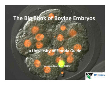
The Big Book of Bovine Embryosa University of Florida GuideVersion March 7, 2013
The purpose of this collection of images of bovine oocytes and embryos is toassist new bovine embryologists with identification of structures encounteredunder the microscope. Unless otherwise stated, all images are of oocytes andembryos from in vitro maturation, fertilization and culture procedures performedin the laboratory of Peter J. Hansen, University of Florida.The images are of oocytes and embryos viewed under inexpensive microscopes,usually dissecting scopes. By collecting typical views as might be seen in manylaboratories, it is hoped to facilitate the process of learning to identify structures.Where known, the name of the person providing the image is on the lower rightcorner of each image.Each image is shown twice – once without a label and a second time with importantfeatures identified. There is no particular order to the images and, in general, themagnification is not shown.The Big Book of Bovine Embryos will be updated as more images are collected.
Let the Viewing Begin
First, oocytes and embryos in developmental sequence(special thanks to Firdous Khan)
Cumulus-Oocyte Complexes at the End of MaturationKatherine Hendricks
Zona pellucida
Matured oocytes(after treatment with hyaluronidase)Zona pellucidaFirdous Khan
Two-cell EmbryosBlastomeresCleavage furrowFirdous Khan
3-4-cell embryosFirdous Khan
5-8 cell embryosFirdous Khan
9-16 cell embryosFirdous Khan
MorulaeMorulaCompact morulaFirdous Khan
BlastocystsBlastocoelFirdous Khan
Expanded Blastocystsner cell massTrophectodermFirdous Khan
Expanded blastocystsFirdous Khan
Hatching BlastocystsFirdous Khan
Hatched BlastocystsFirdous Khan
Random images to help hone your skills at embryo identification
Embryo about 12-cellsMorulaFirdous Khan
A Collection of Oocytes at the End of MaturationAline Bonilla
BlastocystsexpandedNon-expandedMorula/early blastocystAmber Brad
gBlastocystshatchedhatchedhatchedexpandedKatherine Hendricks
Hatching BlastocystsExpanded and justbeginning to hatchHatchingAline Bonilla
Two-cell embryoFour-cell embryoFirdous Khan
Blastocystsexpanded are labeled with EEEEEAline Bonilla
Putative Zygotesfollowing fertilization and removal of cumulus cellsAline Bonilla
Eight-cell embryoFirdous Khan
Expanded BlastocystsAline Bonilla
2-cell embryosFirdous Khan
Hatched blastocystsAline Bonilla
Embryos at Day 7after tblastblastAbnormallyshaped morulaOne cell or unfertilizedAnna Denicol
Hatched blastocyst at Day 8 – this embryowas the abnormal morula in thePrevious slideAnna Denicol
And now, some embryos that have been labeled with specificmarkers .
Differential immunostaining using FITC for TE (CDX2 ) cells and Hoescht 33342 for allnucleiBlue cell with nogreen – ICMGreen cell – TE(note the green cellsare blue alsobut it is difficult tovisualize)Anna Denicol
ContributorsP.J. Hansen (text)Aline BonillaAmber BradAnna DenicolKatherine HendricksFirdous Khan
The Big Book of Bovine Embryos a University of Florida Guide Version March 7, 2013. The purpose of this collection of images of bovine oocytes and embryos is to assist new bovine embryo