Transcription
Applied anatomy of the wrist, thumband handCHAPTER CONTENTSJoints, joint capsules and ligaments . . . . . . . . . . e102Distal radioulnar joint . . . . . . . . . . . . . . . . e102Wrist joint . . . . . . . . . . . . . . . . . . . . . . e103Carpometacarpal joints . . . . . . . . . . . . . . . e104Trapezium–first metacarpal joint . . . . . . . . . . e104Metacarpophalangeal joints . . . . . . . . . . . . . e105Interphalangeal joints . . . . . . . . . . . . . . . . e105Muscles and tendons . . . . . . . . . . . . . . . . . . e105Extrinsic muscles and tendons . . . . . . . . . . . e105Intrinsic muscles . . . . . . . . . . . . . . . . . . e107Nerve structures . . . . . . . . . . . . . . . . . . . . . e109Median nerve . . . . . . . . . . . . . . . . . . . . e109Ulnar nerve . . . . . . . . . . . . . . . . . . . . . e110Radial nerve . . . . . . . . . . . . . . . . . . . . . e111The anatomy of wrist, thumb and hand is complex because ofthe presence of many different functional joints: the distalradioulnar joint, the wrist joint (containing the radiocarpaland the intercarpal joints, the carpometacarpal joints, thetrapezium–first metacarpal joint), the metacarpophalangealjoints and the interphalangeal joints. The contractile structurescan be divided into extrinsic and intrinsic muscles. The former– the long tendons – are quite regularly affected, while thelatter are clinically of less importance.Joints, joint capsules and ligamentsDistal radioulnar jointThe joint is a uniaxial pivot joint. The articular surfaces arebetween the convex head of the ulna and the concave ulnar Copyright 2013 Elsevier, Ltd. All rights reserved.notch of the radius. A fibrocartilaginous articular disc binds thedistal end of the ulnar notch on the radius to the ulnar styloidprocess. This disc is part of the so-called triangular fibrocartilage complex (TFCC) and sits between the ulnar head and theulnar carpus (lunate and triquetrum). Therefore the distalradioulnar joint can be seen as L-shaped: the short leg isbetween radius and ulna and the longer leg between the distalulna and the articular disc (see Putz, Fig. 318). It has a loosecapsule, reinforced with some ligaments, that allows rotationmovement of the radius about the ulna. The two bones areheld together mainly by the triangular fibrocartilage complex(TFCC) and to a lesser degree by the interosseous membraneand the pronator quadratus muscle (Fig. 1).The TFCC contains different parts: (a) the triangular fibrocartilage, which has a central component – the articular disc– and two adjoining ligaments, the dorsal and palmar radioulnarligaments. These take their origin at the dorsal and volar edgesof the sigmoid notch and insert onto the ulnar styloid base(b) a meniscus homologue. This thickened ulnar part of theTFCC, inserts into the triquetral and hamate bones and thebase of the fifth metacarpal; (c) the ulnar collateral ligament;(d) the ulnolunate and ulnotriquetral ligaments; and (e) thesheath of the extensor carpi ulnaris tendon, which is stronglyattached to the posterolateral aspect of the TFCC. The TFCCstabilizes the radioulnar and ulnocarpal joints, transmits anddistributes load from the carpus to the ulna and facilitatescomplex movements to the wrist (Fig. 2).This distal radioulnar joint allows pronation–supinationmovements of the forearm, inevitably in combination withmovement at the upper radioulnar joint, around an axis whichruns through the head of the ulna. The distal end of the radiusmakes a circumferential gliding movement around and in frontof the head of the ulna (see online chapter Applied anatomyof the elbow). Pronation has an amplitude of 85 and isstopped by the radius impacting against the ulna and by tensionin the dorsal radioulnar ligament and the interosseous membrane. Consequently, there is an elastic end-feel. Supinationceases at an angle of 90 when the posterior aspect of the ulnar
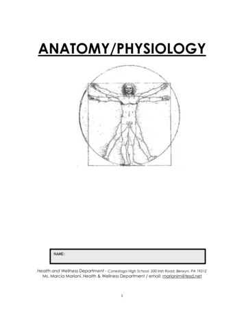
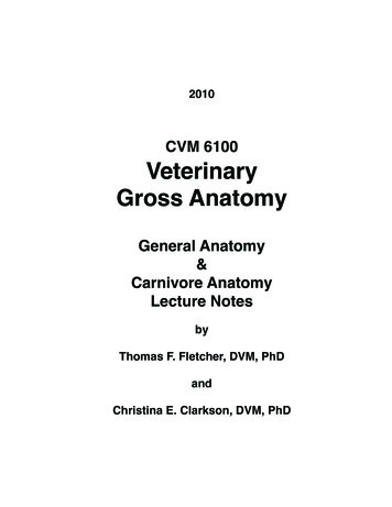
Applied anatomy of the wrist, thumb and k ofhamateTrapeziumCapitatePisiformTubercle of scaphoidFig 1 Right distal radioulnar joint in supination. T, triquetrum; H,hamate; L, lunate; S, scaphoid; U, ulna; UCL, ulnocapitate ligament.TriquetrumLunateFig 3 Palmar carpus of the left hand. From Standring, Gray’s Anatomy,40th edn. Churchill Livingstone/Elsevier, Philadelphia, 2009 with eniscus homologueUlnolunate ligamentPalmar radioulnarligamentArticular discDorsal radioulnarligamentExtensor carpiulnaris sheathRadiusUlnaFig 2 Right distal radioulnar joint in pronation.notch of the radius is brought into contact with the styloidprocess of the ulna through the extensor carpi ulnaris tendon.Its end-feel is also elastic (see Standring, Fig. 50.17).Wrist jointThe wrist joint consists of two rows of carpal bones. Theproximal row contains (from radius to ulna) the scaphoid, Copyright 2013 Elsevier, Ltd. All rights reserved.lunate, triquetral and pisiform bones, and the distal row thetrapezium, trapezoid, capitate and hamate (Fig. 3, see Standring, Fig. 50.6A).The wrist joint is complex: there are two components,proximal and distal (see Putz, Fig. 326). Proximally the distalpart of the radius and the articular disc articulate with theproximal row of carpal bones to form a condylar joint (radiocarpal joint) which has its concave surface at the radial side.The joint moves along two axes: anteroposterior for ulnarand radial deviation, and transverse for flexion and extension(Fig. 4).Distally, the intercarpal joint is between the proximal anddistal rows of bones (Fig. 5), is an open S-shape and acts as ahinge. It should not be considered as an independent jointbecause its function is to augment the mobility of the carpalbones and thus allow greater mobility at the wrist.Movements are restricted by collateral (Fig. 5), palmar anddorsal ligaments (Fig. 6).Radial and ulnar collateral ligamentsThe radial collateral ligament, between the styloid process ofthe radius and the scaphoid bone, is taut when the hand isbrought into ulnar deviation. The ulnar collateral ligament,between the styloid process of the ulna and the triquetraland pisiform bones, is under tension during radial deviation ofthe hand.Dorsal ligamentsThe dorsal wrist ligaments are comparatively thin. They arereinforced by the floor and septa of the fibrous tunnels for thesix dorsal compartments (see below) and have a ‘Z-shaped’configuration. The fibres of the dorsal radiocarpal ligaments arealigned more or less in the same axis as the forearm, those ofe103
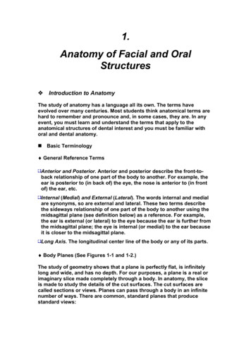
The Wrist, Thumb and Hand21Fig 4 The two axes of movement in the wrist joint:1, anteroposterior axis along which ulnar/radial deviation takesplace; 2, transverse axis along which flexion/extension takes place.Fig 6 The dorsal ligaments. The arrows emphasize the directionof the ligamentous fibres at the joint lines.9ulna. These palmar ligaments are of little clinical importanceand are taut during extension of the wrist.101312143B25A678C11Fig 5 The wrist and collateral ligaments (right hand dorsal view):1, pisiform; 2, triquetrum; 3, lunate; 4, scaphoid; 5, hamate;6, capitate; 7, trapezoid; 8, trapezium; 9, ulna; 10, radius;11, metacarpal; 12, ulnar collateral ligament; 13, radial collateralligament; A, radiocarpal joint line; B, intercarpal joint line;C, carpometacarpal joint line.the intercarpal ligaments are more transverse to the carpusand the carpometacarpal ligaments are longitudinal again (seeStandring, Fig. 50.15B). The pattern and shape of these ligaments is important for treatment with deep transverse frictionwhich should be oblique to the direction of the fibres.Palmar ligamentsOn the palmar aspect of the wrist, a complex system of ligaments joins the different carpal bones to the radius and thee104MovementsThe amplitude of radial deviation is only 15 , whereas ulnardeviation has an amplitude of approximately 45 , the wristbeing held in the neutral position between flexion and extension. Flexion and extension both have a range of 85 , withthe wrist in the neutral position between radial and ulnardeviation.Flexion–extension has an end-feel of an elastic, capsulartype. Radial and ulnar deviation has an end-feel that is quitehard but is clinically of less importance.Carpometacarpal jointsThe joints between the distal row of carpal bones and thesecond to fifth metacarpal bones are scarcely mobile, exceptfor the joint with the fifth metacarpal, which permits slightmovement in a palmar direction necessary to allow oppositionof the fifth finger. They are provided with dorsal and palmarcarpometacarpal ligaments, the fibres of which are in line withthe axis of the metacarpal bones. Intercarpal ligaments ‘tie’ thebones together.Trapezium–first metacarpal jointThe shape of the articular surfaces of both the trapezium andthe first metacarpal bone (‘saddle’) allows movement in two Copyright 2013 Elsevier, Ltd. All rights reserved.
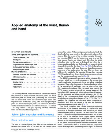
Applied anatomy of the wrist, thumb and handHead offirstmetacarpalShaftBaseFig 8 Flexion and extension of the metacarpophalangeal joint.TrapeziumNutrientforaminaDistally the amplitude of flexion varies between 80 andGroove for 90 , the little finger again being the most mobile. The jointflexor carpi capsules are reinforced by palmar and collateral ligaments.radialisdAPprFig 7 Palmar view of the left trapezium–metacarpal joint. FromThe slight rotatory movements possible at the interphalangeal joints are not important from a clinical point of view.Muscles and tendonsGosling et al, Human Anatomy, 5th edn. Mosby/Elsevier, Philadelphia, 2008 withpermission.directions: abduction–adduction and opposition–reposition(Fig. 7). The joint capsule is slack so as to allow free movementbut strong ligaments control the range.Metacarpophalangeal jointsThe metacarpophalangeal joint of the thumb is different fromthat of the other fingers. It is a hinge and allows only a flexionmovement (80–90 ) and in hypermobile individuals someextension. Two sesamoid bones are often embedded in thepalmar part of the capsule.The metacarpophalangeal joint of each of the second to fifthfingers is a ‘ball-and-socket’ joint with a slack joint capsule,which is reinforced by strong palmar ligaments and collateralligaments (see Gosling, Fig. 3.102). The collateral ligamentsare taut when the joint is flexed.The possible movements are flexion (90 , Fig. 8) and extension (sometimes to 90 , dependent on the laxity of the jointcapsule), as well as abduction and adduction (the index fingerhaving the greatest mobility). The rotatory movements are notclinically important.Interphalangeal jointsThe interphalangeal joints are hinges that allow flexion withoutextension, except in some individuals for the interphalangealjoint of the thumb and the distal interphalangeal joint of thefingers (see Gosling, Fig. 3.102). In the proximal interphalangeal joints, the range of flexion is more than 90 and increasesfrom the index to little finger in which flexion can be 135 ,which allows a ‘fist’ to be made. Copyright 2013 Elsevier, Ltd. All rights reserved.Clinically, the long tendons of the wrist, thumb and fingers –the tendons of the extrinsic muscles – are of more importancethan the small muscles of hand, thenar and hypothenar – theintrinsic muscles.Extrinsic muscles and tendonsAs lesions are usually well localized, a topographical description of the tendinous structures is necessary. An understandingof exactly where they lie and where they insert is important.Dorsal aspectOn the dorsal aspect of the wrist six osteofibrous tunnels canbe distinguished (Fig. 9, see Standring, Fig. 50.14), whichcontain the tendon sheaths of the extensors of the wrist andfingers (Table 1) and the abductor of the thumb.Tunnel 1This contains the tendons of the abductor pollicis longus andthe extensor pollicis brevis (see Standring, Fig. 50.38): The abductor pollicis longus inserts partly at the base ofthe first metacarpal bone, radial to the extensor pollicisbrevis and partly into the trapezium. Its main function isabduction of the thumb, a movement that takes placebetween the trapezium and the first metacarpal. The extensor pollicis brevis inserts at the base of theproximal phalanx of the thumb and is responsible forextension of the thumb. Additionally, because of its closerelationship with the abductor longus, it helps inabduction.Both muscles lie in a common tendon sheath and have a furtherfunction: radial deviation and palmar flexion of the wrist.e105
The Wrist, Thumb and Hand The extensor carpi radialis brevis attaches to the dorsaland radial aspect of the base of the third metacarpalbone. It extends the wrist and brings it back from ulnardeviation into a neutral position.34Tunnel 3Dorsal tubercle5126The third tunnel contains the tendon of the extensor pollicislongus, which runs along the ulnar aspect of the dorsal tubercleof the radius and then deviates 45 towards the thumb. Levelwith the carpus it passes over the tendons of the second tunneland so forms the ulnar border of the anatomical snuffbox. Itthen continues over the dorsal aspect of the thumb towardsthe base of the distal phalanx. Its main function is to extendthe thumb, although is also helps in extension of the wrist andabduction of the thumb.Tunnel 4Five different tendons are found here: the tendon of the extensor indicis proprius and the four tendons of the common extensor digitorum:Fig 9 The extensor tendons and their sheaths (numbers indicatethe tunnels described in the text).Table 1 The extensorsMuscleInnervation The extensor indicis proprius: the fibres of the extensorindicis proprius blend with the dorsal aponeurosis of theindex finger. It extends the index finger. The extensor digitorum: the tendons of the extensordigitorum merge with the dorsal aponeurosis of thesecond to fifth fingers and extend the fingers, especiallythe proximal phalanges. Their secondary functions areextension and ulnar deviation of the wrist.Tunnel 5Peripheral nerveNerve rootAbductor pollicis longusRadialC7–C8Extensor pollicis brevisRadialC7–T1Extensor carpi radialis longusRadialC6–C7This contains just one tendon, the extensor digiti minimi,which runs over the distal radioulnar joint and then parallel tothe fifth tendon of the extensor digitorum into the dorsalaponeurosis of the fifth finger. Its function is identical to thatof the extensor digitorum.Extensor carpi radialis brevisRadialC7Tunnel 6Extensor pollicis longusRadialC7–C8Extensor indicis propriusRadialC6–C8Extensor digitorum communisRadialC6–C8Extensor digiti minimiRadialC6–C8Extensor carpi ulnarisRadialC7–C8The extensor carpi ulnaris lies in a groove at the ulnar aspectof the head of the ulna. It then crosses the lateral aspect ofthe carpus to insert in the dorsal and ulnar aspect of the baseof the fifth metacarpal bone. On account of its position, itfunctions mainly as a strong ulnar deviator of the wrist and alsoacts as an opponent to the abductor pollicis longus. Its extension function is very much secondary.Palmar aspectTogether they form the radial border of the ‘anatomicalsnuffbox’.Tunnel 2The second tunnel contains the tendons of extensor carpiradialis longus and brevis. They diverge level with the carpus: The extensor carpi radialis longus inserts into the dorsaland radial aspect of the base of the second metacarpalbone and combines with the extensor carpi ulnaris toperform dorsiflexion of the wrist. In conjunction with theflexor carpi radialis muscle, it produces radial deviation.e106On the palmar aspect of the wrist lie the tendons of the flexorsof wrist and fingers (Table 2). These are enclosed by the carpaltunnel and the tunnel of Guyon, which are of greatimportance.The carpal tunnelThe palmar aspect of the carpus is concave and covered withthe transverse carpal ligament, also called the flexor retinaculum. An osteofibrous canal is thus formed, of which the palpable boundaries are: proximally the scaphoid and pisiformbones and distally the trapezium and hamate bones (Fig. 10). Copyright 2013 Elsevier, Ltd. All rights reserved.
Applied anatomy of the wrist, thumb and handTable 2 The flexorsMuscleInnervationPeripheral nerveNerve rootFlexor carpi radialisMedianC7–T1Palmaris longusMedianC7–T1Flexor digitorum superficialisMedianC7–T1Flexor digitorum profundusMedianUlnarC7–T1Flexor carpi ulnarisUlnarC7–C8Flexor pollicis longusMedianC7–C82143tendons in two. Their functions are mainly flexion of theproximal phalanges of the fingers and ulnar deviation ofthe wrist. The tendons of the deep flexors of the fingers runthrough the carpal tunnel in a common tendon sheathwith the superficial extensor digitorum and insert at thebase of the distal phalanges of the second to fifth fingers.The muscle flexes the fingers and also the wrist. The flexor carpi ulnaris attaches to the pisiform, whichsome consider a sesamoid bone in the tendon of the flexorcarpi ulnaris. Distal to the pisiform, the tendon dividesinto two ligamentous structures: the pisohamate ligament(which covers the tunnel of Guyon) and the pisiform–fifthmetacarpal ligament. Together with the flexor carpiradialis, the flexor carpi ulnaris flexes the wrist and, withthe extensor carpi ulnaris, performs ulnar deviation of thewrist. The flexor pollicis longus originates with a radial headat the anterior aspect of the radius, just distal to theinsertion of the supinator brevis muscle and also from theinterosseous membrane. There is an additional humeralhead at the medial epicondyle. It runs lateral to the flexorcarpi radialis muscle under which it crosses proximal tothe wrist and then runs in a separate tendon sheaththrough the carpal tunnel to insert at the base of thedistal phalanx of the thumb. It flexes the thumb andslightly flexes and radially deviates the wrist.The tunnel of GuyonFig 10 Boundaries of the carpal tunnel: 1, trapezium;2, scaphoid; 3, hamate; 4, pisiform.This tunnel lies between the pisiform and the hamate bone andis covered by the pisohamate ligament, which is a continuationof the flexor carpi ulnaris tendon. The ulnar nerve and arteryare also enclosed within this tunnel (Fig. 13, see Standring,Fig. 50.46).Intrinsic musclesThe canal lies thus more distally and more towards the ulnaraspect than is often thought – at the heel of the hand.Within the carpal tunnel are found: the median nerve;the tendons of the flexor carpi radialis and the flexor pollicislongus, in separate sheaths; and the superficial and deepflexors of the digits, within a common tendon sheath(Fig. 11).The tendinous structures (Fig. 12, see Standring, Fig. 50.31)are the following: The flexor carpi radialis traverses the carpal tunnel alongthe scaphoid bone and inserts at the palmar aspect of thebase of the second metacarpal. It flexes the wrist andhelps in radial deviation. The palmaris longus muscle is absent in 15% ofindividuals. It is inserted into the palmar aponeurosis inthe palm and helps in palmar flexion of the wrist as wellas tensing the palmar fascia. The tendons of the superficial flexors of the fingers run, in acommon sheath, through the carpal tunnel and insert intothe middle phalanges of the second to fifth fingers; indoing this they separate each of the deep flexor digitorum Copyright 2013 Elsevier, Ltd. All rights reserved.(Table 3)Apart from the dorsal interossei, the intrinsic muscles of thehand (see Putz, Fig. 374) are seldom strained: The thenar muscles: these are clinically less important andinclude the abductor pollicis brevis, opponens pollicis,flexor pollicis brevis with its superficial and deeper headand the adductor pollicis with its transverse and obliquehead. The hypothenar muscles are the abductor digiti minimi,flexor digiti minimi brevis and opponens digiti minimi. The lumbrical muscles: there are four lumbrical muscles.They originate at the radial aspect of the tendons of thedeep flexor digitorum muscle and insert at the dorsalaponeurosis of the fingers and at the joint capsulesof the metacarpophalangeal joints. They flex themetacarpophalangeal joints and extend the interphalangealjoints. The palmar interossei: the three palmar interossei adductthe fingers towards the middle finger, flex themetacarpophalangeal joints and extend the interphalangealjoints of the fingers (Fig. 14).e107
The Wrist, Thumb and HandUlnarnerve andarteryThenarmusclesHypothenarmusclesMedian xortendonsADigitalextensortendonsmlaPFig 11 Proximal view of the right carpal tunnel. From Gosling et al, Human Anatomy, 5th edn. Mosby/Elsevier, Philadelphia, 2008 with permission.The dorsal interosseiThe palmar aponeurosisMore important are the four dorsal interossei. They originatefrom the sides of the five metacarpal bones, each having twoheads, each in the side of two adjacent metacarpal bones (Fig.15). They course towards the proximal phalanges, where theyattach into the extensor aponeurosis of the fingers. The firstdorsal interosseus goes to the radial aspect of the index finger.The second dorsal interosseus goes to the radial aspect of themiddle finger, the third to the ulnar aspect of the middle finger.The fourth dorsal interosseus goes to the ulnar aspect of thefourth finger.The dorsal interossei abduct the fingers away from themiddle finger (Fig. 14). They also flex the metacarpophalangealjoints and extend the interphalangeal joints.The palmar aponeurosis of the hand is a continuation of theflexor retinaculum (the reinforcement of the distal part ofthe antebrachial fascia, also called transverse ligament of thecarpus) and consists of transverse and longitudinal fibres (seeStandring, Fig. 50.4A). The latter are part of the tendonsheaths of the flexor tendons (deep transverse metacarpal ligaments) and also connect to the capsules of the metacarpophalangeal joints. The aponeurosis is also connected to thedeep fascia of the hand, which is attached to its skeleton.Together with the ligaments, septa and fasciae, the palmaraponeurosis forms a functional unit. During a strong grip itfixes the skin of the palm to the metacarpal bones and protectsthe soft tissues of the mid-hand.e108 Copyright 2013 Elsevier, Ltd. All rights reserved.
Applied anatomy of the wrist, thumb and handTable 3 The intrinsic musclesMuscleFig 12 The flexor tendons: 1, flexor carpi radialis; 2, palmarislongus; 3, superficial flexors of the fingers (4 tendons); 4, deepflexors of the fingers (4 tendons); 5, flexor carpi ulnaris; 6, flexorpollicis longus.InnervationPeripheral nerveNerve rootAbductor pollicis brevisMedianC8–T1Oppens pollicisMedianC6–C7Flexor pollicis brevisSuperficial headDeep headMedianUlnarC8–T1C8–T1Adductor pollicisUlnarC8–T1Abductor digiti minimiUlnarC8–T1Flexor digiti minimi brevisUlnarC8–T1Opponens digiti minimiUlnarC8–T1LumbricalsRadial two lumbricalsUlnar two lumbricalsMedianUlnarC8–T1C8–T1Dorsal interosseiUlnarC8–T1Palmar interosseiUlnarC8–T1123Fig 14 (Left) Abduction, four dorsal interossei; (right) adduction,three palmar interossei.nerve becomes compressed in the region of Guyon’s tunnel(see Standring, Fig. 50.45A). Lesions of the radial nerve at thewrist are less common.Fig 13 The tunnel of Guyon: 1, pisiform; 2, hamate; 3, ulnarnerve.Nerve structuresThe three most important nerves in the wrist and hand are themedian, ulnar and radial nerves. The median nerve is mostfrequently affected in the carpal tunnel, whereas the ulnar Copyright 2013 Elsevier, Ltd. All rights reserved.Median nerveThe median nerve enters the hand through the carpal tunnel,in between the tendons of the flexor pollicis longus and flexordigitorum superficialis. Distal to the transverse ligament, thenerve divides into two branches (Fig. 16). A short motorbranch goes to the thenar eminence, where it usually suppliesthe abductor pollicis brevis and opponens pollicis muscles, andsometimes the flexor pollicis brevis and the first and seconde109
The Wrist, Thumb and HandFig 17 The ulnar nerve: dorsal cutaneous branch.Fig 15 The dorsal interossei.Fig 16 The median nerve.Fig 18 The ulnar nerve: palmar branch.lumbrical muscles. The sensory palmar digital branches innervate the palmar surface and the dorsal aspect of the distalphalanges of the thumb, the second and third fingers and theradial half of the fourth finger.distally, the dorsal cutaneous branch arises and supplies theulnar side of the dorsum of the hand (Fig. 17), the dorsalaspect of the fifth finger and the ulnar half of the fourthfinger.The ulnar nerve, together with the ulnar artery, passesbetween the pisiform and the hook of the hamate through thetunnel of Guyon. As it leaves the tunnel it divides into a mainlysensory superficial terminal branch, which supplies the distalulnar border of the palm of the hand and the palmar surfacesof the fifth and ulnar half of the fourth finger (Fig. 18) and adeep terminal branch, which is entirely motor and innervatesnearly all of the small muscles of the hand.Ulnar nerveProximal to the wrist, the palmar cutaneous branch of the ulnarnerve arises and runs across the palmar aspect of the forearmand wrist, outside the tunnel of Guyon, to supply the proximalpart of the ulnar side of the palm. A few centimetres moree110 Copyright 2013 Elsevier, Ltd. All rights reserved.
Applied anatomy of the wrist, thumb and handRadial nerveAt the elbow, the radial nerve divides into the posterior interosseous nerve (a deep motor branch), which innervates theextensor muscles of the wrist and fingers, and the superficialradial nerve (a superficial sensory branch), which lies under thetendon of the brachioradialis muscle. In the hand, the superficial radial nerve divides into terminal digital branches thatsupply the dorsolateral aspect of the hand and the dorsal aspectof the first three and a half fingers, except the distal phalanges(Fig. 19).Fig 19 Terminal digital branches of the superficial radial nerve. Copyright 2013 Elsevier, Ltd. All rights reserved.e111
Gosling et al, Human Anatomy, 5th edn. Mosby/Elsevier, Philadelphia, 2008 with permission. d P A pr. Head of first metacarpal Shaft Base Trapezium Nutrient foramina Groove for flexor carpi radialis. Fig 8