Transcription
Diabetologia (2018) 1-4ARTICLEEpigenetic modifications of the Zfp/ZNF423 gene control murineadipogenic commitment and are dysregulated in humanhypertrophic obesityMichele Longo 1 & Gregory A. Raciti 1 & Federica Zatterale 1 & Luca Parrillo 1 &Antonella Desiderio 1 & Rosa Spinelli 1 & Ann Hammarstedt 2 & Shahram Hedjazifar 2 &Jenny M. Hoffmann 2 & Cecilia Nigro 1 & Paola Mirra 1 & Francesca Fiory 1 &Pietro Formisano 1 & Claudia Miele 1 & Ulf Smith 2 & Francesco Beguinot 1Received: 10 May 2017 / Accepted: 8 September 2017 / Published online: 24 October 2017# The Author(s) 2017. This article is an open access publicationAbstractAims/hypothesis Subcutaneous adipocyte hypertrophy is associated with insulin resistance and increased risk of type 2diabetes, and predicts its future development independent ofobesity. In humans, subcutaneous adipose tissue hypertrophyis a consequence of impaired adipocyte precursor cell recruitment into the adipogenic pathway rather than a lack of precursor cells. The zinc finger transcription factor known as zincfinger protein (ZFP) 423 has been identified as a major determinant of pre-adipocyte commitment and maintained whiteadipose cell function. Although its levels do not change duringadipogenesis, ectopic expression of Zfp423 in non-adipogenicmurine cells is sufficient to activate expression of the geneencoding peroxisome proliferator-activated receptor γ(Pparγ; also known as Pparg) and increase the adipogenicpotential of these cells. We investigated whether the Zfp423gene is under epigenetic regulation and whether this plays aMichele Longo, Gregory A. Raciti and Federica Zatterale contributedequally to this study.Ulf Smith and Francesco Beguinot are joint senior authors.Electronic supplementary material The online version of this article(https://doi.org/10.1007/s00125-017-4471-4) contains peer-reviewed butunedited supplementary material, which is available to authorised users.* Francesco Beguinotbeguino@unina.it1URT Genomics of Diabetes-IEOS, CNR & Department ofTranslational Medicine, Federico II University of Naples,Via Pansini 5, 80131 Naples, Italy2Lundberg Laboratory for Diabetes Research, Department ofMolecular and Clinical Medicine, Sahlgrenska Academy, Universityof Gothenburg, Gothenburg, Swedenrole in the restricted adipogenesis associated with hypertrophic obesity.Methods Murine 3T3-L1 and NIH-3T3 cells were used asfibroblasts committed and uncommitted to the adipocyte lineage, respectively. Human pre-adipocytes were isolated fromthe stromal vascular fraction of subcutaneous adipose tissue of20 lean non-diabetic individuals with a wide adipose cell sizerange. mRNA levels were measured by quantitative real-timePCR, while methylation levels were analysed by bisulphitesequencing. Chromatin structure was analysed by micrococcalnuclease protection assay, and DNA-methyltransferases werechemically inhibited by 5-azacytidine. Adipocyte differentiation rate was evaluated by Oil Red O staining.Results Comparison of uncommitted (NIH-3T3) and committed (3T3-L1) adipose precursor cells revealed that Zfp423 expression increased (p 0.01) in parallel with the ability of thecells to differentiate into mature adipocytes owing to bothdecreased promoter DNA methylation (p 0.001) and nucleosome occupancy (nucleosome [NUC] 1 p 0.01; NUC2p 0.001) in the 3T3-L1 compared with NIH-3T3 cells.Interestingly, non-adipogenic epigenetic profiles can bereverted in NIH-3T3 cells as 5-azacytidine treatment increased Zfp423 mRNA levels (p 0.01), reduced DNA methylation at a specific CpG site (p 0.01), decreased nucleosome occupancy (NUC1, NUC2: p 0.001) and induced adipocyte differentiation (p 0.05). These epigenetic modifications can also be initiated in response to changes in the preadipose cell microenvironment, in which bone morphogeneticprotein 4 (BMP4) plays a key role. We finally showed that, inhuman adipocyte precursor cells, impaired epigenetic regulation of zinc nuclear factor (ZNF)423 (the human orthologue ofmurine Zfp423) was associated with inappropriate subcutaneous adipose cell hypertrophy. As in NIH-3T3 cells, the normal
370ZNF423 epigenetic profile was rescued by 5-azacytidineexposure.Conclusions/interpretation Our results show that epigeneticevents regulate the ability of precursor cells to commit anddifferentiate into mature adipocytes by modulating ZNF423,and indicate that dysregulation of these mechanisms accompanies subcutaneous adipose tissue hypertrophy in humans.Keywords Adipose tissue differentiation . Basic science .DNA methylation . Epigenetic regulation . Human . Insulinsensitivity and resistance . Pathogenic mechanisms .Transcription factors . Weight regulation and obesityAbbreviationsBMP4Bone morphogenetic protein 4MNaseMicrococcal nucleaseMSCMesenchymal stem cellNUC1/2 Nucleosome 1/2qPCRQuantitative real-time PCRSATSubcutaneous adipose tissueSVFStromal vascular fractionWISP2WNT-inducible secreted protein 2ZFPZinc finger proteinIntroductionThe worldwide increase in obesity is a major cause of thecurrent epidemic of type 2 diabetes [1, 2]. However, obesityis not a homogeneous condition. Approximately 10–30% ofobese individuals do not show metabolic complications.These individuals typically have an increased number of smalladipocytes in their subcutaneous adipose tissue (SAT) and lowvisceral and other ectopic fat depots [3–5]. In addition, a similar proportion of non-obese individuals exhibit reduced insulin sensitivity and altered glucose metabolism [6]. At the molecular level, these human phenotypes remain incompletelycharacterised, generating uncertainties on how fat tissue expansion impacts the trajectory to type 2 diabetes.Adipose tissue expansion is usually caused by an increasein adipocyte size (hypertrophy) and/or recruitment of newadipocytes from multipotent mesenchymal stem cells(MSCs) already in the stromal vascular compartment(hyperplasia) [7, 8]. Limited expandability and recruitmentof new cells in SAT leads to prominent adipocyte hypertrophy,which is associated with ectopic accumulation of fat, functional dysregulation of SAT, low-grade chronic inflammation, decreased insulin sensitivity and enhanced oxidative stress[9–11]. In humans, SAT hypertrophy appears to be a consequence of impaired adipocyte precursor cell recruitment intoDiabetologia (2018) 61:369–380the adipogenic pathway rather than a lack of precursor cells[12–15]. Although the underlying molecular mechanismshave only been partially elucidated, current evidence indicatesthat restricted adipogenesis in SAT predicts future development of type 2 diabetes independent of obesity [16]. The present understanding of SAT expansion in human obesity anddiabetes is limited by incomplete understanding of the molecular basis of pre-adipocyte determination [17]. Recently, thezinc finger transcription factor known as zinc finger protein(ZFP) 423 was identified as a major determinant of preadipocyte commitment [17] and maintained white adipose cellfunction [18]. Zfp423 expression is enriched in a number ofadipogenic fibroblast cell lines compared with fibroblasts uncommitted to the adipocyte lineage. Although Zfp423 levelsare essentially unchanged during adipogenesis, ectopic expression of Zfp423 in non-adipogenic murine cells is sufficientto activate expression of the gene encoding peroxisomeproliferator-activated receptor γ (Pparγ; also known asPparg) and increase the adipogenic potential of these cells[17, 19]. Zfp423 knockout mice feature impaired developmentof both white and brown adipose tissue [17, 19].The activity of ZFP423 in adipose precursor cells is repressed by the intracellular and secreted mediator WNTinducible secreted protein 2 (WISP2). WISP2 production issignificantly upregulated in the SAT of individuals with hypertrophic obesity, and is positively correlated to adipose cellsize [20]. In the cytoplasm, WISP2 protein forms a complexwith ZFP423 and prevents its translocation into the nucleus.Bone morphogenetic protein 4 (BMP4), a secreted protein andkey regulator of the commitment of multipotent MSCs to theadipocyte lineage, dissociates this complex, allowing nuclearentry of ZFP423, thereby activating Pparγ transcription andcommitment of precursor cells into the adipocyte lineage [12,20].Several studies have reported that epigenetic regulatorymechanisms are involved in the determination of multipotentprecursor cells to form committed pre-adipocytes and the differentiation of pre-adipocytes to mature adipocytes [21].Bioinformatic analysis of CpG islands in the promoter regionsof obesity-related genes has identified regions with a highdensity of CpGs implicated in adipogenesis and inflammation,such as Pparγ, phosphatase and tensin homologue, leptin andtumour necrosis factor-α [22, 23]. Methylation of these CpGislands influences local chromatin structure and function, andparticipates in regulation of transcriptional activation of genes[24, 25].Elucidation of the molecular mechanisms responsible fortranscriptional regulation of Zfp423 may improve the understanding of restricted adipogenesis in hypertrophic obesity.Here, we investigated whether Zfp423 is epigenetically regulated and whether these events are involved in the restrictedadipogenesis seen in humans with expanded subcutaneousadipose cells.
Diabetologia (2018) 61:369–380MethodsMedia, sera, insulin, TRIzol and SuperScript III were obtainedfrom Invitrogen (San Diego, CA, USA), rosiglitazone fromAlexis (Grünberg, Germany) and 5-azacytidine, 3-isobutyl-1methylxanthine and dexamethasone from Sigma-Aldrich (StLouis, MO, USA). pCpGfree-Lucia, Escherichia coli GT115cells, and Luciferase reporter assay kit were from InvivoGen(San Diego, CA, USA), SYBR Green from Bio-Rad(Hercules, CA, USA) and the DNA Methylation Kit fromZymo Research (Orange, CA, USA). Micrococcal nuclease(MNase), Dam /Dcm Escherichia coli cells andHpyCH4IV, M.SssI, HhaI and HpaII enzymes were obtainedfrom New England Biolabs (Ipswich, WI, USA). The DNAPurification Kit and pGEM-T Easy Vector were fromPromega (Madison, WI, USA), the PCR Purification kit fromQiagen (Hilden, Germany), and the Big Dye Terminatorv3.1Cycle Sequencing Kit from Applied Biosystems (FosterCity, CA, USA).Cell culture and adipocyte differentiation Mouse embryonic fibroblasts (3T3-L1, NIH-3T3) were obtained from theAmerican Type Culture Collection (Manassas, VA, USA).These mycoplasma-free cell lines were grown in DMEM with10% FCS. For adipocyte differentiation, see electronic supplementary material (ESM) Methods.Participants This study is a secondary analysis of participantsfrom the European network on Functional Genomics of Type2 Diabetes (EUGENE2) consortium [26]. Adipose tissuederived stromal vascular fraction (SVF) cells were obtainedfrom 20 healthy, non-obese individuals whose recruitment andclinical phenotyping has previously been described [26]. Thestudy was approved by the appropriate Institutional ReviewBoards. All participants gave informed consent.Adipose tissue biopsies were obtained from abdominalSAT. Following careful dissection, adipose cells were digestedwith collagenase for 45 min at 37 C. After digestion, the suspension was centrifuged to obtain two phases: an upper (mature adipocytes) and a lower (SVF cells) phase. Adipocytesize was measured according to previously described procedures [13, 16]. SVF cells, in which we analysed ZNF423expression, were cultured in DMEM and Ham’s F-12 supplemented with 10% FBS and 0.002 mol/l glutamine as previously reported [13], in order to remove erythrocytes and inflammatory cells.RNA isolation and quantitative real-time PCR RNA wasisolated by TRIzol reagent according to the manufacturer’s protocol. RT-PCR of 1 μg of RNA was performed usingSuperScript III. The cDNA obtained was used as a templatefor quantitative real-time PCR (qPCR), performed in triplicateusing iQ SYBR Green Supermix on an iCycler real-time371detection system (Bio-Rad). Relative quantification of gene expression was relative to the control (equal to 1) and was calculated according to the comparative 2 ΔΔCt method based on thecycle threshold (Ct) values of the target and housekeeping genes.The primers used for Ppia (also known as Cypa), Pparγ2,Fabp4 (also known as Ap2), Glut4 (also known as Slc2a4),Adipoq, Zfp423 and ZNF423 gene expression in qPCR arereported in the ESM Table 1.MNase protection assay Nuclei were isolated from bothNIH-3T3 (5 105 cells) and 3T3-L1 (5 105 cells) anddigested with 200 U of MNase for 20 min at 37 C. The purified DNA was subsequently amplified by qPCR. The percentage of nucleosome occupancy across the analysed regions wasquantified by the 2 ΔCt method using the undigested input as anormalising control, using NuPoP software (available fromhttps://rdrr.io/bioc/NuPoP/; accessed November 2015).DNA methylation assessment Genomic DNA was extractedusing a DNA Purification Kit (Promega). Bisulphite treatmentof extracted genomic DNA was performed using the EZ DNAMethylation Kit (Zymo Research). The bisulphite-convertedgenome was amplified by PCR using bisulphite-specificprimers for Zfp423 and ZNF423 (see ESM Table 1 for theprimers used). Bisulphite genomic sequencing was performedas previously reported [27]. DNA sequencing was performed onan ABI 3500 Automatic Sequencer using Big Dye Terminatorv3.1 (Applied Biosystems). Bioinformatic analysis was carriedout using EMBOSS CpGplot (available from www.ebi.ac.uk/Tools/seqstats/emboss cpgplot/; accessed November 2015).In vitro methylation and luciferase reporter assay The 5′flanking region of Zfp423 ( 1324 to 764) was amplified byPCR and cloned into pCpGfree-Lucia (InvivoGen) luciferasereporter vector. Amplification of the reporter construct wasperformed using Dam /Dcm E. coli cells. These cells werepurchased from the New England Biolabs and are mycoplasma-free. The luciferase reporter vector was in vitro methylatedby incubation with 1 unit/μg of M.SssI enzyme (which methylates all CpGs) or 1 unit/μg of HhaI (which methylates thecytosines of the sequence GCGC) and HpaII enzymes (whichmethylate the cytosines in the sequence CCGG) at 37 C for1 h. Fully methylated, unmethylated and partially methylatedZfp423 reporter vectors were transfected in NIH-3T3 cells. Tonormalise luciferase activity, a control plasmid encoding aRenilla luciferase gene was cotransfected into the cells. After48 h, firefly and Renilla luciferase activity were assayed usinga luciferase reporter assay kit (InvivoGen), according to themanufacturer’s instructions.Site-directed mutagenesis and luciferase reporter assayZfp423 promoter ( 1037/ 1002) was amplified by PCR and
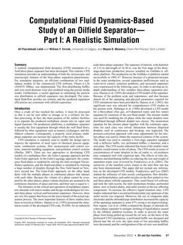
372cloned into the firefly luciferase reporter pCpGfree-promoterLucia vector (InvivoGen). A one-step PCR-based mutagenesis technique was used to generate site-specific mutation [28]and produce a mutated construct. One complementary pair ofprimers was designed that contained the desired mutation,replacing the cytosine at position 1016 with adenine. Thewild-type and mutated constructs were transformed intoE. coli GT115 cells. These cells were purchased fromInvivoGen and are mycoplasma-free. In vitro methylationwas performed using M.SssI methyltransferase following themanufacturer ’s protocol (New England BioLabs).Unmethylated wild-type and mutated constructs were obtained in the absence of M.SssI. Methylation was confirmed byresistance to HpyCH4IV digestion (New England Biolabs).After 48 h, firefly and Renilla luciferase activity were assayedusing a luciferase reporter assay kit, as reported in the previousparagraph.Statistical analysis All experiments were performed threetimes for each determination, and results are shown as mean SD. Values for p between datasets were determined by twotailed, unpaired Student’s t test. Significant p values are indicated as ***p 0.001, **p 0.01 and *p 0.05. Correlationanalysis was calculated using Pearson’s correlationcoefficient.ResultsPromoter methylation reduces Zfp423 expression in NIH3T3 cells Using qPCR, we compared Zfp423 mRNA expression in NIH-3T3 and in 3T3-L1 cells and found it to be barelydetectable in the former and high in the latter (p 0.01) (Fig.1a). Importantly, there was no sequence variation of theZfp423 promoter in NIH-3T3 and 3T3-L1 cells (data notshown), suggesting that the differential expression observedhad to be attributed to other mechanisms, including differentepigenetic profiles. Furthermore, the expression of other keyadipogenic marker genes was strongly silenced in NIH-3T3cells (ESM Fig. 1).To explore this, we subjected the Zfp423 promoter regionto bioinformatic analysis. EMBOSS CpGplot revealed a large560 bp CpGi upstream Zfp423 transcription start site, providing a potential basis for methylation control of Zfp423 expression. We analysed the methylation status of the Zfp423 CpGiby bisulphite sequencing in NIH-3T3 and 3T3-L1 cells, andfound massive demethylation in the latter (methylation:15.3%vs 90.2% in NIH-3T3 cells; p 0.001; Fig. 1b, c).We then cloned the Zfp423 promoter into the luciferasereporter vector (pCpGfree-basic-Lucia), which was eithertreated with M.SssI methylase and fully methylated, or partially methylated using HhaI and HpaII methylases. Digestionwith the methylation-sensitive restriction enzyme HpyCH4IVDiabetologia (2018) 61:369–380enabled control of methylation level in the two conditions(data not shown). Luciferase activity in the constructsharbouring the fully and the partially methylated Zfp423 promoters declined by 80% and 40%, respectively, comparedwith the unmethylated promoter (p 0.001) (Fig. 1d). Theseresults demonstrate that methylation regulates Zfp423 promoter function in vitro.Nucleosome occupancy of Zfp423 promoter is increased inNIH-3T3 compared with 3T3-L1 cells Based on NuPoPanalysis, the Zfp423 promoter exhibited several potential regions where nucleosome positioning featured a high prediction score (Fig. 2a), suggesting that differential Zfp423 expression in NIH-3T3 and 3T3-L1 cells is also accompanied by avariation in nucleosome occupancy. To validate this and assess nucleosome occupancy at the best predicted regions, anMNase protection assay was performed and nucleosome positioning checked in mono-nucleosomal DNA by qPCR (Fig.2b). The percentage of nucleosome occupancy at two suchregions of the Zfp423 promoter was significantly higher inNIH-3T3 cells (nucleosome [NUC]1% occupancy: 72.2 vs51.5 in 3T3-L1 cells, p 0.01; NUC2% occupancy: 94.6 vs46.4 in 3T3-L1 cells, p 0.001; Fig. 2c). No significant difference was observed in the CTRL R (negative control) region, where nucleosome positioning featured a low bioinformatic prediction score. Thus, in these cells, nucleosome occupancy of the promoter inversely correlates with Zfp423 expression, suggesting that dynamic chromatin remodellingmay also contribute to transcriptional regulation.5-Azacytidine enhances Zfp423 expression and allows differentiation of non-adipogenic NIH-3T3 cells To assesswhether DNA methylation also regulates Zfp423 expressionin intact cells, we investigated the ability of the DNA methyltransferase inhibitor 5-azacytidine to remove the transcriptional block imposed on Zfp423 in the NIH-3T3 cells. Incubatingthe cells with 5-azacytidine mainly affected methylation levelat CpG position 1016 (40% methylation in exposed vs 90%in unexposed cells, p 0.01; Fig. 3a, b); this was associatedwith a sixfold increase in Zfp423 mRNA expression (p 0.01;Fig. 3c). However, apart from the 1016 CpG, the overallmethylation profile at the Zfp423 promoter did not change in5-azacytidine-treated cells (data not shown), providing a potential explanation for why mRNA expression levels are stilllower than in 3T3-L1 cells (Fig. 3c). MNase protection studiesrevealed that 5-azacytidine significantly reduced nucleosomeoccupancy at the NUC1 and NUC2 regions (p 0.001; Fig.3d), further underlining the potential role of chromatin remodelling of the Zfp423 regulatory region in transcriptionalregulation.In parallel, 5-azacytidine robustly enhanced expression ofPparγ and the differentiation markers Fabp4, Adipoq andGlut4 after induction of differentiation (Fig. 4a), accompanied
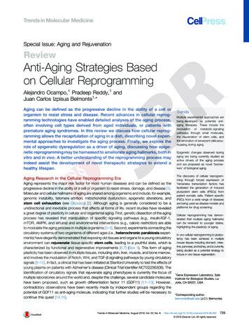
Diabetologia (2018) 61:369–380373Fig. 1 Zfp423 mRNA expression and promoter methylation in NIH-3T3and 3T3-L1 cells. (a) Total RNA was isolated from the cells, and Zfp423mRNA levels were assessed by qPCR. Data normalisation was achievedusing the housekeeping Ppia gene as internal control. Results are themean SD from three independent experiments. Statistical significancewas tested by two-tailed Student’s t test (**p 0.01 vs NIH-3T3). (b, c)Bisulphite sequencing analysis and assessment of Zfp423 promoter methylation of individual methylated CpG sites were compared in NIH-3T3(b) and 3T3-L1 (c) cells (methylation: 90.2 2.1% NIH-3T3,15.3 2.7% 3T3-L1). Each PCR product was subcloned, and ten cloneswere analysed by bisulphite sequencing. Individual CpG sites at theZfp423 promoter, either methylated (black circles) or unmethylated(white circles), were aligned to their sequence position as indicated abovethe panels. Results are the mean SD from three independent experiments. Statistical significance was tested by two-tailed Student’s t test(p 0.001 vs NIH-3T3). (d) Luciferase activity of the in vitrounmethylated, methylated or partially methylated Zfp423 promoter reporter constructs in NIH-3T3 cells. Relative luciferase activity was normalised against the activity of a cotransfected internal vector. Results arethe mean SD of three independent experiments. Statistical significancewas tested by two-tailed Student’s t tests (***p 0.001 vs unmethylatedvector, †p 0.05 vs partially methylated vector)by greater than twofold increased cytoplasmic accumulationof Oil Red O (Fig. 4b, c). Thus, in parallel with transcriptionalactivation, Zfp423 promoter demethylation by 5-azacytidinealso promoted differentiation of the non-adipogenic NIH-3T3cell line.cells enhanced the expression of both Zfp423, by almost fivefold (p 0.001; Fig. 5a), and its downstream target genePparγ (p 0.01; Fig. 5b), while Zfp423 expression was onlyslightly reduced, albeit not reaching significance, by BMP2treatment (p 0.118; Fig. 5a). Bisulphite sequencing revealedthat this BMP4-dependent change was accompanied by analmost threefold reduction in methylation at position 1016in the Zfp423 promoter (p 0.01), reminiscent of that seenwith 5-azacytidine treatment (Fig. 5c, d).We therefore aimed to establish the functional significanceof the 1016 dinucleotide for Zfp423 promoter function bysite-directed mutagenesis. After replacing the cytosine at position 1016 with adenine, the 35 bp fragment ( 1037 to 1002) of the Zfp423 promoter was cloned in a luciferaseBMP4 promotes Zfp423 expression by inducing promoterdemethylationBMPs are members of the transforming growth factor superfamily that play a key role in inducing adipocyte precursor cellcommitment towards the adipogenic lineage; however, themolecular details of their action have been only partially elucidated. Adding BMP4 to the culture medium of NIH-3T3
374Fig. 2 Nucleosome occupancy at the Zfp423 promoter in 3T3-L1 andNIH-3T3 cells. (a) Schematic representation of the regions potentiallyoccupied by nucleosomes at the Zfp423 promoter in NIH-3T3 and 3T3L1 cells. Genomic position: NUC1 chr8:87,960,746–87,960,945; NUC2chr8:87,959,646-87,959,845; CTRL R chr8:87,960,121-87,960,322. (b)Genomic DNA was obtained by lysis of 3T3-L1 and NIH-3T3 cell nucleiand either digested with MNase or left untreated. (c) Percentage of nucleosome occupancy at two bioinformatically identified regions of theZfp423 promoter (NUC1, NUC2) and in the negative control region(CTRL R) was assessed by qPCR. Nucleosome occupancy across theanalysed region was quantified by the 2 ΔCt method using the undigestedinput as normalising control. Black bars, 3T3-L1 cells; white bars, NIH3T3 cells. Results are the mean SD from three independent experiments. Statistical significance was tested by two-tailed Student’s t tests(**p 0.01, ***p 0.001 vs 3T3-L1 cells)reporter vector (pCpGfree-promoter-Lucia). Luciferase activity was assayed in NIH-3T3 cells transfected with either thein vitro methylated or the unmethylated promoter. As shownin Fig. 5e, the 1016 mutation did not affect the activity of theunmethylated Zfp423 promoter. However, it abolished the effect of methylation on promoter silencing, indicating that the 1016 dinucleotide modulates Zfp423 transcription in vitro.Pre-adipocyte ZNF423 expression correlates with maturesubcutaneous adipose cell size in humans To explore thesignificance of Zfp423 expression in the development andfunction of human adipose tissue, we analysed transcriptionof human ZNF423 (the human orthologue of Zfp423) [29] inpre-adipocytes from the SVF of 20 healthy, non-obese individuals. Participants were recruited as previously described[26] (see Table 1 for their clinical characteristics). The sizeDiabetologia (2018) 61:369–380Fig. 3 Effect of 5-azacytidine on Zfp423 mRNA expression and nucleosome occupancy in NIH-3T3 cells. Cells were cultured in medium supplemented with 5-azacytidine (AZA). (a, b) Bisulphite sequencing analysis of DNA methylation and percentages of methylated 1016 CpGpositions in the Zfp423 promoter in NIH-3T3 and NIH-3T3 AZA cells(methylation: 1016 CpG 90 10% NIH-3T3, 40 10% NIH-3T3 AZA). Each PCR product was subcloned, and ten clones were analysedby bisulphite sequencing. The methylation profile of each CpG site in theZfp423 promoter, either methylated (black circles) or unmethylated(white circles), is aligned to its sequence position. Results are the mean SD from three independent experiments. Statistical significance wastested by two-tailed Student’s t tests (p 0.01). (c) Expression ofZfp423 mRNA was measured by qPCR in NIH-3T3, NIH-3T3 AZAand 3T3-L1 cells. Data normalisation was achieved using the housekeeping Ppia gene as internal control. Results are the mean SD from threeindependent experiments. Statistical significance was analysed by twotailed Student’s t tests (**p 0.01, ***p 0.001 vs NIH-3T3 cells). (d)Percentage of nucleosome occupancy was analysed by qPCR in the previously identified NUC1 and NUC2 regions of the Zfp423 promoter.Assessment was performed in NIH-3T3 cells in the absence (black bars)or presence (white bars) of AZA. Results are the mean SD from threeindependent experiments. Statistical significance was tested by two-tailedStudent’s t tests (***p 0.001 vs NIH-3T3 without AZA)of the subcutaneous adipose cells varied over a broad rangeeven in these non-obese individuals, consistent with differentadipogenic potential of the precursor cells. ZNF423 mRNAwas detectable in cells from all donors, and, importantly,
Diabetologia (2018) 61:369–380Fig. 4 Effect of 5-azacytidine on NIH-3T3 cell adipogenic differentiation. NIH-3T3 cells were cultured in the absence (black bars) or presence(white bars) of 5-azacytidine (AZA). Gene expression and lipid accumulation were assessed 8 days after the induction of adipocyte differentiation. (a) The relative mRNA levels of Pparγ, Fabp4, Adipoq and Glut4were determined by qPCR. Black bars, NIH-3T3 cells; white bars, NIH3T3 AZA cells. Data normalisation was performed using the housekeeping Ppia gene as internal control. Results are the mean SD of threeindependent experiments. Statistical significance was established by twotailed Student’s t tests (*p 0.05, **p 0.01, ***p 0.001 vs NIH-3T3without AZA). (b) At day 8, cells were fixed and stained with Oil Red O.Representative microphotographs are shown at two different magnifications ( 20, 10); scale bars, 50 μm. (c) Quantification of Oil Red Ostaining. Results are the mean SD of three independent experiments.Statistical significance was tested by two-tailed Student’s t tests(*p 0.05 vs NIH-3T3 without AZA)expression in pre-adipocytes exhibited a significant negativecorrelation with the size of mature subcutaneous adipose cellsfrom the same individuals (Fig. 6; r 0.5258, p 0.05).Thus, low ZNF423 expression in adipose precursor cells is amarker of the donor’s subcutaneous adipocyte cell size, andthus adipogenesis and the development of inappropriate375adipose cell hypertrophy, and associated with an insulinresistant phenotype.The central enhancer CpGi at the human ZNF423 locusfeatures 80% homology in mammals [29] and, based onsite-specific mutagenesis studies in leukaemia cells, has beenshown to be relevant for the functional regulation of both αand β ZNF423 promoters [29]. In human pre-adipocytes, weobserved that ZNF423α was the predominant isoform, whileZNF423β was barely detectable (ESM Fig. 2).To further explore the mechanisms determining ZNF423expression in SVF pre-adipocytes, we performed bisulphitesequencing of the entire enhancer CpGi in three individualswho had the smallest sized subcutaneous adipocytes, and anequal number of individuals exhibiting the largest adipocytesize. All individuals were non-obese and had a similar BMI.This revealed massively increased methylation levels at twosubregions of the CpG enhancer island in the latter individuals(Fig. 7a). The R2 subregion showed 90% methylation atCpG dinucleotides 30 and 33 in participants with subcutaneous adipocyte hypertrophy, compared with 20% in thosewith smaller adipocytes (Fig. 7b). The R3 subregion revealedthat methylation at CpG nucleotides 54, 55, 56, 57, 60 and 62is 90% and 45% respectively, in individuals with and without adipose cell hypertrophy. In addition, 5-azacytidine treatment of pre-adipocytes from individuals with adipose cellhypertrophy led to a greater than twofold increase in expression of ZNF423 (Fig. 7c) and its downstream target genePPARγ (Fig. 7d). 5-Azacytidine strongly decreased the methylation level at CpG positions 54, 55, 56, 57, 60 and 62 of theR3 subregion, consistent with the important role of theseCpGs in regulating
inducible secreted protein 2 (WISP2). WISP2 production is significantly upregulated in the SAT of individuals with hy-pertrophic obesity, and is positively correlated to adipose cell size [20]. In the cytoplasm, WISP2 protein forms a complex with ZFP423 and prevents its transloca