Transcription
SLS Symposium onComplimentary and ExtendedAnalysesTuesday, July 3, 201210:00 to 12:15, WBGB/01910:00 Light and neutron spectroscopies applied to strongly correlated metalsJohan Chang, N.B. Christensen, S. M. Hayden, C. Monney, J. Mesot, M. Shi, Ch.Niedermayer, H. Rønno and, T. Schmitt10:30 Third Generation of On-axis in situ Optical Spectroscopy: Extendingthe Scope of Macromolecular CrystallographyFlorian Dworkowski, G. Pompidor,V . Thominet, C. Schulze-Briese and M. Fuchs11:00 Coffee11:15 High Spatial Resolution Quantitative Chemical Imaging byComplementary TechniquesH. A. O. Wang, D. Grolimund, L. R. Van Loon, C. N. Borca, J. Shaw-Stewart, P.Karvinen and D. Günther11:45 Accessing reciprocal space with hard X-ray grating interferometryPeter Modregger, M. Kagias, F. Scattarella, B. R. Pinzer, C. David, R. Bellotti,and M. Stampanoni
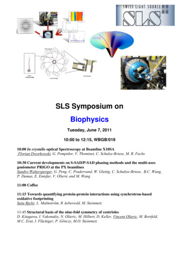
Light and neutron spectroscopies applied tostrongly correlated metalsJ. Chang1,2, N.B. Christensen3, S. M. Hayden4, C. Monney2, J. Mesot1,2, M. Shi2, Ch.Niedermayer5, H. Rønnow2, T. Schmitt2.1Institut de la materière complexe, Ecole Polytechnique Fedérale de Lausanne(EPFL), CH-1015 Lausanne, Switzerland.2Paul Scherrer Institut, Swiss Light Source, CH-5232 Villigen PSI, Switzerland3Department of Physics, Technical University of Denmark, DK-2800 KongensLyngby, Denmark.4H. H. Wills Physics Laboratory, University of Bristol, Bristol, BS8 1TL, UnitedKingdom.5Laboratory for Neutron Scattering, Paul Scherrer Institut, CH-5232 Villigen PSI,Switzerland.Metals have played an important role in the past millenniums of human history.Periods, like the bronze and iron age, have been named after the most importantmetals used at the time. Harnessing semiconductors like silicon has enabled thecreation of computers and revolutionized our daily life. The impact of materialresearch is therefore also reflected in Nobel prize nominations. In the past couple ofdecades, more than a handful of Nobel prizes have been awarded to the discovery orunderstanding of metal physics such as superconductivity (1987, 2003), colossalmagnetoresistance (2007), quantum hall effect (1998), and most recently thediscovery of graphene (2010). Landau theory of Fermi liquids and its notion ofquasiparticles underlies much of our understanding of how electron interactionsaffect the properties of a metal. There is, however, a large class of so-called stronglycorrelated electron systems that defy a description within the Landau Fermi liquidconcept. In this talk I will discuss how new insights into these strongly correlatedmetals can emerge from complementary spectroscopy techniques that can beperformed at the Swiss light source (SLS) and neutron spallation source (SINQ). Inparticular spin and charge excitations of these metallic materials will be discussed.Figure 1: ARPES spectra and analysis (J. Chang et al. PRB 2008) recorded on the stronglycorrelated metal La2-xSrxCuO4 with x 0.145.
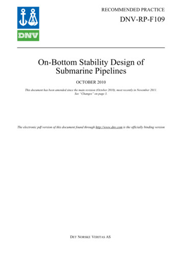
Third Generation of On-axis in situ Optical Spectroscopy -Extending the Scope of Macromolecular CrystallographyFlorian Dworkowski*, Guillaume Pompidor*, Vincent Thominet*, Clemens Schulze-Briese , MartinFuchs** Swiss Light Source, Paul Scherer Institut, 5232 Villigen, Switzerland DECTRIS Ltd, 5400 Baden, Switzerlandflorian.dworkowski@psi.chX-ray diffraction based structure determination of biological macromolecules is one of the fundamentaltools of a structural biologist. However, the electron density maps obtained by this method are limited toelucidate the three-dimensional structure of a macromolecule, but do not yield information on, for example,the chemical state of co-factors, the redox state of metal centers or disulphides, or the identity of boundligands. Many of these observations can be directly or indirectly linked to X-ray radiation induced damageto the sample, one of the central limitations of synchrotron x-ray diffraction data collection [1]. To be able tobetter quantify the extent of this effect and obtain additional complementary data on the sample, the on-axisgeometry for in-situ combination of optical spectroscopic methods with the diffraction experiment has provenhighly effective [2].At beamline X10SA at the Swiss Light Source (SLS) we now are commissioning the third generation of anon-axis multi-mode micro-spectrophotometer (SLS-MS3). It is fully integrated into the new D3 experimentalendstation and designed to remain always online at the beamline, thereby strongly increasing users'acceptance for spectroscopic radiation controls by dramatically reducing setup times. Since the SLS-MS2instrument started user operation in 2011 we now support UV/Vis, Fluorescence and also Raman andResonance Raman spectroscopy. With the new instrument we introduced a new modular concept whichmakes it even more accessible to users with a structural biology background to use all those possibilities.We present quantitative mappings of the on-axis sampling geometry to demonstrate its advantages foraligning the X-ray and optical beams and thereby sampling volumes. Especially in combination with the highsampling rate provided by the PILATUS 6M pixel detector, this setup is also ideal for kinetic crystallographyexperiments with observation of reactions on the minute timescale.We show applications of UV/Visspectroscopy and resonant and non-resonant Raman spectroscopy toward monitoring the photo reductionof metal centers, ligand abstraction and bond breakage. Recently the methodology proved also very usefulfor determination of specific reaction mechanisms inside of proteins, showing unexpected photo-switchingbehavior and metal coordination [3,4].Figure 1: Evolution of the Micro-Spectrophotometer: SLS-MS (UV/Vis), SLS-MS2.6 (UV/Vis, rRaman,Raman, Fluorescence), SLS-MS3 (full endstation integration).[1] Garman, E. F. and M. Weik. Journal of Synchrotron Radiation (2011), 18(3): 313-317[2] R. L. Owen, A. R. Pearson, et al., Journal of Synchrotron Radiation (2009), 16, 173-182[3] Regis Faro, A., P. Carpentier, et al., Journal of the American Chemical Society (2011) 133(41), 16362-16365[4] He, C., M. R. Fuchs, et al., Angewandte Chemie International Edition (2012), 51(18), 4470-4473
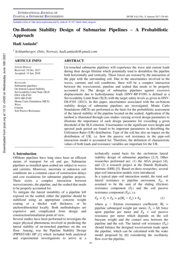
High Spatial Resolution Quantitative Chemical Imaging by ComplementaryTechniquesH. A. O. Wang1,5, D. Grolimund1, L. R. Van Loon2, C. N. Borca1,J. Shaw-Stewart3, P. Karvinen4, D. Günther51microXAS Beamline Project, Swiss Light Source, Paul Scherrer Institute2Laboratory for Waste Management, Paul Scherrer Institute,3Materials Group, General Energies Department, Paul Scherrer Institute4Laboratory for Micro- and Nanotechnology, Paul Scherrer Institute5Trace Element and Micro Analysis Group, ETH ZurichA broad range of scientific and industrial research applications require quantitativechemical images with high spatial resolution, e.g. for the identification of chemicalprocesses or reaction rate quantification. Trace elements quantification is normally adifficult task in X-ray fluorescence analysis (XRF), also inevitable in high fluxSynchrotron-based microXRF (SR-microXRF). However, the high spatial resolution andhigh throughput of SR-microXRF are advantageous over other chemical analysistechniques. In this work, we combined it with laser ablation inductively coupled plasmamass spectrometry (LA-ICPMS), which provided quantitative output on trace elements,but with a reduced spatial resolution.We applied this complementary approach on a test sample to investigate themigration of a Cs contamination plume into a heterogeneous geological material(Opalinus clay rock). Both techniques yield 2D chemical images which correlatedbetween the two techniques (Figure 1). Quantitative LA-ICPMS images were utilized to‘cross-calibrate’ the qualitative SR-microXRF results, which resulted in high spatialFig. 1 High spatial resolution chemical imaging of a Cs contaminationplume in a heterogeneous geological medium.
resolution quantitative 2D chemical images.The importance of micro-/nano- properties and chemical relativities in macroscopicmaterials is gaining more and more recognition. The developing need to give a detailedpicture of such (sub-)microscopic structures and features does thrust the developmentof quantitative chemical analysis with advanced spatial resolution. Recent developmentof hard X-ray SR-nanoXRF allows chemical imaging with approximately 200nanometers spatial resolution by a pair of high-efficiency linear zone plates. In parallel,the aerosol transport systems used for LA-ICPMS were studied in detail and optimizedaccordingly. These modifications provide now the capabilities to ablate materials at aspatial resolution close to 1µm, while maintaining access to trace elementconcentrations. The complementary application of these two “sub-micron” techniqueswas tested on micro-structured thin film pattern. Highly correlated chemical imagesprovide evidence of successful high-resolution 2D quantitative chemical imaging basedon cross-calibration using microLA-ICPMS and SR-nanoXRF.References:H.A.O. Wang, D. Grolimund, L.R. Van Loon, K. Barmettler, C.N. Borca, B. Aeschlimann,D. Günther, Quantitative Chemical Imaging of Element Diffusion into Heterogeneous Media Using LaserAblation Inductively Coupled Plasma Mass Spectrometry, Synchrotron Micro-X-ray Fluorescence, andExtended X-ray Absorption Fine Structure Spectroscopy, Anal. Chem., 83, (2011) 6259-6266.H.A.O. Wang, D. Grolimund, L.R. Van Loon, K. Barmettler, C.N. Borca, B. Aeschlimann,D. Günther, High Spatial Resolution Quantitative Imaging by Cross-calibration Using Laser AblationInductively Coupled Plasma Mass Spectrometry and Synchrotron Micro-X-ray Fluorescence Technique,CHIMIA, 66, No 4, (2012) 223-228.
Тема, Тип, UIDAccessing reciprocal space with hard X-ray grating interferometryP. Modregger1,2, M. Kagias3, F. Scattarella4,5, B. R. Pinzer1, C. David6, R. Bellotti4,5, and M.Stampanoni1,31Swiss Light Source, Paul Scherrer Institut, Villigen, SwitzerlandSchool of Biology and Medicine, University of Lausanne, Lausanne, Switzerland3Institute for Biomedical Engineering, UZH/ETH Zürich, Zürich, Switzerland4Università degli Studi di Bari “Aldo Moro”, Bari, Italy5Istituto Nazionale Fisica Nucleare, Sezione di Bari, 70125 Bari, Italy6Laboratory for Micro- and Nanotechnology, Paul Scherrer Institut, Villigen, Switzerland2peter.modregger@psi.chX-ray grating interferometry (GI) constitutes phase contrast imaging technique that provides aparticular high sensitivity towards electron density variations in the sample. This characteristicrenders GI especially suitable for imaging soft tissue material and, thus, GI has experienced anincreasing interest from researchers in the bio-medical field [1]. Up to now, X-ray imaging with GIwas regarded to deliver information only in real space. Recently, we have demonstrated that theultra-small angle X-ray scattering (USAXS) distribution [2] and, thus, reciprocal space informationcan be accessed with GI as well, and this even simultaneously with real space sensing. This wasperformed by introducing an alternative approach to the contrast formation process of GI and usingthe appropriate data analysis procedures for the experimental data. In doing so, we increased thenumber of complementary image modalities provided by GI from previously three to hundreds.Compared to established techniques for reciprocal space mapping such as scanning smallangle X-ray scattering (SAXS), USAXS with GI is able to finely resolve reciprocal space around itsorigin. This implies that USAXS with GI delivers information, which is complementary totechniques like SAXS. However, due to the small sample to detector distance the relationshipbetween the USAXS signal, which we defined as the scatter signal at the detector, and thereciprocal space of the sample is not straight forward. As a consequence we have experimentallyobserved the occurrence asymmetric scattering distributions. Further, the comparably smallmomentum transfer indicates that multiple scatter events have to be taken into account if GIUSAXS is to be combined with tomography. In the presentation, we will discuss our recenttheoretical investigations into these challenges and show our first experimental examples.Fig. 1: Scatter images of a human calcified heart valvederived nodule. Each image shows the relative scatteringstrength for different scattering angles.References[1] B. R. Pinzer, M. Cacquevel, P. Modregger, S.A. McDonald, J.C. Bensadoun, T. Thuering, P.Aebischer, M. Stampanoni, NeuroImage (2012) 61, 1336-1346.[2] P. Modregger, F. Scattarella, B. R. Pinzer, C. David, R. Bellotti, and M. Stampanoni , Phys.Rev. Lett. (2012) 108, 048101.1
High Spatial Resolution Quantitative Chemical Imaging by Complementary Techniques H. A. O. Wang 1,5, D. Grolimund 1, L. R. Van Loon 2, C. N. Borca 1, J. Shaw-Stewart3, P. Karvinen 4, D. Günther 5 1microXAS Beamline Project, Swiss Light Source, Paul Scherrer Institute 2Laboratory for Waste Management, Paul