Transcription
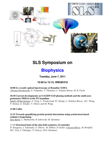
SLS Symposium onBiophysicsTuesday, June 7, 201110:00 to 12:15, WBGB/01910:00 In crystallo optical Spectroscopy at Beamline X10SAFlorian Dworkowski, G. Pompidor, V. Thominet, C. Schulze-Briese, M. R. Fuchs10:30 Current developments on S-SAD/P-SAD phasing methods and the multi-axesgoniometer PRIGO at the PX beamlinesSandro Waltersperger, G. Peng, C. Pradervand, W. Glettig, C. Schultze-Briese, B.C. Wang,P. Dumas, E. Ennifar, V. Olieric and M. Wang11:00 Coffee11:15 Towards quantifying protein-protein interactions using synchrotron-basedoxidative footprintingSaša Bjeliċ, L. Malmström, R Aebersold, M. Steinmetz11:45 Structural basis of the nine-fold symmetry of centriolesD. Kitagawa, I. Vakonakis, N. Olieric, M. Hilbert, D. Keller, Vincent Olieric, M. Bortfeld,M.C. Erat, I. Flückiger, P. Gönczy, M.O. Steinmetz
In crystallo optical Spectroscopy at Beamline X10SA.F. Dworkowski1, G. Pompidor1, V. Thominet1, C. Schulze-Briese1,2, M. R. Fuchs112Paul Scherrer Institute, Swiss Light Source, MX Group, CH-5232 Villigen PSI, SwitzerlandDECTRIS Ltd., Neuenhoferstrasse 111, CH-5400 Baden, Switzerlandflorian.dworkowski@psi.chX-ray diffraction based structure determination of biological macromolecules is one of thefundamental tools of a structural biologist. However, this method is limited to elucidatethe atomic structure of a macromolecule, but does not yield any information about itselectronic structure. By combining optical spectroscopy with X-ray diffraction, it becomespossible to investigate not only atomic but also vibrational and electronic parameters ofthe molecule of interest, yielding important complementary data. This data can beutilized, for example, to verify the redox state of a metal center contained in the protein,observe the amount of radiation damage the sample is subjected to during diffractionmeasurements, confirm correct binding of ligands or even selectively trap enzymereaction intermediates in the crystal [1].This approach, however, is only useful when both methods are applied to the samesingle protein crystal. At the SLSpectroLab at beamline X10SA we thus integrated amultimode, on-axis, on-line micro-spectrophotometer into the high resolution X-raydiffraction end station [2]. This setup allows for closely interweaved acquisition ofdiffraction on spectroscopic data without compromising sample integrity. Currently wesupport measurement of UV-Visible absorption and fluorescent spectroscopic data aswell as resonant and non-resonant Raman scattering.Here we show recent improvement of the instrument and the capabilities it offers on theexample of monitoring specific photo-reduction of disulphide bonds in protein crystalsduring diffraction measurements.References1.2.Pearson, A.R. and R.L. Owen, Combining X-ray crystallography and single-crystalspectroscopy to probe enzyme mechanisms. Biochemical Society Transactions, 2009. 037(2):p. 378-381.Owen, R.L., et al., A new on-axis multimode spectrometer for the macromolecularcrystallography beamlines of the Swiss Light Source. J Synchrotron Radiat, 2009. 16(Pt 2): p.173-82.
Current developments on S-SAD/P-SAD phasing methods and the multi-axesgoniometer PRIGO at the PX beamlinesSandro Waltersperger1), Guanya Peng1), Claude Pradervand1), Wayne Glettig5), ClemensSchultze-Briese2), B.C. Wang3), Philippe Dumas4), Eric Ennifar4), Vincent Olieric1) and MeitianWang1)1) Swiss Light Source at Paul Scherrer Institute, 5232 Villigen PSI, Switzerland2) DECTRIS Ltd., 5400 Baden, Switzerland3) Dept. of Biochemistry and Molecular Biology and SER-CAT, Univ. of Georgia, Athens, GA306024) Insitut de Biologie Moleculaire et Cellulaire, Strasbourg, France5) CSEM, Neuchâtel, SwitzerlandThe usage of the small but significant anomalous signal of intrinsic sulfur atoms of proteins can offerdistinct advantages over derivatization based phasing methods and has become very popular during thepast years (S-SAD). The same applies for the structure determination of nucleic acids but usingphosphorous atoms. Studies on data-redundancy, data-quality, X-ray wavelength and resolution cut-offshave been performed to date and state the current limitations of this method. The requirement of highlyredundant data usually collected with relatively low-energy X-rays entails strong absorption effects andradiation damage, thereby causing inaccuracy in data processing, scaling and phasing.We aim to further develop S-SAD and P-SAD protocols for crystals showing mid-range diffractionbetween 2.3 and 2.8 Å resolution. Promising results applying optimized measurement protocols havebeen obtained and will be presented as well as new hardware beamline developments. These invoce thenovel multi-axes goniometer PRIGO (Parallel Robotics Inspired Goniometer, Figure 1) that allows tocollect data with true redundancy and Bijvoet pairs collected at the same image (to minimize radiationdamage effects) and the PILATUS detector to reduce the signal-to-noise ratio to detect very smallintensities.These optimized measurement protocols will be very useful for general users at macromolecularcrystallography beamlines to deal with challenging S-SAD/P-SAD datasets.Figure 1: The multi-axes goniometer PRIGO installed at X06DA
YNCHROTRON-BASEDOXIDATIVEFOOTPRINTINGS. Bjeliċ1, L. Malmström2,, R Aebersold2 ,M. Steinmetz11Biomolecular Research, Paul Scherrer Institute, Villigen2Department of Biology, ETHZ, ZurichProtein-protein interactions are central in all of biology butare difficult to study quantitatively under native conditions.Synchrotron-based oxidative footprinting is a proxy for measuringsolvent exposure and splits water into hydroxyl-radicals that oxidizethe solvent-accessible amino acids. Changes in solvent-exposure canhence be detected by measuring the relative level of oxidation, whichin turn make it possible to in interaction.We used synchrotron-based oxidativefootprinting to oxidize solvent exposedamino acids of a homo-dimer, EB1c, in adose-dependent manner. The samples weredigested using trypsin and the resultingpeptides were analyzed using s technology.The gproteinproteininteractionsquantitatively as wellas protein motions in ahigh-throughput mode as the method is highly automated usingrobotics. Sample demand is 2-3 order smaller compared to X-ray or NMRmethods. It works in minimally perturbed systems, as no chemicalsbesides water are needed and the proteins are measured in solution.
Structural basis of the nine-fold symmetry of centriolesDaiju Kitagawa1,5, Ioannis Vakonakis2,4,5, Natacha Olieric3,5, Manuel Hilbert2,3,5,Debora Keller1, Vincent Olieric2, Miriam Bortfeld3, Michèle C. Erat4,Isabelle Flückiger1, Pierre Gönczy1,5, Michel O. Steinmetz3,51SwissInstitute for Experimental Cancer Research (ISREC), EPFL, Lausanne, SwitzerlandLight Source, Paul Scherrer Institut, Villigen PSI, Switzerland3Biomolecular Research, Paul Scherrer Institut, Villigen PSI, Switzerland4Department of Biochemistry, University of Oxford, Oxford, United Kingdom5These authors contributed equally to this work2Swissvincent.olieric@psi.chThe centriole and the related basal body is an ancient organelle characterized by auniversal nine-fold radial symmetry and which is critical for generating cilia, flagella andcentrosomes. The mechanisms directing centriole formation are not understood andrepresent a fundamental open question in biology since its discovery almost fifty years agowith the advent of electron microscopy. Here, we demonstrate that the centriolar proteinSAS-6 forms rod-shaped homodimers that interact through their N-terminal domains toform oligomers. We establish that such oligomerization is essential for centriole formationin C. elegans and human cells. We further generate a structural model of the relatedprotein Bld12p from C. reinhardtii, in which nine homodimers assemble into a ring fromwhich nine coiled-coil rod domains radiate outwards. Moreover, we demonstrate thatrecombinant Bld12p self-assembles into structures akin to the central hub of the cartwheel,which serves as a scaffold for centriole formation. Overall, our findings establish astructural basis for the universal nine-fold symmetry of centrioles [1].Reference:[1] Kitagawa D., Vakonakis I., Olieric N., Hilbert M., Keller D., Olieric V., Bortfeld M., Erat M.C., Flückiger I.,Gönczy P., Steinmetz M.O. (2011) Structural basis of the nine-fold symmetry of centrioles. Cell, 144(3):364-75 (Must read article in Faculty of 1000)
In crystallo optical Spectroscopy at Beamline X10SA. F. Dworkowski1, G. Pompidor1, V. Thominet1, C. Schulze-Briese1,2, M. R. Fuchs1 1 Paul Scherrer Institute, Swiss Light Source, MX Group, CH-5232 Villigen PSI, Switzerland 2 DECTRIS Ltd., Neuenhoferstrasse 111, CH-5400 Baden, Switzerland florian.dworkowski