Transcription
JOURNAL OF ORAL & MAXILLOFACIAL RESEARCHBaziar et al.Endodontic Management of a Mandibular First Molar withFour Canals in a Distal Root by Using Cone-Beam ComputedTomography: a Case ReportHani Baziar1, Farzaneh Daneshvar1, Abbas Mohammadi2, Hamid Jafarzadeh3Department of Endodontics, Faculty of Dentistry, Zahedan University of Medical Sciences, Zahedan, Iran.Department of Oral and Maxillofacial Radiology, Faculty of Dentistry, Zahedan University of Medical Sciences, Zahedan,Iran.3Dental Research Center, Department of Endodontics, Faculty of Dentistry, Mashhad University of Medical Sciences,Mashhad, Iran.12Corresponding Author:Hamid JafarzadehFaculty of Dentistry and Dental Research CenterP.O. Box: 91735-984, Vakilabad Blvd, MashhadIranPhone: 98-511-8829501Fax: 98-511-8829500E-mail: hamid j365@yahoo.com; JafarzadehBH@mums.ac.irABSTRACTBackground: Aberrations in the root canal anatomy are clinically challenging for clinicians. Mandibular first molars usuallyhave 2 roots and 3 or 4 canals, but various combinations may exist. A distal root with three canals is rare and its incidencein literature is about 0.2 - 3%. As a diagnostic tool, cone-beam computed tomography (CBCT) may be a better choice fordiagnosis of extra roots or canals comparing to conventional radiography.Methods: An endodontic management of a mandibular first molar with six canals was performed. CBCT was used to confirmthe diagnosis and to understand the morphology of the canals.Results: Evaluation of the axial and coronal slices of CBCT images confirmed the presence of 2 roots and 6 canals. The distalroot had four distinct root canal orifices with two apical foramens, being described as type XIV canal configuration.Conclusions: Dentists should be aware of unexpected canal morphology when performing endodontic treatment. The presentcase demonstrated the use of CBCT in diagnosis and negotiation of extra canals in a mandibular first molar.Keywords: anatomy; cone-beam computed tomography; molar; tooth root; tooth canal.Accepted for publication: 17 March 2014To cite this article:Baziar H, Daneshvar F, Mohammadi A, Jafarzadeh H. Endodontic Management of a Mandibular First Molar with Four Canalsin a Distal Root by Using Cone-Beam Computed Tomography: a Case Report.URL: 5ht.pdfdoi: chives/2014/1/e5/v5n1e5ht.htmJ Oral Maxillofac Res 2014 (Jan-Mar) vol. 5 No 1 e5 p.1(page number not for citation purposes)
JOURNAL OF ORAL & MAXILLOFACIAL RESEARCHINTRODUCTIONThe main objective of the root canal therapy isthorough debridement of the root canal spacefollowed by complete obturation for creating thethree-dimensional seal [1]. According to Nair [2],one of the main reasons associated with unsuccessfultreatment is the survival of microorganisms within theroot canal system. Vertucci [3] reported a substantialnumber of failures related to aberrant anatomy,such as missing canals. Therefore, a comprehensiveknowledge of the root canal anatomy and itsmorphological variations is crucial for successfultreatment [4].Over the years, there have been numerous studiesdescribing the internal morphology of teeth, includingmandibular first molar [5-10]. The majority ofmandibular first molars are two-rooted with twomesial and one or two distal canals. The major variantof the root canal system of mandibular first molar isthe presence of a middle mesial canal with 1 - 15%incidence [6]. However, three canals have also beenreported in the distal root [7,8] with an incidence of0.2 - 3% [9]. The management of a mandibular firstmolar with four distal canals in two separate distalroots was reported by Ghoddusi et al. [10].Since the introduction of CBCT, this threedimensional technique has been established asimportant tool for diagnosis and treatment planningin an increasing number of fields in dentistry [11,12].CBCT has the ability of overcoming the limitations ofconventional radiography such as three-dimensionalevaluation of the complex canal anatomy duringendodontic treatment [13]. An important benefit ofCBCT is in diagnosis of extra roots or canals. Tu etal. [14] showed a higher prevalence of extra rootsin the mandibular first molars assessed by CBCT incomparison to conventional radiography.This case report presents the successful, non-surgicalendodontic treatment of a mandibular first molar withfour canals in a distal root diagnosed by cone-beamcomputed tomographic evaluation.CASE DESCRIPTION AND RESULTSA 42-year-old male patient was referred to the dentaloffice with chief complaint of severe spontaneouspain in the left mandibular first molar. The patient’smedical history was non-contributory.The clinical examination showed extensive amalgamrestoration of this tooth. Vitality tests (cold by icestick, heat by warm instrument, and electric 5n1e5ht.htmBaziar et al.by a pulp tester) were negative; however, the toothwas tender to percussion. Periapical radiographicexamination revealed a deep restoration near distalpulp horn with no signs of periapical radiolucency andaberrant anatomy (Figure 1A). The clinical diagnosisof necrotic pulp with acute apical periodontitis wasmade, and root canal therapy was planned.The patient was anesthetized with 2% lidocainewith 1:80,000 epinephrine. After rubber damisolation, endodontic access cavity was made. Thepulp chamber was repeatedly flushed with 5%sodium hypochlorite to remove necrotic tissue andmicroorganisms. Inspection of the pulp chamberrevealed four canal openings in the distal root andtwo in the mesial root. In the distal root, the thirdand fourth canals were located between distobuccaland distolingual canals. The working lengths wereestablished with an electronic apex locator (Root ZX,Morita, Tokyo, Japan) and a radiograph was taken(Figure 1B). The presence of six canals was confirmedand pulpectomy was performed.To confirm this unusual canal anatomy and tounderstand its configuration, CBCT imaging of thetooth was performed. Calcium hydroxide was placedas temporary dressing and the access cavity wassealed by zinc oxide-eugenol cement. After obtainingthe informed consent from the patient, CBCT of themandible with the focus on the left mandibular firstmolar was performed (Vatech, PaX-Reve 3D plus, 5.5cm field of view, and voxel size of 0.08 mm). Axial,coronal, and sagital CBCT slices revealed six canals(four in the distal root and two in the mesial root) inthe referred tooth (Figure 1C - E).At the next visit, the patient was asymptomatic.Root canal preparation was performed with ProTaperrotary instruments (Dentsply Maillefer, Ballaigues,Switzerland) in crown-down technique. Irrigationwas performed with 2.5% sodium hypochlorite duringinstrumentation, followed by 17% EDTA. After a finalrinse with normal saline, canals were dried with sterilepaper points (Ariadent, Tehran, Iran) and obturatedwith gutta-percha (Ariadent, Tehran, Iran) and AHPlus sealer (Dentsply, Maillefer, Konstanz, Germany)using cold lateral condensation technique (Figure 1F).Then, the patient was refereed for crown restoration.Six months after the endodontic treatment, the patientwas asymptomatic (Figure 1G).DISCUSSIONThe diagnosis and treatment of extra roots or canalsin mandibular first molars is definitely an endodonticchallenge. A comprehensive understanding ofJ Oral Maxillofac Res 2014 (Jan-Mar) vol. 5 No 1 e5 p.2(page number not for citation purposes)
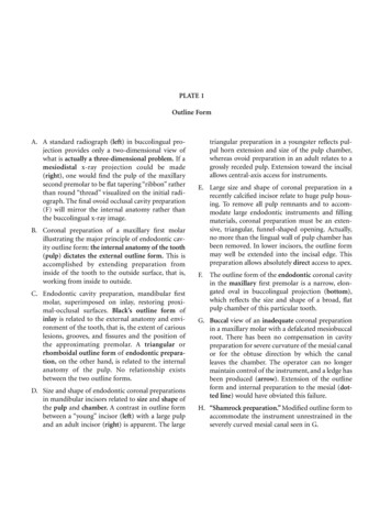
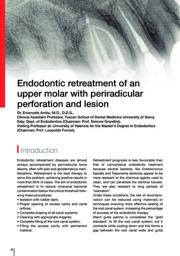
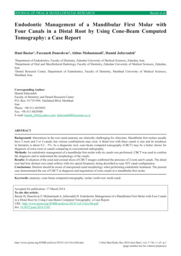
JOURNAL OF ORAL & MAXILLOFACIAL RESEARCHBaziar et al.ABCDEFGFigure 1.A Preoperative periapical radiograph of a left mandibular first molar.B Periapical radiograph showing the working length determination of six root canals.C Enlarged axial CBCT image section at the mid-root level showing 4 canals in thedistal root and 2 canals in the mesial root.D Enlarged coronal section of the distal root showing the configuration of the four canals.E Enlarged sagital section of the distal root showing the overlapping of the four canals.F Postoperative periapical radiograph showing all root canals.G Six months follow-up of the root canal treatment.the most common root canal configuration and itsvariations is essential to achieve long-term success ofthe endodontic treatment. Hoen and Pink [15] reported42% incidence of missed root or canals in the teeththat needed retreatment. Thus, complete debridementand obturation of the root canal system is an utmostimportant procedure in endodontics.Diagnostic imaging is an important tool for locatingcanal orifices including a careful examination of thepulp chamber floor with a sharp explorer, stainingwith 1% methylene blue dye, sodium hypochloritetesting (Champagne bubble test), and visualizingcanal bleeding points. Magnifier loupes and operatingmicroscope would also increase the location of hiddencanals [16].Anatomical variations in the anatomy of the distalroot of mandibular molars may be identified throughcareful evaluation of multiple angled pretreatmentradiographs. Periapical radiographs produce only atwo-dimensional image. Thus, they are decreased invalue in cases of aberrant anatomy [17]. However,it should be noted that significant constraint inconventional periapical radiography is that it producesa two-dimensional image of a three-dimensionalobject. So, periapical radiographs are of limited valuein cases with complex anatomy [18]. Recently, CBCThas been used in endodontics for the evaluation 1e5ht.htmthe root canal anatomy. An advantage of the computedtomography (CT) scanning over the conventionalradiograph is that it permits the operator to look atmultiple sections of the roots and their canals [19].Nance et al. [20] reported that the detection of canalsincreased significantly by CT scan compared withconventional radiography.In this case report, CBCT imaging was used fora better understanding of the complex root canalanatomy. Evaluation of the axial and coronal slices ofCBCT images confirmed the presence of 2 roots and6 canals. The distal root had four distinct root canalorifices with two apical foramens, which could bedescribed as type XIV canal configuration, accordingto Sert and Bayirli [21].Treatment of extra canals may be challenging toeach endodontist or general practitioner; however,the inability to find and properly treat the canals maycause failure. With advance diagnostic aids such asCBCT, these challenges may be overcome.CONCLUSIONSDentists should be aware of unexpected canalmorphology when performing endodontic treatment.The present report demonstrated the use of a cone-J Oral Maxillofac Res 2014 (Jan-Mar) vol. 5 No 1 e5 p.3(page number not for citation purposes)
JOURNAL OF ORAL & MAXILLOFACIAL RESEARCHbeam computed tomography examination as a tool forthe diagnosis and negotiation of extra canals in thedistal root of a mandibular first molar.Baziar et al.ACKNOWLEDGESTATEMENTANDDISCLOSUREThe authors wish to thank the members of theDepartment of Oral and Maxillofacial Radiology andDepartment of Endodontics (Faculty of Dentistry,Zahedan University of Medical Sciences) for theirassistance. The authors report no conflicts of interestrelated to this 15.16.17.18.19.20.Vertucci FJ. Root canal morphology and its relationship to endodontic procedures. Endod Topics 2005;10:3-29.[doi: 10.1111/j.1601-1546.2005.00129.x]Nair PN. On the causes of persistent apical periodontitis: a review. Int Endod J. 2006 Apr;39(4):249-81. Review.[Medline: 16584489] [doi: 10.1111/j.1365-2591.2006.01099.x]Vertucci FJ. Root canal anatomy of the human permanent teeth. Oral Surg Oral Med Oral Pathol. 1984 Nov;58(5):589-99.[Medline: 6595621]Krasner P, Rankow HJ. Anatomy of the pulp-chamber floor. J Endod. 2004 Jan;30(1):5-16. [Medline: 14760900][doi: 10.1097/00004770-200401000-00002]Sperber GH, Moreau JL. Study of the number of roots and canals in Senegalese first permanent mandibular molars. IntEndod J. 1998 Mar;31(2):117-22. [Medline: 9868938] [doi: 10.1046/j.1365-2591.1998.00126.x]Baugh D, Wallace J. Middle mesial canal of the mandibular first molar: a case report and literature review. J Endod. 2004Mar;30(3):185-6. Review. [Medline: 15055441] [doi: 10.1097/00004770-200403000-00015]Stroner WF, Remeikis NA, Carr GB. Mandibular first molar with three distal canals. Oral Surg Oral Med Oral Pathol.1984 May;57(5):554-7. [Medline: 6587303]Beatty RG, Interian CM. A mandibular first molar with five canals: report of case. J Am Dent Assoc. 1985 Nov;111(5):76971. [Medline: 3864840]Kottoor J, Sudha R, Velmurugan N. Middle distal canal of the mandibular first molar: a case report andliterature review. Int Endod J. 2010 Aug;43(8):714-22. Epub 2010 May 19. Review. [Medline: 20491988][doi: 10.1111/j.1365-2591.2010.01737.x]Ghoddusi J, Naghavi N, Zarei M, Rohani E. Mandibular first molar with four distal canals. J Endod. 2007 Dec;33(12):14813. Epub 2007 Oct 22. [Medline: 18037064] [doi: 10.1016/j.joen.2007.08.018]Jeger FB, Lussi A, Bornstein MM, Jacobs R, Janner SF. [Cone beam computed tomography in endodontics: a review fordaily clinical practice]. Schweiz Monatsschr Zahnmed. 2013;123(7-8):661-8. Review. German. [Medline: 23966013]Hassan BA. Reliability of periapical radiographs and orthopantomograms in detection of tooth root protrusion in themaxillary sinus: correlation results with cone beam computed tomography. J Oral Maxillofac Res. 2010 Apr 1;1(1):e6.[Medline: 24421962] [doi: 10.5037/jomr.2010.1106] [PMC free article: 3886038]Durack C, Patel S. Cone beam computed tomography in endodontics. Braz Dent J. 2012;23(3):179-91. Review.[Medline: 22814684] [doi: 10.1590/S0103-64402012000300001]Tu MG, Liu JF, Dai PW, Chen SY, Hsu JT, Huang HL. Prevalence of three-rooted primary mandibular first molars inTaiwan. J Formos Med Assoc. 2010 Jan;109(1):69-74. [Medline: 20123588] [doi: 10.1016/S0929-6646(10)60023-X]Hoen MM, Pink FE. Contemporary endodontic retreatments: an analysis based on clinical treatment findings. J Endod.2002 Dec;28(12):834-6. [Medline: 12489654] [doi: 10.1097/00004770-200212000-00010]Jain S. Mandibular first molar with three distal canals. J Conserv Dent. 2011 Oct;14(4):438-9 9. [Medline: 22144821][doi: 10.4103/0972-0707.87223] [PMC free article: 3227299]Holtzman L. Root canal treatment of mandibular second premolar with four root canals: a case report. Int Endod J. 1998Sep;31(5):364-6. [Medline: 9823141] [doi: 10.1046/j.1365-2591.1998.00166.x]Gupta S, Jaiswal S, Arora R. Endodontic management of permanent mandibular left first molar with six rootcanals. Contemp Clin Dent. 2012 Apr;3(Suppl 1):S130-3. [Medline: 22629055] [doi: 10.4103/0976-237X.95124][PMC free article: 3354780]Patel S, Dawood A, Whaites E, Pitt Ford T. New dimensions in endodontic imaging: part 1. Conventionaland alternative radiographic systems. Int Endod J. 2009 Jun;42(6):447-62. Review. [Medline: 19298577][doi: 10.1111/j.1365-2591.2008.01530.x]Nance R, Tyndall D, Levin LG, Trope M. Identification of root canals in molars by tuned-aperture computed tomography.Int Endod J. 2000 Jul;33(4):392-6. [Medline: 11307216] [doi: org/JOMR/archives/2014/1/e5/v5n1e5ht.htmJ Oral Maxillofac Res 2014 (Jan-Mar) vol. 5 No 1 e5 p.5(page number not for citation purposes)
JOURNAL OF ORAL & MAXILLOFACIAL RESEARCHBaziar et al.21. Sert S, Bayirli GS. Evaluation of the root canal configurations of the mandibular and maxillary permanentteeth by gender in the Turkish population. J Endod. 2004 Jun;30(6):391-8. [Medline: 15167464][doi: 10.1097/00004770-200406000-00004]To cite this article:Baziar H, Daneshvar F, Mohammadi A, Jafarzadeh H. Endodontic Management of a Mandibular First Molar with Four Canalsin a Distal Root by Using Cone-Beam Computed Tomography: a Case Report.J Oral Maxillofac Res 2014;5(1):e5URL: 5ht.pdfdoi: 10.5037/jomr.2014.5105Copyright Baziar H, Daneshvar F, Mohammadi A, Jafarzadeh H. Published in the JOURNAL OF ORAL &MAXILLOFACIAL RESEARCH (http://www.ejomr.org), 1 April 2014.This is an open-access article, first published in the JOURNAL OF ORAL & MAXILLOFACIAL RESEARCH, distributedunder the terms of the Creative Commons Attribution-Noncommercial-No Derivative Works 3.0 Unported License, whichpermits unrestricted non-commercial use, distribution, and reproduction in any medium, provided the original work and isproperly cited. The copyright, license information and link to the original publication on (http://www.ejomr.org) must /1/e5/v5n1e5ht.htmJ Oral Maxillofac Res 2014 (Jan-Mar) vol. 5 No 1 e5 p.5(page number not for citation purposes)
Endodontic Management of a Mandibular First Molar with Four Canals in a Distal Root by Using Cone-Beam Computed Tomography: a Case Report Hani Baziar1, Farzaneh Daneshvar1, Abbas Mohammadi 2, Hamid Jafarzadeh3 1Department of Endodontics, Faculty of Dentistry, Zahedan University of Medical Sciences, Zahedan, Iran.Cited by: 10Publish Year: 2014Author: Hani Baziar, Farzaneh Daneshvar, Abbas Mohammadi, Hamid Jafarzadeh