Transcription
Clinical Autonomic Research (2021) 03-8RESEARCH ARTICLEAutonomic dysfunction following COVID‑19 infection: an earlyexperienceKamal Shouman1 · Greg Vanichkachorn2 · William P. Cheshire3 · Mariana D. Suarez1 · Shahar Shelly1 ·Guillaume J. Lamotte1 · Paola Sandroni1 · Eduardo E. Benarroch1 · Sarah E. Berini1 · Jeremy K. Cutsforth‑Gregory1 ·Elizabeth A. Coon1 · Michelle L. Mauermann1 · Phillip A. Low1 · Wolfgang Singer1Received: 2 March 2021 / Accepted: 25 March 2021 / Published online: 16 April 2021 Springer-Verlag GmbH Germany, part of Springer Nature 2021AbstractPurpose Post-COVID-19 syndrome is a poorly understood aspect of the current pandemic, with clinical features that overlap with symptoms of autonomic/small fiber dysfunction. An early systematic analysis of autonomic dysfunction followingCOVID-19 is lacking and may provide initial insights into the spectrum of this condition.Methods We conducted a retrospective review of all patients with confirmed history of COVID-19 infection referred forautonomic testing for symptoms concerning for para-/postinfectious autonomic dysfunction at Mayo Clinic Rochester orJacksonville between March 2020 and January 2021.Results We identified 27 patients fulfilling the search criteria. Symptoms developed between 0 and 122 days following theacute infection and included lightheadedness (93%), orthostatic headache (22%), syncope (11%), hyperhidrosis (11%), andburning pain (11%). Sudomotor function was abnormal in 36%, cardiovagal function in 27%, and cardiovascular adrenergicfunction in 7%. The most common clinical scenario was orthostatic symptoms without tachycardia or hypotension (41%);22% of patients fulfilled the criteria for postural tachycardia syndrome (POTS), and 11% had borderline findings to supportorthostatic intolerance. One patient each was diagnosed with autoimmune autonomic ganglionopathy, inappropriate sinustachycardia, vasodepressor syncope, cough/vasovagal syncope, exacerbation of preexisting orthostatic hypotension, exacerbation of sensory and autonomic neuropathy, and exacerbation of small fiber neuropathy.Conclusion Abnormalities on autonomic testing were seen in the majority of patients but were mild in most cases. The mostcommon finding was orthostatic intolerance, often without objective hemodynamic abnormalities on testing. Unmasking/exacerbation of preexisting conditions was seen. The temporal association between infection and autonomic symptomsimplies a causal relationship, which however cannot be proven by this study.Keywords Autonomic diseases · Postural orthostatic tachycardia syndrome · Autoimmune autonomic ganglionopathy ·Orthostatic intolerance · COVID-19Introduction* Wolfgang Singersinger.wolfgang@mayo.edu1Department of Neurology, Mayo Clinic, 200 First Street SW,Rochester, MN 55905, USA2Preventive, Occupational, and Aerospace Medicine, MayoClinic, 200 First Street SW, Rochester, MN 55905, USA3Department of Neurology, Mayo Clinic, 4500 San Pablo Rd,Jacksonville, FL 32224, USAThe novel severe acute respiratory syndrome coronavirus type 2 (SARS-CoV-2) and resulting coronavirus disease 2019 (COVID-19) emerged in late 2019 and quicklyevolved into a pandemic that posed unprecedented challenges to the global health care system. As of the writingof this document, there have been 28,285,544 COVID-19cases and 508,949 deaths reported in the United States alone[1]. Not only has SARS-CoV-2 infection been associatedwith multiple para-infectious neurological and autonomicabnormalities [2, 3], but postinfectious continuation or emergence of such derangements has also plagued many of the13Vol.:(0123456789)
386affected patients, a phenomenon known as “long COVID”[4], referred to as post-COVID syndrome at Mayo Clinic.The latter has attracted much interest due to the significantfunctional limitations experienced by sufferers and hasgarnered the attention of providers interested in autonomicmedicine due to the associated symptoms of orthostaticand exercise intolerance. Case reports have recently beenpublished of postural tachycardia syndrome (POTS) as apostinfectious complication of SARS-CoV-2 [5, 6]. Othershave reported post-COVID-19 POTS associated with othersigns of autonomic dysfunction such as hyperhidrosis [7].Cases of autonomic dysfunction beyond POTS have alsobeen reported, including phenomena such as small fiber neuropathy with orthostatic cerebral hypoperfusion syndrome[8] and post-COVID-19 exacerbation of paroxysmal hypothermia and hyperhidrosis [9]. Post-COVID-19 acute inflammatory demyelinating polyneuropathy (AIDP), known for itspotential association with autonomic dysfunction, has beendescribed as well [10, 11].All of this highlights the need for a more systematicattempt at capturing the bigger picture of autonomic dysfunction in relation to COVID-19 via the investigation ofa larger cohort of patients, utilizing careful neurologicalassessment and standardized autonomic function testing.Our goal here is to share the experience of Mayo Clinic’slarge autonomic laboratories, with combined testing volumes of over 7000 patients a year, in order to provide amore comprehensive and less biased perspective regardingthe autonomic impairment that post-COVID patients experience, in hopes that this report will lead to better understanding and management of autonomic dysfunction followingCOVID-19 infection, and help guide future prospectivestudies.MethodsPatientsThis is a retrospective analysis undertaken with the approvalof the Mayo Clinic Institutional Review Board (IRB). Usingan electronic search of the Mayo Clinic medical record system, we identified all patients who were referred for autonomic function testing at either Mayo Clinic Rochester, MN,or Jacksonville, FL, between March 2020 and January 2021for symptoms concerning for autonomic dysfunction thatemerged or were exacerbated after a laboratory-confirmed(polymerase chain reaction [PCR] and/or antibody testing)SARS-CoV-2 infection. The search results were expandedwith a few additional cases encountered by the authors inthe autonomic laboratory or autonomic clinic that were notcaptured in the original search. Patients were excluded ifthey had a suspected but not laboratory-confirmed infection.13Clinical Autonomic Research (2021) 31:385–394Clinical variablesWe then conducted a detailed medical record review ofidentified cases to derive demographic information (age,sex), COVID-19 infection details (date, symptoms, dateof confirmatory testing), symptoms compatible with autonomic dysfunction (type, onset, duration), and findingson neurological examination. After review of all available clinical information and results of autonomic function testing, the most appropriate autonomic diagnosis wasdetermined [12, 13].Autonomic function testingAll patients underwent standardized autonomic functiontesting [14] with autonomic reflex screen (ARS) and/orthermoregulatory sweat test (TST). The ARS assessespostganglionic sympathetic sudomotor, cardiovagal, andcardiovascular adrenergic function. Postganglionic sympathetic sudomotor function is assessed using quantitativesudomotor axon reflex testing (QSART) [15], cardiovagal function by quantifying heart rate responses to deepbreathing and heart rate responses to the Valsalva maneuver [16], and cardiovascular adrenergic function by assessing blood pressure responses to the Valsalva maneuver andpassive head-up tilt [17]. All indices were compared tonormative values for age and sex. A composite autonomicseverity score (CASS) was derived from the ARS, whichallows for quantification of severity and distribution ofautonomic failure [18]. CASS is a validated instrumentand is composed of three subdomains: sudomotor (scorerange 0–3), cardiovagal (0–3), and adrenergic (0–4). TotalCASS ranges from 0 to 10, with scores from 7 to 10 indicating severe autonomic failure.The TST investigates sudomotor pathways from the hypothalamus to the eccrine sweat gland by quantifying areas ofanhidrosis across the entire anterior body surface [19]. It is astandardized clinical procedure performed in a temperatureand humidity-controlled environment. The body core temperature is raised to 38 C by standardized elevation of theenvironmental temperature while the patient is lying supine.The skin is covered with an indicator powder that changescolor when exposed to sweat. Quantification of the percentage of anhidrosis is performed using digital photography.StatisticsSummary statistics were used to describe demographic,clinical, and autonomic variables including median, range,frequencies, and percentages.
Clinical Autonomic Research (2021) 31:385–394ResultsWe identified 27 patients who met the criteria for confirmedCOVID-19 diagnosis and autonomic symptoms that developed at or after the infection and continued, leading to referral for autonomic nervous system testing.Patient characteristicsPatient characteristics are detailed in Table 1. Sixteenpatients (59%) were female and 11 were male (41%). Agesranged from 21 to 77 years, with a median of 30 years. Timefrom onset of first COVID-19 symptoms to COVID-19diagnosis ranged from 0 to 16 days (median: 0), time fromonset of first COVID-19 symptoms to autonomic symptomsranged from 0 to 122 days (median: 7), and time from onsetof first COVID-19 symptoms to autonomic testing rangedfrom 25 to 267 days (median: 119).Eleven (41%) patients developed autonomic symptomsduring the infection and 16 (59%) after infection. Symptomscompatible with autonomic/small fiber dysfunction includedlightheadedness (93%), orthostatic headache (22%), syncope(11%), hyperhidrosis (11%), burning pain (11%), orthostatic tachycardia (7%), flushing (7%), and weight loss (7%).Hypohidrosis, heat intolerance, sicca syndrome, urinary hesitancy, urinary urgency, early satiety, blurry vision, numbness, and allodynia were each reported by one patient only.Autonomic testing resultsDetails of autonomic function testing are summarized inTable 2. Autonomic function testing was abnormal in 17/27(63%) patients. CASS ranged from 0 to 8, with a median of1 (n 22; five cases were excluded due to possible anticholinergic medication effects). Sudomotor (n 22), cardiovagal(n 22), and cardiovascular adrenergic (n 27) subscoreswere as follows (range, median): (0–3, 0), (0–3, 0), and (0–4,0), respectively. Quantitative sudomotor axon reflex test wasabnormal in 8/22 (36%), cardiovagal testing was abnormalin 6/22 (27%), and cardiovascular adrenergic function wasabnormal in 2/27 (7%) patients.Maximum systolic blood pressure change on tilt rangedfrom 54 to 18 mmHg (median: 8), and maximum diastolic blood pressure change ranged from 20 to 25 mmHg(median: 0), respectively. Maximum heart rate increase ontilt ranged from 5 to 66 beats per minute (median: 23). Values are one-minute averages.Symptoms were reported during head-up tilt by 22/27(81%) patients. Lightheadedness was reported by 17 (63%),headache by seven (26%), tremulousness by four (15%),warmth by four (15%), shortness of breath by seven (26%),387shoulder pain by one (4%), palpitations by two (7%), hyperhidrosis by three (11%), chest pain by five (19%), leg heaviness by three (11%), foot numbness by one (4%), tinnitus byone (4%), and abdominal pain by one (4%).Four patients had a thermoregulatory sweat test; all wereabnormal. Two had widespread anhidrosis (73.2% and78.2%) and two had a length-dependent pattern of sweatloss (4% and 14.1%).DiagnosisThe most commonly encountered clinical scenario wassymptoms of orthostatic intolerance without demonstrableorthostatic tachycardia or orthostatic hypotension (n 11,41%); these patients would best be categorized as havingsubjective orthostatic intolerance (OI). Six patients (22%)fulfilled diagnostic criteria for POTS [20], and an additionalthree patients (11%) had orthostatic symptoms along withborderline tilt findings (borderline heart rate increase; borderline blood pressure drop), consistent with mild orthostatic intolerance. The following diagnoses were made inone patient each: autoimmune autonomic ganglionopathy(AAG), inappropriate sinus tachycardia, vasodepressor syncope, cough/vasovagal syncope, exacerbation of sensorypredominant autonomic neuropathy, exacerbation of smallfiber neuropathy, and exacerbation of neurogenic sentative casesPostural tachycardia syndromePatient 15 A 35-year-old woman developed cervical lymphadenopathy, diarrhea, and nausea. A SARS-CoV-2 testby nasal swab PCR 10 days after onset of symptoms waspositive. Two months later, she started experiencing constant tingling in the feet, orthostatic lightheadedness, andbrain fog. Over the next month, her symptoms continuedto worsen despite a rehabilitative physical therapy program.Activities of daily living became impaired by orthostaticintolerance progressing to presyncope and numbness andburning pain in the lower legs with associated allodynia.She was evaluated by a neurologist, and electromyography(EMG) and magnetic resonance imaging (MRI) of the cervical and thoracic spine were normal. Treatment with tramadol, bupropion, and prednisone was unhelpful.She presented to us with persistent symptoms 2 monthsafter onset. Neurological examination revealed mild distalsensory loss to pinprick and temperature and was otherwisenormal. ARS and TST showed symptomatic orthostatictachycardia on tilt (heart rate increase of 31 beats per minute [bpm] associated with feelings of lightheadedness and13
388Clinical Autonomic Research (2021) 31:385–394Table 1 Patient characteristicsPtSex Age COVID-19 Sxto autonomic Sx(days)COVID-19 SxAutonomic presenting SxAutonomic Dx1F35122LH, hyperhidrosisPOTS (deconditioning andhyperadrenergic)2F330LH, syncope, early satiety, WLPOTS (hypovolemic)34MF2134660LHLH, flushing, hypohidrosis, heatintolerance, urinary hesitancyPOTS (hyperadrenergic)AAG 5F720LHOI67FF4229079LHLH, flushingOIPOTS (deconditioning)8 M9 F10 M3760772020LHLH, orthostatic HALHVasodepressor syncopeOILBD, exacerbation of nOH11 F670Burning pain, LHExacerbation of sensory-predominant autonomic neuropathy12 M3910113 M3631Cough, fever, LH myalgia, sorethroat, anosmia, dysgeusia,congestion, rhinorrheaFatigue, SOB, OI, LH, syncope,anosmia, dysgeusiaHA, anosmia, insomniaPalpitations, flushing, OI, fatigue,hypohidrosis, heat intolerance,urinary hesitancyHA, myalgia, insomnia, anorexia,SOB, cough, fatigue, LH, brainfogAnosmia, dysgeusia, LHFatigue, worsening Sx of ulcerative colitis (diarrhea)SOB, fatigue, CP, coughFever, cough, HA, fatigueGeneralized weakness, LH,tremor, SOB anorexia, WL,coughVision change, foot pain, paresthesia, hyperhidrosis, HA, fatigue,LHAnosmia, dysgeusia, flu-like Sx,fatigue, cough, diarrheaSOB, DVT, tremulousness14 F15 F6035107516 F17 F5659273218 M40319 F37020 M21 F22 F33484500023 M24 M523821025 M26 M27 F4954271577SOB, CPDiarrhea, nausea, SOB, CP, lymphadenopathySore throat, fever, anosmiaCough, SOB, fever, nausea, diarrheaMyalgia, brain fog, WL, generalized weakness, HAFatigue, insomnia, fog, thirst, LH,urinary urgencySOB, fatigue, LHHA, fatigue, LHMyalgia, HA, SOB, fatigue, palpitations, LHFatigue, SOB, CPFever, myalgia, SOB, pain, LH,ear ringing, blurry vision, brainfog, anxietyFever, cough, chillsFatigue, fever, cough, SOB, CPHA, fatigue, SOB, panicSyncope (cough-associated)Cough-associated syncope(vasovagal)Syncope, LH, numbnessInappropriate sinus tachycardia,SFN exacerbationLHOILH, burning pain, sicca symptoms POTS (neuropathic)LH, orthostatic HA, hyperhidrosis OILHOIOrthostatic HA, WL, LH, hyperhidrosisLH, urinary urgencyPOTS (hyperadrenergic)LHOrthostatic HA, LHLH, orthostatic HA and tachycardiaBurning pain, allodyniaLH, orthostatic blurry visionOIOIOISFN exacerbationOILHLH, orthostatic tachycardiaLH, orthostatic HAOIOIOIOIPt patient, Sx symptoms, LH lightheadedness, POTS postural orthostatic tachycardia syndrome, SOB shortness of breath, OI orthostatic intolerance, HA orthostatic headache, AAG autoimmune autonomic ganglionopathy, nOH neurogenic orthostatic hypotension, LBD Lewy body dementia, DVT deep vein thrombosis, CP chest pain, WL weight loss, SFN small fiber neuropathy13
LHLH, syncope, early satiety, weight lossLHLHLHLHLH, flushingLHLH, orthostatic HALHBurning pain, LHSyncope (cough associated)Syncope, LH, numbnessLHLH, Burning pain, sicca symptomsLH, orthostatic HA, hyperhidrosisLHOrthostatic HA, WL, LH, hyperhidrosisLH, urinary urgencyLHOrthostatic HA, LHLH, orthostatic HA and tachycardiaBurning pain, allodyniaLH, orthostatic blurry visionLHLH, orthostatic tachycardiaLH, orthostatic 1031240114 46 18 16 10477 8 54 6 12 2 18 12 10 4 41810 2 34 24 2 10 32 181080 42 88258 142 468 6 20 210141014 2000 14 46617535457SystolicADRSUDCVMax blood pressure Δ Heart rate Δon tiltCASSLH, HA, tremulousnessLH, SOB, CPLH, warmth, HAPalpitations, LH, SOBSOBCPLH, leg heavinessLH, warmth, hyperhidrosisCP, LH, Leg heaviness,HANoneNoneNoneLH, leg heavinessNoneLH, hyperhidrosis, warmthLH, HASOB, tremulousnessHyperhidrosis, palpitations,HA, CP, SOB, LH, warmth, tinnitusLH, CP, foot numbness,tremulousnessSOB, LHWarmth, HALH, HA, tremulousness, SOBShoulder painAbdominal pain, LH, malaiseLHInitial LHNoneTILT espreadPattern78.3%73%TSTPt patient, Sx symptoms, CASS composite autonomic severity score, SUD sudomotor function, CV cardiovagal function, ADR cardiovascular adrenergic function, TST thermoregulatory sweattest, LH lightheadedness, SOB shortness of breath, CP chest pain, HA headacheAutonomic presenting SxPtTable 2 Autonomic testingClinical Autonomic Research (2021) 31:385–39438913
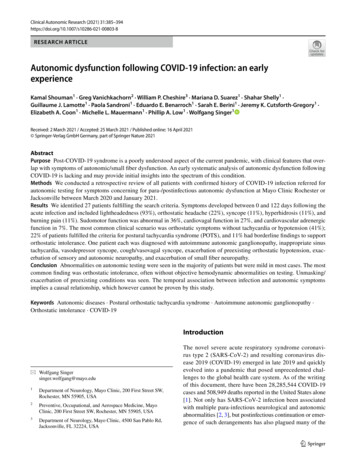
390Clinical Autonomic Research (2021) 31:385–394This case represents POTS with features of both deconditioning and a hyperadrenergic state. The patient was treatedwith a rehabilitative reconditioning program, cognitivebehavioral therapy, and low-dose beta-blocker with subsequent improvement and was able to return to work part-timeafter 3 months.Mild orthostatic intoleranceFig. 1 Thermoregulatory sweat test using alizarin red powder showing length-dependent anhidrosis of the fingers, left lower leg, andbilateral feet. The findings are consistent with length-dependent autonomic (small fiber) neuropathy. Hidrotic areas are colored in purpleand anhidrotic areas in yellow; the face was not powderedwarmth) and length-dependent anhidrosis of the feet andfingertips (Fig. 1).This case represents neuropathic POTS. The etiology islikely a postinfectious form of a limited autonomic (smallfiber) neuropathy.Patient 1 A 35-year-old woman with obesity and anxietycontracted SARS-CoV-2 with multiple symptoms includingfatigue, chest pain, and tachycardia. She quarantined witha limited level of activity until symptoms improved, andreturned to work after several weeks. At that time, she wasin contact with a co-worker who tested positive for COVID19 and had to quarantine again. While she did not get reinfected, she continued to have fatigue, brain fog, chest pain,insomnia, and decreased level of activity. She presentedabout 4 months after the initial infection with orthostaticlightheadedness, tachycardia, and, later, hyperhidrosis.Examination was normal except for signs of deconditioning.Her chest pain was diagnosed as costochondritis.Autonomic reflex screen revealed excessive heart rateacceleration during tilt meeting POTS criteria, with hyperadrenergic characteristics including an increase in bloodpressure on tilt and resting sweat activity on QSART.13Patient 24 A 38-year-old man developed dyspnea, fever,and myalgia. He tested positive for SARS-CoV-2. Concomitant with his diagnosis, he experienced fragmented sleep,postural lightheadedness, blurred vision, tinnitus, brain fog,visceral discomfort, and anxiety. He was worried that hewould not wake up the next day and would leave behind hisyoung son. This led to multiple emergency department visits. He presented a month after his diagnosis with worseninglightheadedness and fatigue that prevented him from working or exercising. Extensive testing for his visceral discomfort revealed no organic etiology. His neurological examination was normal. ARS revealed normal autonomic reflexes,low-normal baseline blood pressure, and a generous heartrate increase (28 bpm) on tilt table testing associated withlightheadedness.The case signifies that orthostatic symptoms can happenpara-infectiously and be perpetuated by disease anxiety anddeconditioning that outlive the acute phase of the infection.Treatment included reassurance, anxiolytics, and a rehabilitative reconditioning program. However, the patient continued to seek evaluation for his visceral pain and remainedsymptomatic 2 months after evaluation.Small fiber/autonomic neuropathyPatient 23 A 52-year-old man previously of excellent healthcontracted SARS-CoV-2. He experienced moderate respiratory symptoms of shortness of breath and productive coughand was managed in the outpatient setting. About 3 weekslater, he began to experience burning pain in the feet thatspread up to the knees that was associated with imbalanceand falls. The pain would wake him at night, impacted hisfunctional capacity, and was associated with allodynia. Neurological examination revealed high arches, moderate distalsensory loss to pinprick, and antalgic gait, but was otherwise normal including all large fiber sensory modalities.ARS was normal apart from a relatively reduced QSARTresponse at the foot compared to proximal responses, compatible with distal small fiber neuropathy (Fig. 2). Gabapentin and a topical lidocaine cream improved his neuropathicsymptoms.The patient’s presentation is most compatible with asmall fiber-predominant sensory neuropathy unmaskedby COVID-19 infection. The high arches suggest an
Clinical Autonomic Research (2021) 31:385–394391Fig. 2 Quantitative sudomotor axon reflex test showing relative distal (foot site) anhidrosis indicating distal autonomic (small fiber)neuropathy. Sweat output (µL): forearm site (red) 1.03 (5th percen-tile 0.25), proximal leg (blue) 2.11 (5th percentile 0.48), distal leg(green) 2.30 (5th percentile 0.62), and foot (yellow) 0.26 (5th percentile 0.15 and 10th percentile 0.33)otherwise—until the infection—asymptomatic hereditary/genetic form of neuropathy.tionally, the patient experienced worsening early satietybut improvement of her chronic constipation. She unintentionally lost 10 pounds. She had no symptoms of upperrespiratory infection or gastroenteritis and hence did nothave COVID-19 PCR testing. However, since other familymembers had tested positive for COVID-19, she underwentantibody testing a month and a half after symptom onset andwas positive for immunoglobulin (Ig) M but negative forIgG COVID-19 antibodies.The patient was assessed locally, suspected of having POTS, and started on propranolol. She also had aparaneoplastic autoantibody evaluation, and her ganglionic acetylcholine receptor antibody was 111 pmol/L[normal 53 pmol/L], which prompted referral to ourinstitution.She presented to us 5 months after symptom onset. Neurological examination was normal. Her ARS showed prominently labile blood pressure (meeting criteria for orthostatichypotension) and tachycardia (Fig. 3a), and her TST revealedpatchy widespread anhidrosis of 73% (Fig. 3b). Given herpresentation, pattern of anhidrosis, and history of antibodypositivity, the patient was diagnosed with a forme fruste ofAAG and started on intravenous methylprednisolone. Shehad initial improvement followed by a relapse in symptomsafter a COVID-19 reinfection.The case likely represents a forme fruste of autonomicganglionopathy. The history of similar, fluctuating symptoms in the past may indicate a chronic form that was exacerbated by COVID-19 infection. The autoantibody positivityand improvement with immunotherapy support a primaryautoimmune etiology.Patient 11 A 67-year-old woman presented with longstanding biopsy-proven length-dependent small fiber neuropathy in the setting of psoriatic arthritis. Her symptomsof mild acral tingling and burning pain had been stable for10 years, until she contracted SARS-CoV-2. Concomitantwith her viral illness, she developed severe burning painin the hands and feet. She presented 6 months later withpersistent symptoms and occasional orthostasis. Her neurological examination revealed hyperalgesia in the hands andfeet. ARS revealed patchy loss of QSART responses in thelower extremity, mild to moderate cardiovagal dysfunction,and mild adrenergic impairment. The patient’s pain management was optimized, and she was started on a rehabilitative program.This is an example of a chronic premorbid sensory andsmall fiber-predominant autonomic neuropathy exacerbatedby COVID-19 infection.Autoimmune autonomic ganglionopathyPatient 4 A 33-year-old right-handed woman with a historyof episodes of labile, paroxysmal hypertension and hypotension in the setting of stressful events (including pregnancy and delivery), mild heat intolerance, early satiety, andchronic constipation abruptly developed lightheadedness,facial flushing, shaking, prominent labile hypertension andhypotension, and labile heart rate, tinnitus, vision changes,brain fog, and heat intolerance with frank anhidrosis. Addi-13
392Clinical Autonomic Research (2021) 31:385–394Fig. 3 a Hemodynamic profile on head-up tilt study. Note prominentorthostatic tachycardia 129–42 bpm (green) and labile orthostatichypotension (red) with systolic blood pressure change ranging from 14 to 34 mmHg. b Thermoregulatory sweat test showing widespread anhidrosis (73%) in a patchy pattern compatible with autonomic ganglionopathyFor all the cases above, alternative etiologies of autonomic dysfunction were investigated and were absent orinsufficient to explain the patients’ complaints.of autonomic neuropathy have been reported after a varietyof infections [21, 22]. Despite the limited experience, thereis evidence that patients with existing neurological diseasemay develop worsening of their neurological symptoms withCOVID-19 and have a more severe course [23].Patients infected with SARS-CoV-2 can develop a widerange of clinical manifestations including fatigue, myalgias,and gastrointestinal dysfunction [24–26]. These in turn maylead to deconditioning, weight loss, and hypovolemia, whichare known to predispose to orthostatic intolerance. It wouldtherefore be expected that patients with a protracted courseof COVID-19 infection or with lingering postinfectioussymptoms are at increased risk of developing orthostaticintolerance related to those factors. Furthermore, healthrelated anxiety and insomnia in relation to COVID-19 itselfor disease-related measures such as quarantining [27] arelikely risk factors for worsening or emergence of a hyperadrenergic state and deconditioning, further increasing therisk of resulting autonomic symptoms.As expected, many patients with OI had clinical features consistent with deconditioning, hypovolemia, and/ora hyperadrenergic state. Although the methodology used inour report does not allow an accurate measure of the frequency of OI following COVID-19 infection, symptomscompatible with OI are anecdotally commonly encounteredin Mayo Clinic’s post-COVID clinic, which may suggestthat postinfectious OI is more common following COVID19 compared to other viral infections. The limited currentlyavailable data do not suggest that OI following COVID-19differs from that encountered following other viral disorders,DiscussionIn this study, we report the largest cohort to date of patientswith symptoms concerning for autonomic dysfunctionarising in the para-infectious or postinfectious period ofCOVID-19. All the patients had thorough, validated autonomic testing and careful clinical assessments to explore theassociation of often vague, nonspecific postinfectious symptoms with objective abnormalities on autonomic functiontesting. Remarkably, 17/27 (63%) patients had abnormalitieson autonomic function testing, ranging from symptomaticorthostatic tachycardia to severe autonomic failure. The mostcommon autonomic presentation post-COVID-19 was OI.While some of these patients fulfilled criteria for POTS orhad milder changes in orthostatic heart rate and blood pressure providing objective signs of orthostatic intolerance, themajority of patients experiencing orthostatic symptoms hada normal tilt study.Schondorf and Low first described postural orthostatictachycardia syndrome in 1993 [20]. Seven out of the 16reported patients in this landmark study were classified aspost-viral, as they had symptoms of viral upper respiratorytract infection prior to the onset of their symptoms. It wasproposed that these patients experience a form of postinfectious limited autonomic neuropathy. Similarly, other types13
Clinical Autonomic Research (2021) 31:385–394but further research into this phenomenon is needed to solidify this impression. Symptoms induced by the upright position without a clear cardiovascular correlate is not uniqueto this cohort, but the high percentage of patients in thiscategory may indicate an increased level of anxiety, overlapwith chronic fatigue and central sensitization, or a conditionin the category of persistent postural perceptual dizziness.We also identified patients with autonomic and smallfiber neuropathies, which may not be surprising considering that postinfectious neuropathy has been reported after avariety of viral infections such as herpes simplex and infectious mononucleosis [21, 22]. Interestingly, while one ofour patients developed de novo limited autonomic and smallfiber neuropathy with POTS following COVID-19 infection,other cases were characterized by postinfectious worseningof preexisting autonomic/small fiber neuropathies. That wasalso true for
Methods We conducted a retrospective review of all patients with conrmed history of COVID-19 infection referred for autonomic testing for symptoms concerning for para-/postinfectious autonomic dysfunction at Mayo Clinic Rochester or Jacksonville between March 2020 and January 2021. Resu