Transcription
Use of this content is subject to the Terms and Conditions of the MD Consult web site.Cardiology ClinicsVolume 18 Number 3 August 2000Copyright 2000 W. B. Saunders CompanyDIASTOLIC FUNCTION AND DYSFUNCTIONTHE ECHO-DOPPLER EVALUATION OF LEFT VENTRICULAR DIASTOLICFUNCTIONA Current PerspectiveChristopher P. Appleton 1 MDMichael S. Firstenberg 2 MDMario J. Garcia 2 MDJames D. Thomas 2 MD12Division of Cardiovascular Diseases, Mayo Clinic Scottsdale, Arizona (CPA)Cleveland Clinic Foundation, Cleveland, Ohio (MSF, MJG, JDT)Supported in part by grants from the American Heart Association, Scottsdale, Arizona (CPA), Ohio Affiliates, Cleveland Ohio (MSF,MJG), and the National Aeronautics Space Administration and National Institutes of Health (JDT).The role of left ventricular (LV) diastolic function in health and disease is still incompletely understoodand underappreciated by most primary care physicians and many cardiologists. This is not surprisingbecause diastole is a complex phenomenon with many determinants that are difficult to individuallystudy, and has several phases that encompass the relaxation and then filling of the ventricle. [6] [38] [42] [65]Physical examination, electrocardiogram (ECG), and chest radiographs are unreliable in making thediagnosis of LV diastolic dysfunction in most individuals, and invasive measurements of cardiacpressures, rates of LV relaxation, and LV compliance are costly, clinically impracticable as they carryincreased risk, and require special catheters and software analysis programs. [66]This situation changed with the development of echocardiography. Because of its noninvasive nature,large numbers of normal individuals and patients were studied and different LV filling patterns weredescribed, first with M-mode echocardiography [35] [36] [43] and later using pulsed-wave (PW) Dopplertechnique interrogating mitral inflow. [45] [46] [53] [73] [110] [112] Validation of these mitral filling patternsagainst radionuclide and angiographic techniques soon followed; [96] [104] however, enthusiasm forrelating LV filling patterns to diastolic function was dampened by reports that the velocity andproportion of early and late diastolic filling and their peak velocities were affected by preload, [16] [106]afterload, [77] and heart rate. [4] [77]
In 1988, hemodynamic pressure measurements were related to individual LV filling patterns,independent of disease state. [9] Three basic abnormal filling patterns were described and were soonfound to have clinical significance and prognostic value regardless of cardiac disease type. [7] [56] [84] [90][117] [124] The field of diastology using echo-Doppler evaluation was born [20] and steady progresscontinued. Today this LV diastolic evaluation includes interrogation of mitral and pulmonary venousflow velocities, the rate of mitral inflow velocity by color Doppler flow propagation, and the evaluationof mitral annular motion by tissue Doppler imaging (TDI). In addition, manipulation of preload andafterload assesses how sensitive abnormal LV filling patterns are to changes in loading conditions. [21][47] [92] Although all echo-Doppler indices remain imperfect and much remains to be learned, theaggregate sum of this information remains our best and most practical way to assess LV diastolicfunction, and to objectively follow serial changes after medical intervention or with diseaseprogression.To begin to use echo-Doppler information for patient evaluation and management requires a basicunderstanding of cardiac physiology, particularly LV diastolic properties and LV filling patterns, andreproducible high-quality Doppler flow velocity recording. [10] The routine performance of a diastolicfunction examination on every patient referred for echocardiography is recommended for acquiringexperience to eliminate technical and interpretive pitfalls. This article explains a practical way forapproaching the echo-Doppler analysis of LV diastolic function, and how the information obtainedmay be used clinically to aid patient diagnosis and therapy.LEFT VENTRICULAR DIASTOLIC DYSFUNCTIONDefinitionAbnormal diastolic function is a disorder of LV filling. As systolic function effects LV relaxation and oftenLV compliance, all patients with a decrease in LV ejection fraction have diastolic abnormalities. Manypatients with symptoms of congestive heart failure (CHF) or reduced exercise capacity, however, have anormal LV ejection fraction or isolated LV diastolic dysfunction as the etiology of their cardiac problem. Adefinition for LV diastolic dysfunction includes: 1) an inability to fill the left ventricle, during rest orexercise, to a normal end-diastolic volume without an abnormal increase in LV end-diastolic or mean leftatrial (LA) pressure; or 2) a failure to increase LV end-diastolic volume, and therefore cardiac output duringexercise (Fig. 1) . In its earliest stages, diastolic dysfunction may cause only a mild slowing of LV relaxationwithout elevated pressures and the patient may be asymptomatic.
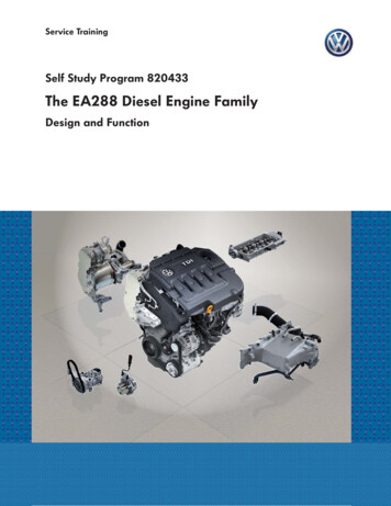
Figure 1. Pulmonary wedge pressure (PWP) versus left ventricular (LV) end-diastolic volume in normal subjects ( square) and sevenpatients ( circle) with congestive heart failure (CHF) but normal LV ejection fraction, and no significant coronary or valvular heartdisease. Compared with the normal subjects, patients with diastolic CHF were unable to increase their LV end-diastolic volume andalso had a marked increase in PWP. This led to marked exercise intolerance with approximately 50% reduction in peak oxygenconsumption, primarily because of the reduction in cardiac index. ( From Kitzman DW, Higginbotham MB, Cobb FR, et al: Exerciseintolerance in patients with heart failure and preserved left ventricular systolic function: Failure of the Frank-Starling mechanism. JAm Coll Cardiol 17:1065-1072, 1991; with permission.)DIASTOLEA Historical PerspectiveOne of the first attempts to explain ventricular filling was provided by Galen in 100 BC, who proposed thatthe heart is filled by dilation of the right ventricle. Centuries later, in 1628, William Harvey recognized theheart was the central pump in a circulatory system containing arteries and veins. This discovery was followedby recognition that most cases of CHF were caused by damage or weakening of the heart muscle and adecrease in LV pumping function. Diastole was largely ignored as simply the interval in which the cardiacchambers passively filled between each pumping cycle.Gradually, clues emerged that LV diastolic dysfunction alone could cause symptoms, and that diastolic andsystolic LV function were interrelated. One discovery was the Frank-Starling mechanism whereby LV enddiastolic volume helps regulate LV stroke volume on a beat-to-beat basis. Another landmark observation wasmade by Katz, who observed that after mitral valve opening in mammalian hearts LV pressure continues todecrease while volume is increasing, therefore demonstrating that the heart acts as a suction pump. [50] It wasalso recognized that a limited filling capacity was the cardinal feature of constrictive pericarditis that reducedcardiac output and resulted in marked peripheral edema and generalized wasting.With the advent of cardiac catheterization in the 1960s, the study of cardiovascular biomechanics accelerated.
Although most research continued to focus on LV systolic function, cardiac diseases with thickened andnoncompliant ventricles (i.e., restrictive and hypertrophic cardiomyopathies) were reported. [105] Soon,angiographic differences in LV filling patterns between normals and patients with various heart diseaseswere noted. [42] Methods to quantitate the rate of LV relaxation [122] and LV compliance [27] were described,and it became apparent that patients with the same LV ejection fraction could have markedly differentdiastolic properties and LV filling characteristics. At the same time, the importance of LV systolic functionin determining LV restoring forces and rate of LV relaxation was also appreciated. Gradually, systole anddiastole came to be viewed more as an intertwined continuum, with each part affecting the other.In the mid-1980s, echocardiographic studies helped show that 20% to 40% of patients with symptoms ofCHF had normal LV ejection fractions and, therefore, presumably isolated diastolic dysfunction as theetiology for their heart failure. [103] [115] Because of the difficulties in quantitating individual LV diastolicproperties, the clinical study of LV diastolic dysfunction proceeded slowly through the analysis of digitizedM-mode, angiographic, or radionuclide LV filling patterns. [35] [36] [43] In 1982, the use of PW Doppler mitralflow velocities to study LV filling was described. [53] Because of its ease of use, noninvasive nature, andability to study changes in LV filling after interventions and over time, this technique revolutionized thestudy of LV diastolic function. Correlations between LV filling pressures and mitral flow velocity patternswere performed, [9] normal age-related changes in LV filling were established, [33] [55] [73] and three basicabnormal LV filling patterns independent of underlying cardiac disease were recognized as part of the"natural history of LV filling." [7] Shortly thereafter, pulmonary venous flow velocity-derived variables weredescribed that characterized LA filling and also aided the interpretation of LV filling patterns and pressures.[7] [52] [59] [63] Today, Doppler mitral flow velocity-derived variables are recognized as powerful prognostic toolsin patients with various cardiac diseases, including CHF [94] and dilated [90] [124] and restrictive [57]cardiomyopathies. At the same time, newer methods such as TDI mitral annular motion (MAM), the rate ofcolor Doppler mitral inflow propagation (Vp), and model-based image processing continue to advance thefield of diastology. * As a result, the clinical syndromes caused by LV diastolic dysfunction and LV fillingdisorders are now more readily recognized by healthcare practitioners, and treatment strategies for thesepatients are being developed. [120]Epidemiology of Left Ventricular Diastolic DysfunctionAlthough isolated LV diastolic dysfunction can be seen in all age groups, it is chiefly a disease of theelderly. In a meta-analysis of the problem, the overall incidence was 30% to 35% with a range varying from13% to 17%. [118] This prevalence is strikingly age-related, with patients younger than 65 years of age havinga incidence of approximately 15%, whereas patients older than 70 years of age have an incidence ofsymptomatic CHF with normal ejection fraction as high as 40% to 50%. [118] [119] This higher incidencereflects the most common cause of isolated diastolic dysfunction, hypertensive heart disease of the elderly.This problem is associated with increasing systolic blood pressures with age and the development of LVhypertrophy. It is estimated that approximately 50% of women over the age of 70 have LV hypertrophy onechocardiograms, with only a slightly smaller prevalence in males. Patients greater than or equal to 70 yearsof age with CHF and normal LV ejection fraction have an increased 1-year mortality associated withworsening Doppler echo-derived diastolic function variables. [94]The prognosis for patients with isolated LV diastolic dysfunction is more favorable than for patients withsystolic dysfunction; however, when compared with age- and gender-matched normal subjects, the mortalityrisk is increased fourfold. [119] The disease also causes considerable morbidity with its symptoms ofpulmonary congestion and decrease in functional capacity owing to reduced exercise capacity. At the presenttime, prospective studies looking at the epidemiology of LV diastolic dysfunction are underway.Diastolic Properties of the Left Ventricle
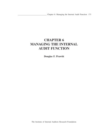
Diastole has traditionally been divided into four phases: isovolumic relaxation, early diastolic filling,diastasis in mid-diastole, and atrial contraction (for additional details see the article by Yellin and Meisner,elsewhere in this issue). Part of the difficulty in studying LV diastolic function has been that themajordeterminants of LV diastolic performance occur during different phases of diastole, often overlap intheir timing and are affected not only by each other but also by LV systolic function, heart rate, and thecardiac conduction system. [66]Although there are numerous independent factors that affect LV diastolic properties and the filling of the leftventricle, all factors work through their combined, resultant effects on the transmitral pressure gradient(TMPG), which is the actual physical determinant of LV filling (Fig. 2) . The effects of two key diastolicproperties, the rate of LV relaxation and LV compliance (which affects LA filling pressure), are especiallyimportant in understanding LV filling patterns in health and disease.Figure 2. Numerous factors affect LV diastolic properties and filling; however, all factors work through their effects on the transmitralpressure gradient (TMPG), which is the actual determinant of LV filling. An early positive TMPG (E TMPG ) relates to early diastolicLV filling as assessed by pulse-wave Doppler mitral flow velocity. A reverse pressure gradient in mid-diastole decelerates earlyfilling, with late filling (L TMPG ) occurring as a result of the increase in the left atrial pressure owing to atrial contraction.LV relaxation describes the rate of LV pressure decline during isovolumic relaxation. It is controlled bycellular events that regulate cytosolic Ca concentration in myocytes. [81] In contrast to LV systole in whichCa is passively released from the sarcoplasmic reticulum and interacts with the actin-troponin myosincontractile elements, LV relaxation is energy (adenosine triphosphate [ATP]) dependent and requires thisCa to be returned to the sarcoplasmic reticulum by Ca ATPase-dependent pumps against a 10,000:1
concentration gradient. These enzymes and their endogenous regu
Definition Abnormal diastolic function is a disorder of LV filling. As systolic function effects LV relaxation and often LV compliance, all patients with a decrease in LV ejection fraction have diastolic abnormalities. Many patients with symptoms of congestive heart failure (CHF) or reduced exercise capacity, however, have a normal LV ejection fraction or isolated LV diastolic dysfunction as .