Transcription
Lang et al. Scandinavian Journal of Trauma,Resuscitation and Emergency Medicine (2017) 25:82DOI 10.1186/s13049-017-0427-4ORIGINAL RESEARCHOpen AccessThe role of whole-body computedtomography in the diagnosis of thoracicinjuries in severely injured patients – aretrospective multi-centre study based onthe trauma registry of the German traumasociety (TraumaRegister DGU )Patricia Lang1†, Martin Kulla2*† , Fabian Kerwagen1, Rolf Lefering3, Benedikt Friemert1, Hans-Georg Palm1and TraumaRegister DGUAbstractBackground: Thoracic injuries are a leading cause of death in polytrauma patients. Early diagnosis and treatmentare of paramount importance. Whole-body computed tomography (WBCT) has largely replaced traditional imagingtechniques such as conventional radiographs and focused computed tomography (CT) as diagnostic tools in severelyinjured patients. It is still unclear whether WBCT has led to higher rates of diagnosis of thoracic injuries and thus to achange in outcomes.Methods: In a retrospective study based on the trauma registry of the German Trauma Society (TraumaRegister DGU ),we analysed data from 16,545 patients who underwent treatment in 59 hospitals between 2002 and 2012 (ISS 9). The 3years preceding and the 3 years following the introduction of WBCT as a standard imaging modality for the investigationof severely injured patients were assessed for every hospital. Accordingly, patients were assigned to either the pre-WBCTor the WBCT group. We compared the numbers of thoracic injuries and the outcomes of patients before and after theroutine use of WBCT.Results: A total of 13,564 patients (pre-WBCT: n 5005, WBCT: n 8559) were included. Relevant thoracic injuries weredetected in 47.8%. There were no major differences between the patient groups in injury severity (pre-WBCT: median ISS21; WBCT: median ISS 22), injury patterns and demographics. After the introduction of WBCT, only minor changes wereobserved regarding the rates of most thoracic injuries. Clinically relevant injuries were pulmonary contusions (pre-WBCT:18.5%; WBCT: 28.7%), injuries to the lung parenchyma (pre-WBCT: 12.6%; WBCT: 5.9%), multiple rib fractures (pre-WBCT: 10.6%; WBCT: 21.6%), and pneumothoraces (pre-WBCT: 17.3%; WBCT: 21.6%). The length of stay in the intensive care unit(pre-WBCT: 10.8 days; WBCT: 9.7 days) and in hospital (pre-WBCT: 26.2 days; WBCT: 23.3 days) decreased. There was nodifference in overall mortality (pre-WBCT: 15.5%; WBCT: 15.6%).(Continued on next page)* Correspondence: martin.kulla@uni-ulm.de†Equal contributors2Department of Anaesthesiology and Intensive Care Medicine, GermanArmed Forces Hospital of Ulm, Oberer Eselsberg 40, 89081 Ulm, GermanyFull list of author information is available at the end of the article The Author(s). 2017 Open Access This article is distributed under the terms of the Creative Commons Attribution 4.0International License (http://creativecommons.org/licenses/by/4.0/), which permits unrestricted use, distribution, andreproduction in any medium, provided you give appropriate credit to the original author(s) and the source, provide a link tothe Creative Commons license, and indicate if changes were made. The Creative Commons Public Domain Dedication o/1.0/) applies to the data made available in this article, unless otherwise stated.
Lang et al. Scandinavian Journal of Trauma, Resuscitation and Emergency Medicine (2017) 25:82Page 2 of 11(Continued from previous page)Conclusions: The routine use of WBCT in the trauma room setting has led to changes in patient management that arenot reflected in the rates of diagnosis of severe thoracic injuries (e.g. tension pneumothoraces, cardiac injuries, arterialinjuries). By contrast, there was a relevant increase in the rates of diagnosis of minor thoracic injuries, which, however, didnot result in an improvement in survival prognosis.Keywords: Tomography, X-ray computed, Multiple trauma, Thoracic injuries, Mortality, Survival rate, Trauma centresBackgroundThoracic injuries account for 25% of deaths in polytrauma patients and are thus a common cause of deathamong these patients [1, 2]. In recent years, a traumaroom algorithm has become widely accepted inGermany which is based on Advanced Trauma Life Support (ATLS ) and the European Trauma Course [3–5].Nevertheless, the imaging technique to be used in thetrauma room setting continues to be a matter of debateon account of time constraints, the level of radiation exposure, the patient’s overall condition, and other factors[6–8]. Computed tomography (CT), and in particularmodern multi-slice computed tomography (MSCT), hasbecome the standard imaging modality for diagnosingthoracic injuries [6–8].After the usefulness of focused CT in addition to chestradiography had been demonstrated, the introduction ofMSCT in 1998 led to a further substantial improvementin imaging techniques in the trauma room setting [9–13].The technical advances associated with MSCT providedthe basis for integrating whole-body computed tomography (WBCT), which is also referred to as a trauma scan,into the management of severely injured patients. In a recent review of the literature, Donaubauer et al. showedthat the use of WBCT as a diagnostic tool had positive effects. They as well as other authors, however, did not specify the indications for WBCT [14, 15]. It is still unclearwhether the replacement of traditional imaging (conventional radiography of the cervical spine, chest and pelvisand subsequent focused CT) by the trauma scan as thestandard diagnostic imaging modality led to an improvement in the diagnosis of thoracic injuries. We conducted aretrospective analysis of the trauma registry of the German Trauma Society (TraumaRegister DGU ) in order toassess the number of diagnosed thoracic injuries beforeand after the introduction of WBCT as a standard imagingmodality and to investigate whether the trauma scan ledto a change in patient outcomes.MethodsThe trauma registry of the German Trauma Society(TraumaRegister DGU ) provided the data used. TheTraumaRegister DGU of the German Trauma Society(Deutsche Gesellschaft für Unfallchirurgie, DGU) wasfounded in 1993. The aim of this multi-centre databaseis the pseudonymised and standardised documentationof care for severely injured patients. Data are collectedprospectively from the site of the accident until discharge from hospital. Included are patients who are admitted to hospital via the emergency room andsubsequently receive intensive or intermediate care andpatients who arrive at hospital with vital signs and diebefore admission to the intensive care unit. The infrastructure for documentation, data management, anddata analysis is provided by the Academy for TraumaSurgery, which is affiliated to the German Trauma Society. Scientific data analysis is approved according to apeer review procedure established by the Committee onEmergency Medicine, Intensive Care and Trauma Management of the German Trauma Society. The participating hospitals are mainly located in Germany. Currently,approximately 25,000 cases from more than 600 hospitals are entered into the database per year. For hospitalsassociated with TraumaNetzwerk DGU , the entry of atleast a basic data set is obligatory for reasons of qualityassurance [3, 16, 17].Patient groups and definitionsDuring the study period from 2002 to 2012, we analysedall cases of patients who were admitted to the traumaroom with an Injury Severity Score (ISS) greater than orequal to 9. Patients who did not undergo immediate surgery or were not admitted to ICU were not included inthis analysis of TR-DGU data.For cases in which the box for ‘Whole-body CT’ in theTraumaRegister DGU data collection form was checked,we assumed that a whole-body trauma scan was performed as a primary diagnostic procedure. The TR-DGUdefines WBCT as a combination of CT studies that produce images (or slices) of the body in a continuous manner and cover at least the region from the skull base tothe pelvis. Neither the type of equipment used nor otherdetails such as table feed per rotation are specified forthis imaging procedure. In addition, data about thescanner type and the application of contrast agentsare not provided.The other diagnostic approach consists of traditionalimaging that involves conventional radiography of thecervical spine, the chest and the pelvis, often followed byfocused CT (e.g. cranial CT).
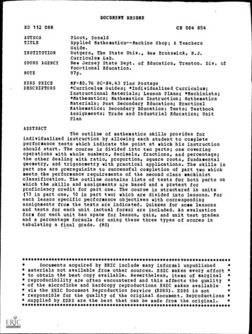
Lang et al. Scandinavian Journal of Trauma, Resuscitation and Emergency Medicine (2017) 25:82The year in which the trauma scan replaced traditionalimaging as the standard diagnostic imaging approach inthe trauma room setting was determined individually forevery hospital by two independent examiners (HP andMK). When the two examiners disagreed, the opinion ofa specialist in biometrics (RL) was obtained and was thefinal determination. The 3 years before (pre-WBCTgroup) and the 3 years after the introduction of thetrauma scan (WBCT group) as a standard imaging procedure were analysed and compared (Fig. 1). The year inwhich the trauma scan was introduced was excludedfrom analysis since this year was usually a period oftransition associated with a mixture of both imagingapproaches. A maximum variation of 30% in both thepre-WBCT and the WBCT group provided the basis fordecision. In addition, there had to be an increase in theMSCT rate by at least 50% or to at least 60% in the yearfollowing the introduction of WBCT when compared tothe year preceding the introduction of the trauma scan.Inclusion and exclusion criteriaOnly patients who underwent primary treatment at a regional (Level II) or supraregional trauma centre (Level I)(as defined by the TraumaRegister DGU ) were included[3, 16]. In addition, continuous documentation over aperiod of at least five consecutive years was required forinclusion. Hospitals were excluded when there wereyears for which no documentation was available or whenthe change in the number of cases was too drastic. Atrauma scan rate of 50% or more in the first documented year was a further exclusion criterion since sucha high rate suggested that this might not have been theyear in which the trauma scan was introduced.Page 3 of 11Statistical analysisData that were obtained before and after the introductionof the trauma scan as a standard imaging modality for theassessment of trauma room patients were compared on thebasis of percentages and means. Data were analysed usingSPSS (version 22, IBM Corp., Armonk, NY, United States).In consultation with a biostatistician affiliated to theTraumaRegister DGU , we decided not to test betweengroup differences for statistical significance since evensmall differences that were of no clinical relevance werelikely to be statistically significant on account of thelarge number of cases. For this reason, differences ofmedical importance were termed “relevant”. Trauma severity was assessed using the Injury Severity Score (ISS)and the New Injury Severity Score (NISS) [18]. RevisedInjury Severity Classification (version II) (RISC II) scoreswere calculated in order to predict mortality, and thusto assess the probability of survival, at the time ofhospital admission [19].We presented the results in a descriptive manner inorder to generate, but not test, a hypothesis. Continuousvariables were presented as means (MV) and standarddeviations (SD). Contingency tables were used to displayfrequency distributions presented as percentages. The95% confidence intervals (95% CI) were calculated inorder to assess the uncertainty of the means and percentages. In order to give a better insight into the distribution of the data, we calculated median value as well asthe interquartile range in order to estimate the skewness,or symmetry, of the distribution where appropriate.Ethics and study registrationThe present study is in line with the publication guidelines of the TraumaRegister DGU and is registered asFig. 1 Example of a hospital to illustrate the use of WBCT on an annual basis. The bars show the percentage of patients whose data wereentered into the TraumaRegister DGU and who underwent trauma scans (WBCT). Data for the years 3 to 1 were used to form the pre-WBCTgroup (traditional imaging) and data for the years 1 to 3 following the introduction of the trauma scan as a standard imaging modality were usedto form the WBCT group. The year 0 is the year in which WBCT was introduced
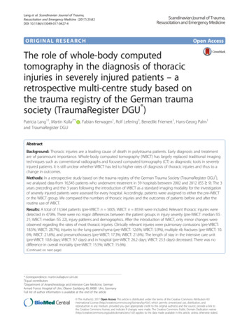
Lang et al. Scandinavian Journal of Trauma, Resuscitation and Emergency Medicine (2017) 25:82TR-DGU Project ID 2013–053 (http://www.traumaregister-dgu.de/fileadmin/user upload/traumaregister-dgu.de/docs/Downloads/TR-DGU - Publikationsrichtlinie.pdf ).As register data are assessed anonymously for scientificdata analysis, individual informed consent is not required.ResultsWe were able to include 16,545 cases from 59 hospitalsin our analysis. Of these cases, 5005 patients (30.3%)underwent traditional diagnostic imaging (pre-WBCTgroup) and 8559 patients (51.7%) had a trauma scan(WBCT group). The patients who underwent an imagingprocedure in the year in which the trauma scan was introduced for routine use (n 2981; 18.0%) were not included in this study because of a wide variety ofimplementation rates (Fig. 2). Table 1 provides an overview of demographic data, prehospital data, injury patterns, and injury severity.With the introduction of the trauma scan in the traumaroom setting, the percentage of patients who underwentCT scanning increased from 73.7 to 92.8%. Once thetrauma scan had become routine practice, 80.1% of alltrauma room patients underwent whole-body MSCT –compared to 10.0% in the preceding years. At the sametime, the percentage of patients who underwent conventional radiography of the chest decreased from 85.2 to59.8% whereas the percentage of patients who had a Focused Assessment with Sonography in Trauma (FAST)Fig. 2 The figure shows a marked increase in the number of traumascans after the introduction of WBCT. The change in the standardimaging approach is further demonstrated by the consecutivedecrease in (conventional) chest radiography. A high proportion ofpatients underwent FAST. This proportion remained almostunchanged during the study period. The whiskers indicate the upperand lower limits of the 95% confidence intervalPage 4 of 11examination remained almost unchanged (pre-WBCTgroup: 88.4%; WBCT group: 86.6%).There were no major differences between the two patient groups in terms of injury severity, injury patternsand demographics. In both groups, the majority of patients were male (pre-WBCT group: 73.5%, 95% CI 72.2–74.7; WBCT group: 72.8%, 95% CI 71.9–73.8). The meanage of the patients in the pre-WBCT group (43.0 years,95% CI 42.5–43.6) was slightly lower than that of the patients in the WBCT group (45.7 years, 95% CI 45.2–46.1).The mean ISS was 23.9 (95% CI 23.5–24.2) before and24.5 (95% CI 24.2–24.7) after the introduction of thetrauma scan as a standard imaging modality. The medianISS was 21 (IQR 14–29) versus 22 (IQR 14–29). The meanNISS was 29.4 (95% CI 28.9–29.8) in the pre-WBCTgroup and 30.2 (95% CI 29.9–30.6) in the WBCT group.The median NISS was 25 (IQR 17–35) versus 27 (IQR17–38). In both patient groups, road traffic accidents werethe main cause of injury (60.0% before and 58.2% after theintroduction of WBCT). The vast majority of patients sustained a blunt thoracic trauma (94.8% in the pre-WBCTgroup and 94.5% in the WBCT group).During the study period, the number of documentedthoracic injuries increased (Table 2). Whereas the percentage of severely injured patients without thoracic injurieswas higher in the pre-WBCT group (45.8%) than in theWBCT group (40.2%), the percentage of patients withrelevant thoracic injuries (Abbreviated Injury Score AISthorax 3) was lower before the introduction of thetrauma scan as a routine imaging modality (44.1%) thanafter the routine use of WBCT (50.0%). Relevant changeswere observed for the following thoracic injuries: injuriesto the lung parenchyma (pre-WBCT: 12.6%; WBCT5.9%), pulmonary contusions (pre-WBCT: 18.5%; WBCT:28.7%), haemothoraces (pre-WBCT: 15.6%; WBCT:14.0%), pneumothoraces (pre-WBCT: 17.3%; WBCT:21.6%), multiple rib fractures and flail chest (pre-WBCT:10.6%; WBCT: 21.6%), and injuries to the thoracic spine(AIS 2) (pre-WBCT: 10.9%; WBCT 13.2%).The mortality of patients who were managed before theroutine use of the trauma scan was 15.5% (95% CI 14.5–16.5). Following the introduction of WBCT as a routineimaging modality, the mortality rate remained almost unchanged (15.6%, 95% CI 14.9–16.4). There was no relevantdifference between the two groups in the probability ofsurvival or mortality as predicted at the time of hospitaladmission. The RISC II score was 17.6% for the preWBCT group of patients and 17.3% for the WBCT group.The Standardised Mortality Ratio (SMR) was 0.88 beforeand 0.90 after the introduction of the trauma scan as astandard primary diagnostic imaging modality.A relevant change in the percentage of patients withorgan failure (pre-WBCT: 43.5%, 95% CI 42.0–45.0;WBCT: 43.9%, 95% CI 42.7–45.1) and multi-organ
Lang et al. Scandinavian Journal of Trauma, Resuscitation and Emergency Medicine (2017) 25:82Page 5 of 11Table 1 Demographic data, injury severities and injury patterns for the two patient groups. Mean and upper and lower limits of the95% confidence intervals (CI) are given. Where appropiate, the median as well as the interquartile range (IQR) are given tooParameterPre-WBCT group (n 5002)WBCT group (n 8559)Demographic dataMale patients [%]mean (95% CI)73.5 [72.2–74.7]73.0% [71.9–73.8]Patient age [years]mean (95% CI)median (IQR)43.0 [42.5–43.6]41 (25–59)45.7 [45.2–46.1]45 (26–62)Patients with blunt trauma [%]mean (95% CI)94.8 [94.2–95.2]94.5 [94.2–95.0]GCSmean (95% CI)median (IQR)11.0 [10.8–11.1]14 (7–15)11.1 [11.0–11.2]14 (8–15)Systolic blood pressure [mmHg]mean (95% CI)median (IQR)119.9 [119.0–120.9]120 (100–140)121.2 [120.5–122.0]120 (100–140)SpO2 [%]mean (95% CI)median (IQR)92.5 [92.2–92.9]96 (92–98)92.7 [92.4–93.0]96 (92–98)Patients with a GCS 8 [%]mean (95% CI)30.3 [29.0–31.6]28.9 [28.0–29.6]Patients with a systolic blood pressure 90 mmHg [%]mean (95% CI)18.2 [17.2–19.4]17.9 [17.0–18.7]17.617.348.7 [47.6–49.8]Prehospital settingPrognosisRISC II (predicted mortality) [%]Patterns of injuryPatients with an AIShead 3 [%]mean (95% CI)48.9 [47.5–50.3]Patients with an AISface 3 [%]mean (95% CI)2.4 [2.0–2.9]4.2 [3.8–4.6]Patients with an AISthorax 3 [%]mean (95% CI)44.1 [42.8–45.5]50.0 [48.9–51.0]Patients with an AISabdomen 3 [%]mean (95% CI)17.5 [16.4–18.5]15.8 [15.0–16.6]Patients with an AISextremities 3 [%]mean (95% CI)37.2 [35.9–38.6]34.0 [33.0–35.0]Patients with an AISsoft tissues 3 [%]mean (95% CI)0.9 [0.6–1.1]2.0 [1.7–2.3]Patients without thoracic injuries [%]mean (95% CI)45.8 [44.4–47.2]40.2 [39.2–41.3]ISSmean (95% CI)median (IQR)23.9 [23.5–24.2]21 (14–29)24.5 [24.2–24.7]22 (14–29)NISSmean (95% CI)median (IQR)29.4 [28.9–29.8]*25 (17–35)30.2 [29.9–30.6]*27 (17–38)Injury severityGCS Glasgow Coma Scale, AIS Abbreviated Injury Scale, (N)ISS (New) Injury Severity Score, RISC Revised Injury Severity Classificationfailure (pre-WBCT: 26.5%, 95% CI 25.2–27.8; WBCT:26.9%, 95% CI 25.8–27.9) was not observed. The duration of ventilation (days of intubation), however, waslonger before the routine use of the trauma scan thanafter the introduction of WBCT (pre-WBCT: 6.7, 95%CI 6.6–7.2; WBCT: 5.6, 95% CI 5.4–5.8). The same applies to the length of ICU stay (pre-WBCT: 10.8, 95% CI10.5–11.2; WBCT: 9.7, 95% CI 9.4–10.0). The preWBCT group had fewer ventilator-free days during thefirst 30 days of hospital stay (19.8 days, 95% CI 19.4–20.1) than the WBCT group (20.8 days, 95% CI 20.6–21.0). The length of hospital stay decreased by approximately 3 days from 26 days (95% CI 25.5–26.9) in thepre-WBCT group to 23 days (95% CI 22.7–23.9) in theWBCT group. The percentage of patients who requireda chest drain in the trauma room or operating room settings dropped from 20.0% before the routine use ofWBCT to 18.5%. The length of stay in the trauma roomdecreased by 14 min from 78 min (95% CI 76.0–79.0) inthe pre-WBCT group to 64 min (95% CI 63.0–65.0) inthe WBCT group. Table 3 provides an overview of theresults obtained as well as median and interquartilerange where appropriate.DiscussionThe objective of our study was to investigate whether theintroduction of the trauma scan for the diagnostic evaluation of polytrauma patients in the trauma room led to achange in the rates of diagnosis of thoracic injuries. For thispurpose, we compared the trauma scan approach and conventional radiography with or without focused CT for thediagnostic assessment of polytrauma patients. We placedparticular emphasis on determining the year in which thetrauma scan replaced conventional techniques as the standard diagnostic imaging approach individually for every hospital and formed the two groups of patients that were
Lang et al. Scandinavian Journal of Trauma, Resuscitation and Emergency Medicine (2017) 25:82Page 6 of 11Table 2 Thoracic injuries in the two patient groupsParameterPre-WBCT group (n 5002)WBCT group (n 8559)Injury to the lung parenchyma [%]12.6 [11.7–13.5]5.9 [5.4–6.4]Pulmonary contusion [%]18,5 [17.4–19.6]28,7 [27.7–29.7]]Pneumothorax [%]17.3 [16.3–18.4]21.6 [20.7–22.5]Tension pneumothorax [%]2.9 [2.4–3.4]2.5 [2.2–2.8]Haemothorax [%]15.6 [14.6–16.6]14.0 [13.3–14.7]Multiple rib fractures and flail chest [%]10.6 [9.7–11.4]21.6 [20.7–22.5]Fractures of two ribs [%]6.4 [5.7–7.1]6.6 [6.1–7.1]Fracture of one rib [%]3.4 [2.9–3.9]3.3 [2.9–3.7]Arterial injury (thorax) [%]1.5 [1.2–1.8]1.5 [1.2–1.8]Type of thoracic injuriesDiaphragmatic injury [%]1.0 [0.7–1.3]1.1 [0.9–1.3]Thoracic spine injury AIS 2 [%]10.9 [10.0–11.8]13.2 [12.5–13.9]Thoracic spinal cord injury [%]1.7 [1.3–2.1]1.9 [1.6–2.2]Cardiac injury [%]0.4 [0.2–0.6]0.5 [0.4–0.7]No thoracic injuries [%]45.8 [44.4–47.2]40.2 [39.2–41.3]Severity of thoracic injuriesAISthorax 1 [%]2.8 [2.3–3.2]2.0 [1.7–2.3]AISthorax 2 [%]9.2 [8.4–10.0]9.3 [8.7–9.9]AISthorax 3 [%]24.9 [23.7–26.1]28.2 [27.3–29.2]AISthorax 4 [%]12.4 [11.5–13.3]14.6 [13.8–15.3]AISthorax 5 [%]4.7 [4.1–5.3]5.3 [4.8–5.8]AISthorax 6 [%]0.2 [0.1–0.3]0.3 [0.2–0.4]AIS Abbreviated Injury Scalecompared on the basis of these hospital-specific data. Ourstudy showed that the routine use of the trauma scan didnot lead to a change in the rates of diagnosis of severe thoracic injuries requiring immediate treatment (e.g. tensionpneumothoraces, cardiac injuries, arterial injuries). By contrast, there was a relevant increase in the rates of diagnosisof minor thoracic injuries (e.g. pulmonary contusions, pneumothoraces, multiple rib fractures). The additional information provided by WBCT, however, did not lead to animprovement in survival prognosis. This and the followingresults regarding the diagnosed injuries must be interpretedcarefully, because we could not rule out a change in the incidence of thoracic injuries during the study period.Our findings are particularly noteworthy as the year2002 has so far been used as the cut-off year for theintroduction of WBCT in similar studies that were performed on the basis of the TR-DGU [20, 21]. In ourstudy, however, the year in which the trauma scan wasused as a standard imaging approach for the management of severely injured patients in the trauma roomsetting was determined individually for 59 hospitals anddata for a total of 13,545 patients were analysed.Previous studies showed that chest-focused CT detected clinically relevant injuries that had been missedwith conventional imaging and that led to a change intheir management. Both focused CT and conventionalradiography are traditional imaging procedures that areused in the trauma room setting. Our study demonstrated limited superiority of the trauma scan over traditional imaging as a tool for diagnosing thoracic injuriesin the trauma room. It is important to note that, unlikeother studies that compared WBCT and chest radiography, our study compared a WBCT group with a preWBCT group that underwent both conventional chestradiography and organ-specific CT (chest CT). MSCTdoes not appear to provide a major diagnostic benefitwhen it comes to the diagnosis of severe acute lifethreatening thoracic injuries such as cardiac injuries andtension pneumothorax. By contrast, the trauma scan appears to be superior to traditional imaging modalities indetecting minor thoracic injuries that are of less clinicalrelevance. Multiple rib fractures, pneumothoraces andpulmonary contusions can apparently be detected farmore easily by a trauma scan. In our opinion, the decrease in the rate of diagnosis of injuries to the lung parenchyma can be explained by the fact that the majorityof hospitals in which the trauma scan was introduced asa standard imaging modality in the trauma room have
Lang et al. Scandinavian Journal of Trauma, Resuscitation and Emergency Medicine (2017) 25:82Page 7 of 11Table 3 In-hospital management and outcome parameters for the two patient groups. Mean and upper and lower limits of the95% confidence intervals (CI) are given. Where appropiate, the median as well as the interquartile range (IQR) are given tooParameterPre-WBCT group (n 5002)WBCT group (n 8559)55.6 [54.2–57.0]49.3 [48.3–50.4]Trauma roomIntubation in the trauma room [%]mean (95% CI)Chest drain in the trauma room [%]mean (95% CI)20,0% [1, 9–21]18,5% [3, 7–19]Time spent in the trauma room [minutes]mean (95% CI)median (IQR)78 [76–79]68 (51–87)64 [63–65]55 (40–77)Chest radiography [%]mean (95% CI)85.2 [84.3–86.2]59.8 [58.7–60.8]Trauma scan [%]mean (95% CI)10.0 [9.2–10.8]80.1 [79.3–81.0]Time to trauma scan [minutes]mean (95% CI)median (IQR)31.3 [29.8–32.9]29 (20–38)23.6 [23.3–24.0]21 (15–30)Discontinuation of trauma room managementbecause of emergency surgery [%]mean (95% CI)6.5 [5.8–7.2]5.6 [5.0–6.1]Surgery before ICU stay [%]mean (95% CI)44.0 [42.5–45.5]40.5 [39.3–41.7]Length of ICU stay [days]mean (95% CI)median (IQR)10.8 [10.5–11.2]8 (3–18)9.7 [9.4–10.0]6 (2–15)Length of intubation/ventilation [days]mean (95% CI)median (IQR)6.9 [6.6–7.2]3 (1–13)5.6 [5.4–5.8]2 (0–9)Length of hospital stay [days]mean (95% CI)median (IQR)26.2 [25.8–26.9]23 (11–37)23.3 [22.7–23.8]17 (9–30)Ventilator-free days [days]mean (95% CI)median (IQR)19.8 [19.4–20.1]24 (2–29)20.8 [20.6–1.1]27 (12–30)Further managementOutcome24-h mortality [%]mean (95% CI)8.9 [8.1–9.7]8.2 [7.6–8.8]Hospital mortality [%]mean (95% CI)15.5 [14.5–16.5]15.6 [14.9–16.4]Organ failure [%]mean (95% CI)43.5 [42.0–45.0]43.9 [42.7–45.1]Pulmonary failure [%]mean (95% CI)26.2 [24.9–27.6]22.2 [21.2–23.2]Multi-organ failure [%]mean (95% CI)26.5 [25.2–27.8]26.9 [25.8–27.9]modern CT scanners that provide images of a betterquality than older devices. Advanced equipment allowsminor lung injuries to be identified more easily. Forexample, a small pneumothorax or a small pulmonarycontusion can today be reliably detected and diagnosed.We believe that the large increase in the rate of diagnosis of multiple rib fractures is a result of the routine useof the trauma scan since undisplaced fractures are notobscured by overlapping anatomical structures and canbe clearly visualised as well. The same applies to theincreased rate of diagnosis of pneumothorax. Higherspatial resolution can explain both the decrease in diagnosed injuries to the lung parenchyma and the increasein diagnosed pulmonary contusions. There is, however,no direct evidence supporting the authors’ assumptionthat there were a high number of false diagnoses ofinjuries to the lung parenchyma in the group of patientswho did not undergo WBCT.The introduction of multi-slice CT and the associatedmarked decrease in scanning time resulted in a considerable reduction of breathing artefacts. In addition, technicaladvances led to an improvement in the overlap betweenadjacent slices and thus to a more accurate diagnosis. Another aspect to consider is that the modality on whichdiagnosis was based in the pre-WBCT group remainedunknown in many cases. For example, the number ofcases in which chest radiography, which has been foundto be generally inferior to CT in detecting thoracic injuries, or chest-focused CT was used is unclear [9].In 2002, Rieger et al. conducted a study comparing therole of the trauma scan with that of conventional chestradiography. Their results underline the superiority ofthe trauma scan. They reported that 18% of thoracic injuries were detected only by WBCT and that WBCTprovided relevant information on the extent of injury in78% of all lesions [22]. In 2004, Albrecht et al. examined50 polytrauma patients using whole-body single-slicespiral CT. Forty-three of these 50 patients had additionalchest radiographs. Whereas conventional chest radiography detected only 20% of all thoracic injuries (12 of 61injuries), CT of the chest, which served as the goldstandard, had a sensitivity of 100% in detecting thoracic
Lang et al. Scandinavian Journal of Trauma, Resuscitation and Emergency Medicine (2017) 25:82injuries (75 of 75 injuries). Conventional chest radiography demonstrated only 2 of 13 pneumothoraces and 6of 18 pulmonary contusions. By contrast, WBCT showedall 14 pneumothoraces and all 21 pulmonary contusions[23]. Although the study by Albrecht et al. was based onsingle-slice CT and not on multi-slice CT, it shows thatCT is superior to chest radiography as an imaging modality and has a higher diagnostic accuracy for injuriesthat are likely to be of potentially less clinical relevance(e.g. a small pneumothorax or pulmonary contusion).In 2007, Weninger et al. too investigated the diagnostic role of whole-body multi-slice CT. They retrospectively studied polytrauma patients (ISS 16) who hadsustained at least one life-threatening injury to the head,chest or abdomen (AIS 4) and survived ICU admission. Two groups of patients were compared. One groupunderwent traditiona
The role of whole-body computed tomography in the diagnosis of thoracic injuries in severely injured patients – a retrospective multi-centre study based on the trauma registry of the German trauma society (TraumaRegister DGU ) Patricia Lang1†, Martin Kulla2*†, Fabian Kerwag