Transcription
Light sheet fluorescence microscopyfor Multiview imaging of living andcleared specimens.ZEISS Lightsheet 7zeiss.com/lightsheet
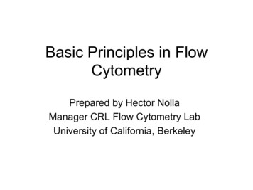
ZEISS Lightsheet 7: Multiview Imaging ofLiving and Cleared Specimens›In Brief›The Advantages›The Applications›The System›Technology and Details› ServiceFlexible. Robust. Easy to Use.Life sciences research can put big demands on your imaging capability, sometimes requiring you to image whole living model organisms, tissues and cells asthey develop. Light sheet fluorescence microscopy (LSFM) with its unique illumination principle is ideal for fast and gentle imaging of such specimens. Theexceptional stability of Lightsheet 7 lets you observe living samples over extendedperiods of time — even days — with less phototoxicity than ever before.What’s more, you can employ this technique to image very large optically clearedspecimens in toto, and with subcellular resolution. Enhance your Lightsheet 7with dedicated optics, sample chambers and sample holders to accurately adjustto the refractive index of your chosen clearing method, and then image yourlarge samples, even whole mouse brains. All of this flexibility comes in the provenand stable boxed light-sheet design from ZEISS.Click here to view this videoMouse brain cleared using CLARITY protocol with final imagingdone in EasyIndex. Label: PV-tdtomato expressed in interneuronsthroughout the brain and in Purkinje cells in the cerebellum.Image data volume: 11 20 8.9 mm. Sample courtesy ofE. Diel and D. Richardson, Harvard University, Cambridge, USA.2
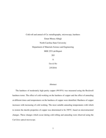
ZEISS Lightsheet 7: Simpler. More Intelligent. More Integrated.›In Brief›The Advantages›The Applications›The System›Technology and Details› ServiceImage Optically Cleared SpecimensGet Best Image Quality and StabilityObserve Real Life – Fast and SensitivelyWhich optical clearing method you choose willTake your LSFM imaging a step further to tackleLightsheet 7 now features the high quantum ef-depend on the type of tissue you are imaging,a broad range of applications and achieve bestficiency of pco.edge sCMOS detectors to enableyour fluorescent labels and the size of the sampleimage quality with your easy-to-use Lightsheet 7.observations of the fastest processes at the lowestitself: Lightsheet 7 is designed to match all ofNewly designed optics and sample chambers letillumination light levels. You'll get a real life viewthese different conditions. You can now imageyou adjust to the perfect refractive index. The newof your samples without the adverse effects ofspecimens at up to 2 cm in size at any refractivesample holder makes mounting larger specimensexcitation light on their biology. For verticallyindex between 1.33 and 1.58, and in almost allsimple. Smart software tools help you adjust im-oriented specimens and highest frame rates, optclearing solutions. Just one stable turnkey systemaging parameters, such as light sheet and samplefor the CMOS detector Axiocam 702: a speciallets you acquire overview images and data withpositions, the right zoom settings, tiles and posi-sample chamber provides heating, cooling andsubcellular resolution. Whether you work with op-tions as well as data processing parameters. All ofCO2 to maintain the perfect environment for yourtically cleared organoids, spheroids, organs, brainsthese new features go hand in hand with the reli-experiments. Add Multiview and triggering op-or other specimens, Lightsheet 7 is your micro-able ZEISS combination of cylindrical lens opticstions to control external devices – Lightsheet 7 isscope of choice for fast, gentle LSFM imaging.and laser scanning to generate the illuminationyour ideal system to observe live processes in anlight sheet. Add the patented Pivot Scan technol-almost unlimited range of organisms.ogy and get artifact-free optical sections with bestimage quality.Mouse kidney cleared with iDISCO, imaged in ethyl cinnamateDedicated optics for your ZEISS Lightsheet 7 allow to tackle awith detection optics 5 / 0.16 foc and Clr 20 / 1.0 nd 1.53broad range of applications with best image quality.Development of Arabidopsis. Red: H2B, mRuby for somatic nuclei.Green: ASY1, eYFP for meiocytes. Courtesy of S. Valuchova,(insert). Red: DyLight 594 for vasculature and glomeruli.P. Mikulkova and K. Riha, Central European Institute of Techno Green: autofluorescence for tissue anatomy.logy (CEITEC), Masaryk University, Brno, Czech Republic.Sample courtesy of U. Roostalu, Gubra, Denmark.3
Your Insight into the Technology Behind It›In Brief›The Advantages›The Applications›The System›Technology and Details› ServiceMaximum Photon Efficiency. Maximum Speed.Maximum Sample Size.Light sheet fluorescence microscopy (LSFM) splits fluorescence excitation anddetection into two separate light paths, with the axis of illumination perpendicular to the detection axis. That means you can illuminate a single thin section of the sample at one time, generating an inherent optical section by exciting only fluorescence from the in-focus plane. No pinhole or image processingis required. Light from the in-focus plane is collected on the pixels of a camera,rather than pixel by pixel as, for example, in confocal or other laser scanningmicroscopes. Parallelization of the image collection on a camera-based detector lets you collect images faster and with less excitation light than you wouldwith many other microscope techniques. In summary, LSFM combines the optical sectioning effect with parallel image acquisition from the complete focalplane. This makes 3D imaging extremely fast and very light efficient.The de-coupling of the detection optics from the illumination optics enablesfluorescence excitation with dedicated lenses at low numerical aperture, without sacrificing detection resolution and sensitivity. This makes LSFM ideal forimaging of samples at the millimeter scale, such as developing organisms orlarge cleared tissue samples.4
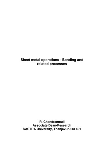
Your Insight into the Technology Behind It›In Brief›The Advantages›The Applications›The System›Technology and Details› ServiceThe Patented Pivot ScannerFigure 1: Without Pivot ScannerDelivers Homogeneous IlluminationWhen the light sheet is passing through thesample, some structures of the specimen, e.g.nuclei, will absorb or scatter the excitation light.This will cast shadows along the illuminationaxis, as you see in Figure 1. This effect occurs inall fluorescence microscopes, but the illumination axis in light sheet fluorescence microscopy isperpendicular to the observation axis and so thiseffect is more obvious.In Lightsheet 7, a patented Pivot Scanner altersthe angle of the light sheet upwards and downwards during image acquisition. By altering theillumination angle the shadows will be cast indifferent directions and excitation light will alsoFigure 2: With Pivot Scannerreach regions behind opaque structures, as yousee in Figure 2. This patented Pivot Scanner is aperfect way to acquire artifact-free images andto improve downstream processing and analysissteps. It is always better to tackle artifacts right attheir origin.5
Expand Your Possibilities›In Brief›The Advantages›The Applications›The System›Technology and Details› ServiceImage Data Processing & AnalysisYour Lightsheet 7 uses ZEN (blue edition) imagingFor the efficient handling of extremely large data is a modular software solution for working withsoftware for data processing, giving you the advan-sets and complex workflows, you can use arivismultichannel 2D, 3D and 4D images of almosttage of the platform's rich portfolio of image pro-Vision4D , bringing you added advantage of pro-unlimited size, independent of available RAM.cessing functionality. This includes ZEISS Multiviewcessing functions like advanced stitching, channelYour Lightsheet 7 generates huge multichannelreconstruction algorithms, deconvolution, fusion ofshift, high resolution volume rendering and muchdatasets that can be handled without constraintsdual side illumination acquisitions and many othermore so you can visualize and quantify your databy arivis Vision4D , which runs on both the ZEISSbenefits. With ZEN (blue edition) you can also stitchin a quick, professional manner. arivis Vision4DStorage & Analysis PC and ACQUIFER HIVE. tiled image datasets with ease.Click here to view this video6
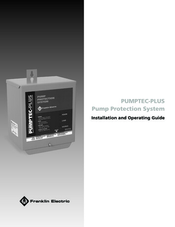
Expand Your Possibilities›In Brief›The Advantages›The Applications›The System›Technology and Details› ServiceLarge Data Storage and ProcessingBecause light sheet fluorescence microscopy(LSFM) can image large three-dimensionalsamples and / or very long time series, it can alsoUsgenerate large datasets in a short period of time.erAs a consequence, you will often be advised to1review your data storage as well as data handlingand processing pipelines when establishing thistechnique in your lab. Storage and computinghardware work best when they are configuredand tuned to the microscope they serve. It'sUsimportant to store the acquired images directly toera safe, fast volume where they can be managed3without further time consuming or expensivecopy processes. In particular, cloud services can-Usernot deal with typical data rates of light sheet and2Usother microscopy techniques, thus making on-erpremise solutions indispensable. Your Lightsheet 74comes with its own ZEISS Storage & Analysis PC,complete with 36 TB storage and computing hardware of a reasonable size for smaller labs.For larger labs, including core facilities andmulti-user environments, you can complementyour Lightsheet 7 with the ACQUIFER HIVE dataIt has proven to be a very good match for ZEISSunit and an autonomous 10 Gbit network infrastruc-platform. This Windows-based, all-in-one solutionLSFM solutions. The basic unit is a 50 terabyteture that connects to microscopy systems. It can beis easy-to-use and includes network and batteries.storage block with a directly-attached processingexpanded or upgraded easily should the need arise.7
Expand Your Possibilities›In Brief›The Advantages›The Applications›The System›Technology and Details› ServiceFlexible Sample Holder DesignCustom designed sample holders can be very beneficial for your LSFM imaging. Your Lightsheet 7is virtually built around your sample, with itsholder designed to best support your experiment's purpose. The new smart sample holderdesign for Lightsheet 7 allows to quickly changethe front end of each holder – depending on yoursample at hand. This interface is open both tocustom design and machining and 3D printing ofyour own sample holder.Visit www.zeiss.com/sampleholder for tricks andtips and to discuss and download custom sampleholders for your most demanding experiments.8
Expand Your Possibilities›In Brief›The Advantages›The Applications›The System›Technology and Details› ServiceMesoscopic ImagingTo make your Lightsheet 7 compatible with evena)larger specimens and low magnification imaging,you can expand your system with the MesoscaleImaging System from Translucence Biosystems.It consists of three components: imaging chamber, specimen holder and safety collar / objectiveadapter. You can now use the excellent optics ofLightsheet 7 for sample volume sizes in the rangeof 3.5 cm3 to perform mesoscopic tissue imaging.The principle of light sheet illumination allowsyou to use widefield low-magnification objectivesto achieve high quality images in a fraction of thetime it would take with other approaches. Forexample, using a Fluar 2.5 / 0.12 objective, youcan image an entire mouse brain with a voxel sizeof 1.8 1.8 12 μm in less than 40 minutes.b)a) shows a sketch of the Translucence Mesoscale ImagingSystem mounted to Lightsheet 7.b) shows a Thy1-EGFP mouse brain cleared and stainedwith a modified version of iDISCO, imaged in a high refractive index solution (RI 1.56) with Fluar 2.5 / 0.12 detectionoptics. Courtesy of S. Gandhi, UC Irvine, USA and TranslucenceBiosystems.1000 µm9
Tailored Precisely to Your Applications›In Brief›The Advantages›The Applications›The System›Technology and DetailsNow you can perform experiments you would never have attempted before. Lightsheet 7 delivers speed in volume imaging. It’s the gentlest way to observe thedevelopment of complete embryos of your model organism and to monitor the fastest physiological processes deep inside the specimen.Furthermore, Lightsheet 7 is the most universal and easy to use microscope to approach a vast range of volume imaging of optically cleared specimens.Typical Applications / Typical SamplesTaskMorphogenesis and Embryogenesis inDevelopmental Biology and Systems BiologyFluorescence imaging of spatio-temporal patterns of gene expression, cell origin and migration, and organogenesis during embryogenesis.Ideal for use with a variety of organisms in developmental biology, providing you with complete imaging of samples such as Drosophila melanogaster,zebrafish, C. elegans and others.Organogenesis and Cell DynamicsFast imaging of cellular dynamics in embryos and small organisms(cell migration, cardiac development, blood flow, vascular development, neuro-development, calcium imaging)3D Cell CultureLive imaging of 3D cell culture, spheroids and cysts, tissue culture, organotypic cultures.Analysis of, e.g., cell migration, expression patterns, cell proliferation.PlantsDevelopmental processes, physiological measurementsImaging of Marine OrganismsFluorescence imaging of marine organisms (e.g., ciona, squid, plankton, flatworms)Structural Imaging of Fixed,Large (mm-sized) SpecimensFluorescence volume imaging of fixed specimens (e.g., early mouse embryos, zebrafish & medaka fish, tissue)Imaging of Optically Cleared SpecimensImaging of fluorescently labeled fixed specimen (tissue sections, mouse brain, embryos, organs, spheroids and biopsies) that are optically cleared withalmost any of the common clearing media with a refractive index between n 1.33 (water) up to n 1.58. Optical properties for high magnification 20 objectives are optimized for Scale A2, (nd 1.38, Hama et al, Nat Neurosci. 2011), FocusClear (by CelExplorer Labs, http://www.celexplorer.com) nd 1.45,the embedding medium for CLARITY (Chung et al, Nature 2013) and U.Clear (nd 1.53, Zhuhao Wu, Icahn School of Medicine, Mount Sinai)› Service10
ZEISS Lightsheet 7 at Work›In Brief›The Advantages›The Applications›The System›Technology and Details› ServiceNephrologyMouse kidney cleared with iDISCO protocol andimaged in ethyl cinnamate with ZEISS Lightsheet 7detection optics 5 / 0.16 foc and Clr 20 / 1.0nd 1.53 (insert). The mouse was perfused withDyLight 594 conjugated tomato lectin to visualizevasculature and glomeruli (red).In green: auto- fluorescence to visualize tissueanatomy. 3D whole organ imaging and computational image analysis of glomerular size andnumber helps to gain a better understandingof the mechanisms of diverse kidney diseases,e.g. diabetic nephropathy. Processed with arivisVision4D on ACQUIFER HIVE.Click here to view this videoClick here to view this videoSample courtesy of U. Roostalu, Gubra, Denmark.11
ZEISS Lightsheet 7 at Work›In Brief›The Advantages›The Applications›The System›Technology and Details› ServiceDevelopmental Biology3D Data set of a P10 mouse trachea displayingthe anatomical organization of mechanosensory nerve fibers. Staining: DAPI, Collagen IV(Alexa 488 antibody), sensorial fibers (reporterstrain expressing tdTomato, Alexa 555 antibody), neurofilament protein NF200 (myelinateds nerve fibers, Alexa 647 antibody). The samplewas cleared in PEGASOS (Jing et al:, 2018, CellResearch) imaged in BB-PEG at RI of 1.54 with5 / 0.16 foc detection optics and Clr 20 / 1.0nd 1.53 respectively. 5 magnification dataset: pixel scaling 0.61 0.61 1.63 micron,3 3 tiles, Zoom 1.5 , 1230 z-sections, volume2.57 2.58 2 mm 20 magnification dataClick here to view this videoClick here to view this videoset: pixel scaling 0.23 μm 0.23 0.58 micron,Sample courtesy of P.-L. Ruffault, C. Birchmeier, Laboratory of Developmental Biology / Signal Transduction; A. Sporbert,1 5 tiles, Zoom 1.0 , 4206 z-sections, volumeM. Richter, Advanced Light Microscopy; M. Delbrück Center for Molecular Medicine, Berlin, Germany2.0 0.45 1.82 mm.12
ZEISS Lightsheet 7 at Work›In Brief›The Advantages›The Applications›The System›Technology and Details› ServiceVertebrate Limb, Spinal Cord RegenerationSalamanders have the remarkable capability toregenerate their limbs and spinal cords. Moleculargenetics tools allow to identify the stem cellsresponsible for this complex regeneration, andthe injury-responsive signals that initiate their proliferation. This axolotl forearm has been clearedin ethyl cinnamate (Masselink, W. et al. Development 146, (2019)) and imaged with 5 / 0.16 focdetection optics at a refractive index of 1.57.The multi-tile data set was aligned, fused andrendered with ZEN imaging software and arivisVision4D software on an ACQUIFER HIVE dataplatform.Click here to view this videoSample courtesy of W. Masselink, Tanaka lab, Research Institute of Molecular Pathology, IMP.Image courtesy of P. Pasierbek, K. Aumayr, IMP BioOptics, Vienna, Austria.13
ZEISS Lightsheet 7 at Work›In Brief›The Advantages›The Applications›The System›Technology and Details› ServiceArabidopis Flower DevelopmentNeuronal MorphologyImmunologyLightsheet 7 allowed to visualize and study theImaging entire cells in the human brain is a taskImaging intact lymphoid organs in 3D allowsstructural development of entire Arabidopsisclose to impossible, due to the tremendouslyto analyze and quantify the immune responseflowers at subcellular resolution over 5 days. Thissophisticated morphology of neurons- and theirto viral infection. T cells were transferred intois an excellent example of gentle long term livespread throughout the entire organ. Organoidswildtype host mice prior to harvest. The node wasimaging with Light Sheet Florescence Microscopyallow the recapitulation of the human brain tocleaned, fixed and cleared using Ce3D (Li et al.(LSFM). Labels: H2B:mRuby2 for visualization ofa certain extent, including the production ofPNAS 163, 2017) prior to imaging at RI 1.49somatic nuclei. ASY1:eYFP expressed specificallyneurons from neuronal stem cell cultures. With(ph 7) with a 5 / 0.16 detection optics (volumein meiocytes. Imaged with W Plan Apo 10 / 0.5ECi clearing, neuronal morphology can be studied2.5 2.5 1.6 mm). The image shows GFP labelledand customized sample mounting and incubationfrom the local to global level, which opens upnative CD8 T cells (yellow), B cell follicles arechamber. Image volume: 587 587 80 μm.fascinating possibilities for the study of neuronalstained using B220 (cyan) and the CD31 vascula-morphology in 3D. 35 day old neuronal organoidsture network (magenta).sparsely labeled with GFP/tdtomato (3% GFP and3% tdtomato) imaged with Clr 20 / 1.0 nd 1.53objective. Pixel scaling: 222 222 567 nm. Image volume: 1.66 0.66 1.6 mm.Click here to view this videoSample courtesy of S. Valuchova, P. Mikulkova and K. Riha,Sample courtesy of D. Reumann and J. Knoblich, IMBA,Sample courtesy of J. Groom, B. Duckworth, The Walter andCentral European Institute of Technology (CEITEC), MasarykVienna, Austria.Eliza Hall Institute of Medical Research, Parkville, Australia.University, Brno, Czech Republic.14
ZEISS Lightsheet 7 at Work›In Brief›The Advantages›The Applications›The System›Technology and Details› ServiceMapping Vasculature of Entire MouseBrain2.5 20 A C57 BL6J mouse was perfused with PBS and4% PFA. The brain was stained perfusing CellTracker CM-DiI Dye – a lipid dye to label thevasculature membranes. The sample was clearedusing iDISCO protocol, equilibrated in ethyl cinnamate as final RIMS. It was then imaged in ethylcinnamate at RI 1.565 with detection opticsFluar 2.5 / 0.12 in a Translucence Mesoscale Imaging Chamber. The high resolution insert imageon the right was acquired with Clr Plan-Neofluar20 / 1.0 Corr nd 1.53.Image volume is 13.1 13.1 6 mm at a pixelresolution of 1.83 1.83 6.77 μm. It was acquiredin about 40 minutes in 4 4 tiles, 866 z-sections.Data volume is 93 GB. Data processed with ZENimaging software and arivis Vision4D on anACQUIFER HIVE data platform.Click here to view this videoClick here to view this videoSample courtesy of E. Diel, D. Richardson, Harvard University, Cambridge, USA.15
ZEISS Lightsheet 7 at Work›In Brief›The Advantages›The Applications›The System›Technology and Details› ServiceMapping Interneurons and Purkinje Cellsof Entire Mouse BrainPV-tdtomato mouse brain was cleared using CLARITY protocol with final imaging donein EasyIndex at a refractive index of RI 1.46.Parvalbumin-Cre yielding expression of tdtomato Parvalbumin is expressed in a population of interneurons throughout the brain and in Purkinje cellsin the cerebellum. The whole brain data set wasacquired on ZEISS Lightsheet 7 with detection optics 5 / 0.16 foc. Image volume is 11 20 8.8 mmat a pixel resolution of 0.91 0.91 5.35 μm(12028 22149 1621 voxels). It was acquired in6 10 tiles, 1621 z-sections. Data volume is 1.2 TB(805 GB after stitching). Data was processed withZEN imaging software and arivis Vision4D on anACQUIFER HIVE data platform.Click here to view this videoSample courtesy of E. Diel, D. Richardson. Harvard University, Cambridge, USA.16
Your Flexible Choice of Components›In Brief›The Advantages›The Applications›The System›Technology and Details541› Service231 Microscope Standalone sealed box system:4 Cameras Clr Plan-Apochromat 20 / 1.0 Corr nd 1.38 Lightsheet 7 detection module “Axiocam”laser safe, no eyepieces, sample chamber, Clr Plan-Neofluar 20 / 1.0 Corr nd 1.45 Lightsheet 7 detection module “pco.edge”sample holder Clr Plan-Neofluar 20 / 1.0 Corr nd 1.53 Selected emission filters and beam splitters Incubation and temperature control options(cooling and heating) Lightsheet 7 detection optics 40 / 1.0(water immersion) CO2-Module5 Software ZEN 3.1 LS (black edition) for image acquisition3 Illumination ZEN 3.1 (blue edition) for image processing2 Objectives Lightsheet 7 illumination optics 5 / 0.1 foc Lightsheet 7 detection optics 5 / 0.16 foc Lightsheet 7 illumination optics 10 / 0.2 foc Lightsheet 7 Multiview Processing Flexible choice of laser lines: 405 nm, 445 nm, 3DXL(water, clearing n 1.33 – 1.58) Lightsheet 7 detection optics 10 / 0.5(water immersion) Lightsheet 7 detection optics 20 / 1.0488 nm, 515 nm, 561 nm, 638 nm Transmission LED for sample positioningand analysis Deconvolution arivis Vision4D and overview(water immersion)17
ZEISS Lightsheet 7: System Overview›››In BriefThe AdvantagesThe Applications›The System›Technology and DetailsSample holder syringe,capillary and universalfor Lightsheet 7Liquid cooling unitLCS-BUshaftHolderplate,pedestal standard,pedestal extendedDetection module"Axiocam"Sample holder setfor large samplesfor Lightsheet 7Detection module"pco.edge"Stemclaw5ConnectorReflector turretfor Laser blockingfilterReflector turretfor Emission selection› Servicedisk4321Holder1 scaffold large w / plate2 scaffold standard w / plate3 pedestal short4 pedestal intermediate5 pedestal longLightsheet7WaterLightsheet 7Detection optics 5x/0.16 foc Clr Plan-NeofluarClearingIncubation CO2-Module Lightsheet 7 TempModule S1 TempModule CZ-LSFM5x/0.1610x/0.520x/1.040x/1.020x/1.0 Corr nd 1.53 Clr Plan-Neofluar20x/1.0 Corr nd 1.45Lightsheet 7Illumination opticsMain system moduleLightsheet 7 5x/0.1 foc 10x/0.2 foc Clr Plan-Apochromat20x/1.0 Corr nd 1.38 2.5x/0.12Sample chamber - large,with carriage,Lightsheet 7Sample chamber - Clearing 20x,with carriage,Lightsheet 7Laserbench rackLightsheet 7ErgoDrivecontrol panelSample chamber - Water,with carriage,Lightsheet 7IncubationPeltier block SLCD TFTflat screen monitor27" or 32"Motioncontroller unit(MCU)Incubationtemperature sensorLightsheet 7PC for system controlLightsheet 7Storage and data analysis PCLightsheet 7System table, small,air-damped,level regulated,pneumatic supply,900 x 750 mm18
Technical Specifications›››In BriefComponentDescriptionIllumination OpticsLightsheet 7 illumination optics 5 / 0.1 focThe AdvantagesThe Applications›The System›Technology and DetailsLightsheet 7 illumination optics 10 / 0.2 focIlluminationTransmission LED for sample positioning and overviewFlexible choice of laser lines: 405 nm, 445 nm, 488 nm, 515 nm, 561 nm, 638 nm at various output power levelsDetection ModulesDetection Module “Axiocam”, Axiocam 702 sCMOS with QE up to 78%, 1216 1920 pixels, pixel size 5.86 µm 5.86 µmDetection Module “pco.edge”, pco.edge 4.2 CLHS with up to 82% QE,sCMOS, 1920 1920 pixels, pixel size 6.5 µm 6.5 µm (requires liquid cooling)Detection Optics› ServiceLightsheet 7 detection optics 5 / 0.16 (water immersion, WD 5.1 mm)Lightsheet 7 detection optics 5 / 0.16 foc (clearing immersion nd 1.33 – 1.58), WD 10.5 mm)Lightsheet 7 detection optics 10 / 0.5 (water immersion, WD 3.7 mm)Lightsheet 7 detection optics 20 / 1.0 (water immersion, WD 2.4 mm)Clr Plan-Apochromat 20 / 1.0 Corr nd 1.38 (clearing immersion, WD 5.6 mm)Clr Plan-Neofluar 20 / 1.0 Corr nd 1.45 (clearing immersion, WD 5.6 mm)Clr Plan-Neofluor 20 / 1.0 Corr nd 1.53 (clearing immersion, WD 6.4 mm)Lightsheet 7 detection optics 40 / 1.0 (water immersion, WD 2.5 mm)Sample Chamber, Sample Holder, ConsumablesStarter kits and all necessary accessories for your experimentsThe following chambers are available: Water chamber (n 1.33) to be used with 5 , 10 , 20 and 40 water immersion lenses 20 Clearing chamber (n 1.35 – 1.58) to be used with Clr 20 immersion lenses Large sample chamber (n 1.33 – 1.58) to be used with 5 foc objective Translucence chamber to be used with 2.5 and 5 detection opticsSoftware ProcessingLightsheet 7 Multiview Processing, Dual side Fusion, Stiching3DXL, arivis Vision4D DeconvolutionSoftware AcquisitionMultidimensional imaging (time, postions, tiles, multiview)Combination of multidimensions possible with the exception of Multiview and Tiling)Semi-automatic z-offset compensationMean and maximum fusion for dual side illumination and multiviewRI compensation for optical sectioning and image size19
Technical Specifications›In Brief›The Advantages›The Applications›The System›Technology and Details› ServiceComponentDescriptionSystem PCHP Z6 G4 workstationChipset: Intel C622Memory: max. 192 GB RAMSSD: 1 512 GB M.2 NVMeHard Drives: 2 4 TB SATA 7200 rpm (configured as 4 TB RAID 1 hard drive); increase capacity from 4 TB (RAID 1) to 8 TB (RAID 10)Processor: Intel Xeon Gold 6134 (3.2 GHz, 24.75 MB cache, 8 cores)Graphics Card: NVIDIA Quadro P4000 8GB DPNetwork Adapter: 2 10 GbE RJ45 (hp Z6); additional network adapter 2 10 GbE RJ45 (hp Z6) e.g. for connection of storage systemsOperating system: Windows 10 IoT Enterprise 2016 LTSC Embedded x64Storage and Data Analysis PCCPU: Intel P XEON E5-2620V3 2,4 GHz LGA2011 L3 15MB BoxGraphics Card: NVIDIA Quadro P4000 8GB DP or NVIDIA Quadro P6000 24GB DPMemory: 64 GB (4 16 GB) included, max. 256 GB RAM;Memory slots: 16 DIMM slotsHard Drives: 6 HDD 8 TB, RAID 5 configured to 36 TB data storage volume; 2 Solid State Drive 240 GB for pagefile and operating system10 Gbit Ethernet on motherboard and 10 GbE cable to connect with PC for system control (high speed data streaming)Network Adapter: LAN: 2 10 GbE5 USB 3.0, 4 USB 2.0 portsOperating system: Windows 10IncubationPeltierblock Sample Chamber with Temperature Sensor with controller TempModule S1 and TempModule CZ-LSFMCO2-ModuleHeating Device HumidityTriggerTrigger-out signal via BNC connector. High level of 3.3 V (nominal value of the high level: 3.2 V 4.0 V,and nominal value of the low level: 0 V 0.4 V). The minimal working resistance is 5 kΩ.20
Technical Specifications›In BriefMicroscopeStandalone box system, sealed, turnkey, laser safe, no eyepieces›The AdvantagesPhysical DimensionsApprox. Width Depth HeightApprox. Weight›The ApplicationsMain System Module Lightsheet 7800 mm 450 mm 500 mm75 kgLaser Rack “LB Rack Lightsheet“600 mm 700 mm 550 mm80 kgSystem Table for main System ModuleLightsheet 7, Level regulated900 mm 750 mm 770 mm90 kgTransmission Contrast for OverviewIR LED illumination, no Köhler Illumination, not specified for high quality imagingSpectral Range of Detection400 – 740 nm›The System›Technology and Details› ServiceDual Camera Port for simultaneous2 Channel DetectionDetection zoom0.36 – 2.5 , continuousFor imaging, the zoom range of 0.7 – 2.5 is recommended,0.36 – 0.7 for sample positioning onlyField of View123 μm to 3.5 mm2.8 mm image diagonal, 5 detection lens, zoom 0.7 ,for sample positioning (zoom 0.36 ) 5 mmEmbedded Specimen SizeFrom 1 µm to 10 10 20 mmSample MountingDedicated sample chambers for live or cleared samples of up to 10 10 20 mm3 in size (approximation: an ellipsoid shape).Universal sample holder for embedded samples;Sample holder with claw and flexible adapters for large or cleared samples.Immersion and Incubation MediaSample chambers and optics designed for aqueous media (nd 1.33) or clearing media nd 1.35 – 1.58 for Clr 20 sample chamber;1.33 – 1.58 for large sample chamber.Light sheet thickness2 μm – approx. 14 μmDepending on sample, at 488 nm21
Technical Specifications›In Brief›The Advantages›The Applications›The System›Technology and DetailsDetection ModulesUp to two detection modules of the same type can be connected to the dual camera portDetection Module “Axiocam“Axiocam 702 mono, Sony MX 174 sensor, aligned on a C-mount for optimized image alignment on dual camera portDetection Module “pco.edge“› ServiceData Acquisition RatePixel size5.86 µmMax. pixel format1216 1920 pixel, (2.3 Megapixel)Bit depth14 bitMax. frame rate QE up to 78%100 fps at 1024 1024 pixel, in continuous z-drive modepco.edge 4.2 CLHS, sCMOS sensor, requires liquid cooling, aligned on a special C-mount for optimized image alignment on dual camera portPi
In summary, LSFM combines the opti-cal sectioning effect with parallel image acquisition from the complete focal plane. This makes 3D imaging extremely fast and very light efficient. . With ZEN (blue edition) you can also stitch