Transcription
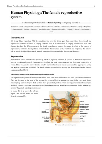
Human Physiology/The female reproductive system1Human Physiology/The female reproductivesystem The male reproductive system — Human Physiology — Pregnancy and birth Homeostasis — Cells — Integumentary — Nervous — Senses — Muscular — Blood — Cardiovascular — Immune — Urinary — Respiratory— Gastrointestinal — Nutrition — Endocrine — Reproduction (male) — Reproduction (female) — Pregnancy — Genetics — Development —AnswersIntroductionAll living things reproduce. This is something that sets the living apart from non-living. Even though thereproductive system is essential to keeping a species alive, it is not essential to keeping an individual alive. Thischapter describes the different parts of the female reproductive system: the organs involved in the process ofreproduction, hormones that regulate a woman's body, the menstrual cycle, ovulation and pregnancy, the female'srole in genetic division, birth control, sexually transmitted diseases and other diseases and disorders.ReproductionReproduction can be defined as the process by which an organism continues its species. In the human reproductiveprocess, two kinds of sex cells ( gametes), are involved: the male gamete (sperm), and the female gamete (egg orovum). These two gametes meet within the female's uterine tubes located one on each side of the upper pelvic cavity,and begin to create a new individual. The female needs a male to fertilize her egg; she then carries offspring throughpregnancy and childbirth.Similarities between male and female reproductive systemsThe reproductive systems of the male and female have some basic similarities and some specialized differences.They are the same in that most of the reproductive organs of both sexes develop from similar embryonic tissue,meaning they are homologous. Both systems have gonads that produce (sperm and egg or ovum) and sex organs.And both systems experience maturation of their reproductive organs, which become functional during puberty as aresult of the gonads secreting sex hormones.In short, this is a known list of sexorgans that evolve from the sametissue in a human life.The human male reproductive system
Human Physiology/The female reproductive system2Cross-sectional diagram of the femalereproductive ullerian ductAppendix testisFallopian tubesMullerian ductProstatic utricleUterus, proximalWolffian ductRete testisRete ovariiMesonephric tubules Efferent ductsEpoophoronWolffian ductEpididymisGartner's ductWolffian ductVas deferensWolffian ductSeminal vesicleWolffian ductProstateSkene's glandsUrogenital sinusBladder, urethraBladder, urethra, distalUrogenital sinusBulbourethral gland Bartholin's glandGenital swellingScrotumLabia majoraUrogenital foldsDistal urethraLabia minoraGenital tuberclePenisClitorisPrepuceClitoral hoodBulb of penisVestibular bulbsGlans penisClitoral glansCrus of penisClitoral cruraDifferences between male and female reproductive systemsThe differences between the female and male reproductive systems are based on the functions of each individual'srole in the reproduction cycle. A male who is healthy, and sexually mature, continuously produces sperm. Thedevelopment of women's "eggs" are arrested during fetal development. This means she is born with a predeterminednumber of oocytes and cannot produce new ones.At about 5 months gestation, the ovaries contain approximately six to seven million oogonia, which initiate meiosis.The oogonia produce primary oocytes that are arrested in prophase I of meiosis from the time of birth until puberty.After puberty, during each menstrual cycle, one or several oocytes resume meiosis and undergo their first meioticdivision during ovulation. This results in the production of a secondary oocyte and one polar body. The meioticdivision is arrested in metaphase II. Fertilization triggers completion of the second meiotic division and the result isone ovum and an additional polar body.
Human Physiology/The female reproductive system3The ovaries of a newborn baby girl contain about one million oocytes. This number declines to 400,000 to 500,000by the time puberty is reached. On average, 500-1000 oocytes are ovulated during a woman's reproductive lifetime.When a young woman reaches puberty around age 10 to 13, a promary oocyte is discharged from one of the ovariesevery 28 days. This continues until the woman reaches menopause, usually around the age of 50 years. Occytes arepresent at birth, and age as a woman ages.Female Reproductive System Produces eggs (ova)Secretes sex hormonesReceives the male spermatazoa duringProtects and nourishes the fertilized egg until it is fully developedDelivers fetus through birth canalProvides nourishment to the baby through milk secreted by mammary glands in the breastExternal GenitalsVulvaThe external female genitalia is referred to as vulva. Itconsists of the labia majora and labia minora (whilethese names translate as "large" and "small" lips, oftenthe "minora" can protrude outside the "majora"), monspubis, clitoris, opening of the urethra (meatus), vaginalvestibule, vestibular bulbs, vestibular glands.The term "vagina" is often improperly used as a genericterm to refer to the vulva or female genitals, eventhough - strictly speaking - the vagina is a specificinternal structure and the vulva is the exterior genitaliaonly. Calling the vulva the vagina is akin to calling themouth the throat.Mons VenerisThe mons veneris, Latin for "mound of Venus"(Roman Goddess of love) is the soft mound at the frontof the vulva (fatty tissue covering the pubic bone). It isalso referred to as the mons pubis. The mons venerisprotects the pubic bone and vulva from the impact ofsexual intercourse. After puberty, it is covered with pubic hair, usually in a triangular shape. Heredity can play a rolein the amount of pubic hair an individual grows.Labia MajoraThe labia majora are the outer "lips" of the vulva. They are pads of loose connective and adipose tissue, as well assome smooth muscle. The labia majora wrap around the vulva from the mons pubis to the perineum. The labiamajora generally hides, partially or entirely, the other parts of the vulva. There is also a longitudinal separation calledthe pudendal cleft. These labia are usually covered with pubic hair. The color of the outside skin of the labia majorais usually close to the overall color of the individual, although there may be some variation. The inside skin isusually pink to light brown. They contain numerous sweat and oil glands. It has been suggested that the scent fromthese oils are sexually arousing.Labia Minora
Human Physiology/The female reproductive systemMedial to the labia majora are the labia minora. The labia minora are the inner lips of the vulva. They are thinstretches of tissue within the labia majora that fold and protect the vagina, urethra, and clitoris. The appearance oflabia minora can vary widely, from tiny lips that hide between the labia majora to large lips that protrude. There is nopubic hair on the labia minora, but there are sebaceous glands. The two smaller lips of the labia minora cometogether longitudinally to form the prepuce, a fold that covers part of the clitoris. The labia minora protect thevaginal and urethral openings. Both the inner and outer labia are quite sensitive to touch and pressure.ClitorisThe clitoris, visible as the small white oval between the top of the labia minoraand the clitoral hood, is a small body of spongy tissue that functions solely forsexual pleasure. Only the tip or glans of the clitoris shows externally, but theorgan itself is elongated and branched into two forks, the crura, which extenddownward along the rim of the vaginal opening toward the perineum. Thus theclitoris is much larger than most people think it is, about 4" long on average.The clitoral glans or external tip of the clitoris is protected by the prepuce, orclitoral hood, a covering of tissue similar to the foreskin of the male penis.However, unlike the penis, the clitoris does not contain any part of the urethra.During sexual excitement, the clitoris erects and extends, the hood retracts,making the clitoral glans more accessible. The size of the clitoris is variable between women. On some, the clitoralglans is very small; on others, it is large and the hood does not completely cover it.UrethraThe opening to the urethra is just below the clitoris. Although it is not related to sex or reproduction, it is included inthe vulva. The urethra is actually used for the passage of urine. The urethra is connected to the bladder. In femalesthe urethra is 1.5 inches long, compared to males whose urethra is 8 inches long. Because the urethra is so close tothe anus, women should always wipe themselves from front to back to avoid infecting the vagina and urethra withbacteria. This location issue is the reason for bladder infections being more common among females.HymenThe hymen is a thin fold of mucous membrane that separates the lumen of the vagina from the urethral sinus.Sometimes it may partially cover the vaginal orifice. The hymen is usually perforated during later fetal development.Because of the belief that first vaginal penetration would usually tear this membrane and cause bleeding, its"intactness" has been considered a guarantor of virginity. However, the hymen is a poor indicator of whether awoman has actually engaged in sexual intercourse because a normal hymen does not completely block the vaginalopening. The normal hymen is never actually "intact" since there is always an opening in it. Furthermore, there is notalways bleeding at first vaginal penetration. The blood that is sometimes, but not always, observed after firstpenetration can be due to tearing of the hymen, but it can also be from injury to nearby tissues.A tear to the hymen, medically referred to as a "transection," can be seen in a small percentage of women or girlsafter first penetration. A transection is caused by penetrating trauma. Masturbation and tampon insertion can, butgenerally are not forceful enough to cause penetrating trauma to the hymen. Therefore, the appearance of the hymenis not a reliable indicator of virginity or chastity.PerineumThe perineum is the short stretch of skin starting at the bottom of the vulva and extending to the anus. It is a diamondshaped area between the symphysis pubis and the coccyx. This area forms the floor of the pelvis and contains theexternal sex organs and the anal opening. It can be further divided into the urogenital triangle in front and the analtriangle in back.The perineum in some women may tear during the birth of an infant and this is apparently natural. Some physicianshowever, may cut the perineum preemptively on the grounds that the "tearing" may be more harmful than a precise4
Human Physiology/The female reproductive systemcut by a scalpel. If a physician decides the cut is necessary, they will perform it. The cut is called an episiotomy.Internal GenitalsVaginaThe vagina is a muscular, hollow tube that extends from the vaginal opening to the cervix of the uterus. It is situatedbetween the urinary bladder and the rectum. It is about three to five inches long in a grown woman. The muscularwall allows the vagina to expand and contract. The muscular walls are lined with mucous membranes, which keep itprotected and moist. A thin sheet of tissue with one or more holes in it, called the hymen, partially covers theopening of the vagina. The vagina receives sperm during sexual intercourse from the penis. The sperm that survivethe acidic condition of the vagina continue on through to the fallopian tubes where fertilization may occur.The vagina is made up of three layers, an inner mucosal layer, a middle muscularis layer, and an outer fibrous layer.The inner layer is made of vaginal rugae that stretch and allow penetration to occur. These also help with stimulationof the penis. microscopically the vaginal rugae has glands that secrete an acidic mucus (pH of around 4.0.) that keepsbacterial growth down. The outer muscular layer is especially important with delivery of a fetus and placenta.Purposes of the Vagina Receives a males erect penis and semen during sexual intercourse.Pathway through a woman's body for the baby to take during childbirth.Provides the route for the menstrual blood (menses) from the uterus, to leave the body.May hold forms of birth control, such as a diaphragm, FemCap, Nuva Ring, or female condom.Clinical Application:Pelvic inflammatory disease (PID) is a widespread infection that originates in the vagina and uterus and spreads tothe uterine tubes, ovaries, and ultimately the pelvic peritoneum. This condition, which occurs in about 10% ofwomen is usually caused by chlamydial or gonorrheal infection, other bacteria infecting the vagina may be involvedas well. Signs and symptoms include tenderness of the lower abdomen, fever, and a vaginal discharge. Even a singleepisode of PID can cause infertility, due to scarring that blocks the uterine tubes. Therefore, patients are immediatelygiven broad-spectrum antibiotics whenever PID is suspected.CervixThe cervix (from Latin "neck") is the lower, narrow portion of the uterus where it joins with the top end of thevagina. Where they join together forms an almost 90 degree curve. It is cylindrical or conical in shape and protrudesthrough the upper anterior vaginal wall. Approximately half its length is visible with appropriate medical equipment;the remainder lies above the vagina beyond view. It is occasionally called "cervix uteri", or "neck of the uterus".During menstruation, the cervix stretches open slightly to allow the endometrium to be shed. This stretching isbelieved to be part of the cramping pain that many women experience. Evidence for this is given by the fact thatsome women's cramps subside or disappear after their first vaginal birth because the cervical opening has widened.The portion projecting into the vagina is referred to as the portio vaginalis or ectocervix. On average, the ectocervixis three cm long and two and a half cm wide. It has a convex, elliptical surface and is divided into anterior andposterior lips. The ectocervix's opening is called the external os. The size and shape of the external os and theectocervix varies widely with age, hormonal state, and whether the woman has had a vaginal birth. In women whohave not had a vaginal birth the external os appears as a small, circular opening. In women who have had a vaginalbirth, the ectocervix appears bulkier and the external os appears wider, more slit-like and gaping.The passageway between the external os and the uterine cavity is referred to as the endocervical canal. It varieswidely in length and width, along with the cervix overall. Flattened anterior to posterior, the endocervical canalmeasures seven to eight mm at its widest in reproductive-aged women. The endocervical canal terminates at the5
Human Physiology/The female reproductive systeminternal os which is the opening of the cervix inside the uterine cavity.During childbirth, contractions of the uterus will dilate the cervix up to 10 cm in diameter to allow the child to passthrough. During orgasm, the cervix convulses and the external os dilates.UterusThe uterus is shaped like an upside-down pear, with a thick lining and muscular walls. Located near the floor of thepelvic cavity, it is hollow to allow a blastocyte, or fertilized egg, to implant and grow. It also allows for the innerlining of the uterus to build up until a fertilized egg is implanted, or it is sloughed off during menses.The uterus contains some of the strongest muscles in the female body. These muscles are able to expand and contractto accommodate a growing fetus and then help push the baby out during labor. These muscles also contractrhythmically during an orgasm in a wave like action. It is thought that this is to help push or guide the sperm up theuterus to the fallopian tubes where fertilization may be possible.The uterus is only about three inches long and two inches wide, but during pregnancy it changes rapidly anddramatically. The top rim of the uterus is called the fundus and is a landmark for many doctors to track the progressof a pregnancy. The uterine cavity refers to the fundus of the uterus and the body of the uterus.Helping support the uterus are ligaments that attach from the body of the uterus to the pelvic wall and abdominalwall. During pregnancy the ligaments prolapse due to the growing uterus, but retract after childbirth. In some casesafter menopause, they may lose elasticity and uterine prolapse may occur. This can be fixed with surgery.Some problems of the uterus include uterine fibroids, pelvic pain (including endometriosis, adenomyosis), pelvicrelaxation (or prolapse), heavy or abnormal menstrual bleeding, and cancer. It is only after all alternative optionshave been considered that surgery is recommended in these cases. This surgery is called hysterectomy. Hysterectomyis the removal of the uterus, and may include the removal of one or both of the ovaries. Once performed it isirreversible. After a hysterectomy, many women begin a form of alternate hormone therapy due to the lack of ovariesand hormone production.6
Human Physiology/The female reproductive systemFallopian TubesAt the upper corners of the uterus are the fallopian tubes. There are two fallopian tubes, also called the uterine tubesor the oviducts. Each fallopian tube attaches to a side of the uterus and connects to an ovary. They are positionedbetween the ligaments that support the uterus. The fallopian tubes are about four inches long and about as wide as apiece of spaghetti. Within each tube is a tiny passageway no wider than a sewing needle. At the other end of eachfallopian tube is a fringed area that looks like a funnel. This fringed area, called the infundibulum, lies close to theovary, but is not attached. The ovaries alternately release an egg. When an ovary does ovulate, or release an egg, it isswept into the lumen of the fallopian tube by the fimbriae.Once the egg is in the fallopian tube, tiny hairs in the tube's lining help push it down the narrow passageway towardthe uterus. The oocyte, or developing egg cell, takes four to five days to travel down the length of the fallopian tube.If enough sperm are ejaculated during sexual intercourse and there is an oocyte in the fallopian tube, fertilization willoccur. After fertilization occurs, the zygote, or fertilized egg, will continue down to the uterus and implant itself inthe uterine wall where it will grow and develop.If a zygote doesn't move down to the uterus and implants itself in the fallopian tube, it is called a ectopic or tubalpregnancy. If this occurs, the pregnancy will need to be terminated to prevent permanent damage to the fallopiantube, possible hemorrhage and possible death of the mother.7
Human Physiology/The female reproductive system8Mammary glandsMammary glands are the organs that produce milk for thesustenance of a baby. These exocrine glands are enlarged andmodified sweat glands.StructureThe basic components of the mammary gland are the alveoli(hollow cavities, a few millimetres large) lined with milk-secretingepithelial cells and surrounded by myoepithelial cells. Thesealveoli join up to form groups known as lobules, and each lobulehas a lactiferous duct that drains into openings in the nipple. Themyoepithelial cells can contract, similar to muscle cells, andthereby push the milk from the alveoli through the lactiferousducts towards the nipple, where it collects in widenings (sinuses)of the ducts. A suckling baby essentially squeezes the milk out ofthese sinuses.The development of mammary glands is controlled by hormones.Cross section of the breast of a human female.The mammary glands exist in both sexes, but they are rudimentaryuntil puberty when - in response to ovarian hormones - they begin to develop in the female. Estrogen promotesformation, while testosterone inhibits it.At the time of birth, the baby has lactiferous ducts but no alveoli. Little branching occurs before puberty whenovarian estrogens stimulate branching differentiation of the ducts into spherical masses of cells that will becomealveoli. True secretory alveoli only develop in pregnancy, where rising levels of estrogen and progesterone causefurther branching and differentiation of the duct cells, together with an increase in adipose tissue and a richer bloodflow.Colostrum is secreted in late pregnancy and for the first few days after giving birth. True milk secretion (lactation)begins a few days later due to a reduction in circulating progesterone and the presence of the hormone prolactin. Thesuckling of the baby causes the release of the hormone oxytocin which stimulates contraction of the myoepithelialcells.The cells of mammary glands can easily be induced to grow and multiply by hormones. If this growth runs out ofcontrol, cancer results. Almost all instances of breast cancer originate in the lobules or ducts of the mammary glands.STRUCTURELOCATION & DESCRIPTIONFUNCTIONBreastsUpper chest one on each side containing alveolarcells (milk production), myoepithelial cells(contract to expel milk), and duct walls (help withextraction of milk).Lactation milk/nutrition for newborn.CervixThe lower narrower portion of the uterus.During childbirth, contractions of the uterus will dilate the cervix up to 10cm in diameter to allow the child to pass through. During orgasm, thecervix convulses and the external os dilatesClitorisSmall erectile organ directly in front of thevestibule.Sexual excitation, engorged with blood.Fallopian tubesExtending upper part of the uterus on either side.Egg transportation from ovary to uterus (fertilization usually takes placehere).HymenThin membrane that partially covers the vagina inyoung females.
Human Physiology/The female reproductive system9Labia majoraOuter skin folds that surround the entrance to thevagina.Lubrication during mating.Labia minoraInner skin folds that surround the entrance to thevagina.Lubrication during mating.MonsMound of skin and underlying fatty tissue, centralin lower pelvic regionOvaries (femalegonads)Pelvic region on either side of the uterus.PerineumShort stretch of skin starting at the bottom of thevulva and extending to the anus.UrethraPelvic cavity above bladder, tilted.Passage of urine.UterusCenter of pelvic cavity.To house and nourish developing human.VaginaCanal about 10-8 cm long going from the cervix tothe outside of the body.Receives penis during mating. Pathway through a womans body for thebaby to take during childbirth. Provides the route for the menstrual blood(menses) from the uterus, to leave the body. May hold forms of birthcontrol, such as an IUD, diaphragm, neva ring, or female condomVulvaSurround entrance to the reproductivetract.(encompasses all external genitalia)EndometriumThe innermost layer of uterine wall.Contains glands that secrete fluids that bathe the utrine lining.MyometriumSmooth muscle in uterine wall.Contracts to help expel the baby.Provides an environment for maturation of oocyte. Synthesizes andsecretes sex hormones (estrogen and progesterone).The Female Reproductive CycleTowards the end of puberty, girls begin to release eggs as part of a monthly period called the female reproductivecycle, or menstrual cycle (menstrual referring to "monthly"). Approximately every 28 days, during ovulation, anovary sends a tiny egg into one of the fallopian tubes. Unless the egg is fertilized by a sperm while in the fallopian inthe two to three days following ovulation, the egg dries up and leaves the body about two weeks later through thevagina. This process is called menstruation. Blood and tissues from the inner lining of the uterus (the endometrium)combine to form the menstrual flow, which generally lasts from four to seven days. The first period is calledmenarche. During menstruation arteries that supply the lining of the uterus constrict and capillaries weaken. Bloodspilling from the damaged vessels detaches layers of the lining, not all at once but in random patches. Endometriummucus and blood descending from the uterus, through the liquid creates the menstruation flow.
Human Physiology/The female reproductive systemThe reproductive cycle can be divided into anovarian cycle and a uterine cycle (compareovarian histology and uterine histology in thediagram on the right). During the uterinecycle, the endometrial lining of the uterusbuilds up under the influence of increasinglevels of estrogen (labeled as estradiol in theimage). Follicles develop, and within a fewdays one matures into an ovum, or egg. Theovary then releases this egg, at the time ofovulation. After ovulation the uterine liningenters a secretory phase, or the ovarian cycle,in preparation for implantation, under theinfluence of progesterone. Progesterone isproduced by the corpus luteum (the follicleafter ovulation) and enriches the uterus with athick lining of blood vessels and capillaries sothat it can sustain the growing fetus. Iffertilization and implantation occur, theembryoproducesHumanChorionicGonadotropin (HCG), which maintains thecorpus luteum and causes it to continueproducing progesterone until the placenta canMenstrual cycletake over production of progesterone. Hence,progesterone is "pro gestational" andmaintains the uterine lining during all of pregnancy. If fertilization and implantation do not occur the corpus luteumdegenerates into a corpus albicans, and progesterone levels fall. This fall in progesterone levels cause theendometrium lining to break down and sluff off through the vagina. This is called menstruation, which marks thelow point for estrogen activity and is the starting point of a new cycle.Common usage refers to menstruation and menses as a period. This bleeding serves as a sign that a woman has notbecome pregnant. However, this cannot be taken as certainty, as sometimes there is some bleeding in earlypregnancy. During the reproductive years, failure to menstruate may provide the first indication to a woman that shemay have become pregnant.Menstruation forms a normal part of a natural cyclic process occurring in healthy women between puberty and theend of the reproductive years. The onset of menstruation, known as menarche, occurs at an average age of 12, but isnormal anywhere between 8 and 16. Factors such as heredity, diet, and overall health can accelerate or delay theonset of menarche.Signs of ovulationThe female body produces outward signs that can be easily recognized at the time of ovulation. The two main signsare thinning of the cervical mucus and a slight change in body temperature.Thinning of the Cervical MucusAfter menstruation and right before ovulation, a woman will experience an increase of cervical mucus. At first, itwill be thick and yellowish in color and will not be very plentiful. Leading up to ovulation, it will become thinnerand clearer. On or around the day of ovulation, the cervical mucus will be very thin, clear and stretchy. It can becompared to the consistency of egg whites. This appearance is known as 'spinnbarkeit'.10
Human Physiology/The female reproductive systemTemperature ChangeA woman can also tell the time of ovulation by taking her basal body temperature daily. This is a temperature takenwith a very sensitive thermometer first thing in the morning before the woman gets out of bed. The temperature isthen tracked to show changes. In the uterine cycle, a normal temperature will be around 97.0 – 98.0. The day ofovulation the temperature spikes down, usually into the 96.0 – 97.0 range and then the next morning it will spike upto normal of around 98.6 and stay in that range until menstruation begins.Both of these methods are used for conception and contraception. They are more efficient in conception due to thefact that sperm can live for two to three days inside of the fallopian tubes. A woman could be off by a couple of daysin her calculations and still become pregnant.Menopause is the physiological cessation of menstrual cycles associated with advancing age. Menopause issometimes referred to as "the change of life" or climacteric. Menopause occurs as the ovaries stop producingestrogen, causing the reproductive system to gradually shut down. As the body adapts to the changing levels ofnatural hormones, vasomotor symptoms such as hot flashes and palpitations, psychological symptoms such asincreased depression, anxiety, irritability, mood swings and lack of concentration, and atrophic symptoms such asvaginal dryness and urgency of urination appear. Together with these symptoms, the woman may also haveincreasingly scanty and erratic menstrual periods.Technically, menopause refers to the cessation of menses; the gradual process through which this occurs, whichtypically takes a year but may last as little as six months or more than five years, is known as climacteric. A naturalor physiological menopause is that which occurs as a part of a woman's normal aging process. However, menopausecan be surgically induced by such procedures as hysterectomy.The average onset of menopause is 50.5 years, but some women enter menopause at a younger age, especially if theyhave suffered from cancer or another serious illness and undergone chemotherapy. Premature menopause is definedas menopause occurring before the age of 40, and occurs in 1% of women. Other causes of premature menopauseinclude autoimmune disorders, thyroid disease, and diabetes mellitus.Premature menopause is diagnosed by measuring the levels of follicle stimulating hormone (FSH) and luteinizinghormone (LH). The levels of these hormones will be higher if menopause has occurred. Rates of prematuremenopause have been found to be significantly higher in both fraternal and identical twins; approximately 5% oftwins reach menopause before the age of 40. The reasons for this are not completely understood. Post-menopausalwomen are at increased risk of osteop
Human Physiology/The female reproductive system 5 cut by a scalpel. If a physician decides the cut is necessary, they will perform it. The cut is called an episiotomy. Internal Genitals Vagina The vagina is a muscular, hollow tube that extends from the va