Transcription
Kong et al. BMC Complementary and Alternative Medicine 2014, 1RESEARCH ARTICLEOpen AccessAngelica sinensis extract inhibits RANKL-mediatedosteoclastogenesis by down-regulated theexpression of NFATc1 in mouse bone marrow cellsLingbo Kong1†, Qinpeng Zhao1†, Xiaodong Wang1, Jinyu Zhu2, Dingjun Hao1* and Chongfei Yang2*AbstractBackground: Destructive erosion of bone or osteolysis is a major complication of inflammatory conditions such asrheumatoid arthritis (RA), periodontal disease, and periprosthetic osteolysis. Natural plant-derived products havereceived recent attention as potential therapeutic and preventative drugs in human disease.Methods: The effect of Angelica sinensis (AS) extract on RANKL-induced osteoclast differentiation was examined inthis study. The osteoclast precursor cell line bone marrow macrophages (BMMs) was cultured and stimulated withRANKL followed by treatment with AS at several doses. Gene expression profiles of c-Fos, c-Jun, NFATc1, TRAP, andOSCAR were sequentially evaluated.Results: AS extract inhibited RANKL-mediated osteoclast differentiation in BMMs in a dose-dependent mannerwithout any evidence of cytotoxicity. AS extract strongly inhibited p38, ERK, JNK, p65 phosphorylation and I-κBdegradation in RANKL-stimulated BMMs. AS extract also inhibited the mRNA expression of c-Fos, c-Jun, NFATc1,TRAP, and OSCAR in RANKL-treated BMMs. Moreover, RANKL-induced c-Fos, c-Jun and NFATc1 protein expressionwas suppressed by AS extract.Conclusions: These results collectively suggested that AS extract demonstrated inhibitory effects on RANKL-mediatedosteoclast differentiation in bone marrow macrophages in vitro, indicating that AS may therefore serve as a useful drugin the prevention of bone loss.Keywords: Angelica sinensis, Osteoclastogenesis, NFATc1, BMMsBackgroundOsteoclasts are derived from hematopoietic stem cells,which are unique multinucleated cells responsible forbone resorption [1]. Osteoblasts and stromal cells expressreceptor activators of the nuclear factor-kB (NF-kB) ligand(RANKL) and macrophage colony-stimulating factor (MCSF) [2]. M-CSF induces expression of RANKL receptor(RANK) as well as supports survival and proliferation inearly stage precursors of osteoclast lineages in mouse bonemarrow cells [3]. RANKL is a member of the tumor necrosis factor (TNF) family and binds to the RANK receptor expressed in osteoclast precursor cells [4]. The binding* Correspondence: dhao.honghui@outlook.com; jinyuzhu@outlook.com†Equal contributors1Hong-Hui Hospital, Xi’an Jiaotong University College of Medicine, 710054Xi’an, China2Institute of Orthopedic Surgery, Xijing Hospital, Fourth Military MedicalUniversity, 710054 Xi’an, Chinaof RANKL and RANK on osteoclast progenitor cells triggers the activation of tumor necrosis factor receptorassociated factor 6 (TRAF6) [5] and subsequently the activation of NF-kB and mitogen-activated protein kinases(MAPKs), such as extracellular signal-regulated kinase 1/2(ERK1/2), p38 and stressactivated protein kinase/c-Jun Nterminal kinase (SAPK/JNK) [6,7]. Although more detailedmechanism is still expected to be unveiled, the majorsignaling events triggered upon RANK ligation include recruitment of TRAF6, the activation of transcription factorsNF-κB, c-Fos, AP-1, and nuclear factor of activated T cells(NFATc1) [2,5,8-11]. NFATc1 is a downstream transcription factor in the RANKL/RANK signal pathwayand as a key molecule of osteoclastogenesis, NFATc1induces a series of osteoclast-specific genes, includingtartrate-resistant acid phosphatase (TRAP), osteoclastassociated receptor (OSCAR) and cathepsin K [12,13]. 2014 Kong et al.; licensee BioMed Central Ltd. This is an Open Access article distributed under the terms of the CreativeCommons Attribution License (http://creativecommons.org/licenses/by/4.0), which permits unrestricted use, distribution, andreproduction in any medium, provided the original work is properly credited. The Creative Commons Public DomainDedication waiver ) applies to the data made available in this article,unless otherwise stated.
Kong et al. BMC Complementary and Alternative Medicine 2014, 1Page 2 of 7c-Fos is also an essential transcription factor for osteoclastogenesis and positively regulates osteoclastogenesis via NFATc1 activation [14].Many plant-derived natural products have been usedin traditional medicine for the treatment of various diseases. Several compounds derived from natural productshave been recently reported to possess inhibitory effectson osteoclast differentiation and function, leading to decreased bone loss in vivo [1]. Angelica sinensis (AS) hasbeen used to regulate menstruation, an inflammatorysyndrome, in Asia for thousands of years. Recently, acomponent of AS extract, ligustilide has been reportedto regulate several extracellular signaling pathways, including ERK1/2, p38 and SAPK/JNK [15]. Additionally,our previous study demonstrated that AS extract couldinhibit wear debris particles-induced bone resorption byattenuating proinflammatory cytokines [16]. However,the effects of AS extract on RANKL and M-CSF functions vital to osteoclast differentiation are not clarified.In this study, we aimed to investigate the effects of ASextract on signaling pathways involved in osteoclast differentiation, activation, and survival in vitro.flushing the femurs and tibiae of 5-week-old ICR micewith α-minimum essential medium (α-MEM; GibcoBRL, Gaithersburg, MD, USA) and suspended in αMEM supplemented with 10% fetal bovine serum (FBS;Gibco BRL, Gaithersburg, MD, USA). Non-adherentcells were collected and cultured for 3 days in the presence of M-CSF (20 ng/ml). Floating cells were discardedand adherent cells on dish bottoms were classified asbone marrow derived macrophages (BMMs). BMMswere seeded at 3.5 104 cells/well in α-MEM/10% FBS,and were cultured in the presence of M-CSF (20 ng/ml)and RANKL (40 ng/ml) for 4 days in the presence or absence of AS extract. Osteoclasts were identified by stainingfor tartrate-resistant acid phosphatase (TRAP) activity, asdescribed below. TRAP-positive multinucleated cells withgreater than three nuclei were counted as osteoclasts.Cytotoxicity assay for AS extract treated BMMs wereplated in 96-well plates at a density of 1 104 cells/well intriplicate. Cells were treated with M-CSF (20 ng/ml) andincreasing concentrations of AS extract were added to themix. Cells were incubated for 3 days. After 3 days, XTTreagent (50 μl) was added to each well. Wells were incubated for 4 h. The optical density at 450 nm was analyzedwith an ELISA reader.MethodsHerb preparationDry root slices of a popular Chinese herb, Angelicasinensis (Dang Gui), was obtained from Tong Ren Tang(Tong Ren Tang Group Co., Ltd.; Beijing, China) and extracted in water (85 C) for 4 h. The water-soluble fraction was cleared sequentially by centrifugation (3300 g,20 min, 4 C) and filtration through a 0.2 mm filter [17].From10 g dry Angelica sinensis root, about 0.8 g yellow,powdery substance was recovered after lyophilization.Reagents and antibodiesHuman RANKL and M-CSF was obtained from Peprotech (London, UK). The XTT assay kit was obtainedfrom Roche (Indianapolis, IN, USA). Antibodies for cFos, c-Jun and nuclear factor of activated T cells 1(NFATc1) were purchased from Santa Cruz Biotechnology (Santa Cruz, CA, USA), and Western blot antibodiesfor phosphor-p65, p-65, phosphor-ERK, ERK, phosphorJNK, JNK, phosphor-p38, p38, and I-κB were from SantaCruz Biotechnology Inc. (Santa Cruz, CA, USA); β-actinantibody was purchased from Sigma-Aldrich, Inc. (St.Louis, MO, USA).Osteoclast differentiationAll the animal work and approach have been approvedby the IACUC of the Hong-Hui Hospital, Xi’an JiaotongUniversity College of Medicine and conducted strictlyfollowed by “the institutional guidelines for the care anduse of laboratory animals at the Jiaotong University College of Medicine”. Bone marrow cells were obtained byClonogenic assayRAW 264.7 cells were seeded in 48-well plates at a density of 3 103cells/well in triplicate and cultured for4 days in the presence of increasing concentrations ofAS extract. After 4 days, the cells were fixed and stainedwith Hematoxylin Sigma-Aldrich, Inc. (St. Louis, MO,USA). Colonies with 50 or greater cells were counted.Real time RT-PCR analysis for c-Fos, c-Jun, NFATc1, TRAP,osteoclast-associated receptor (OSCAR)Total RNA was isolated with TRIzol reagent (InvitrogenInc., USA) per the manufacturer’s instructions. RNA(1 μg) was reverse transcribed using oligo dT primers(10 μg) and dNTPs (10 mM). The mixture was incubatedat 65 C for 5 min, and cDNA was produced by incubatingat 42 C for 50 min with first strand buffer (50 mM Tris–HCl, pH 8.3, 75 mM KCl, 3 mM MgCl2), 100 mM DTT,RNase inhibitor, and Superscript II reverse transcriptase(Invitrogen). The cDNA was amplified using the followingprimer sets: c-Fos, 5′-CTGGTGCAGCCCACTCTGGTC3′ (forward) and 5′-CTTTCAGCAGATTGGCAATCTC3′ (reverse); c-Jun, 5′-ACT CGG ACC TTC TCA CGTCG- 3′ (forward) and 5′-TAG ACC GGA GGC TCACTG TG 3′ (reverse); NFATc1, 5′-CTCGAAAGACAGCACTGGAGCAT-3′ (forward) and 5′-CGGCTGCCTTCCGTCTCATAG-3′ (reverse); TRAP, 5′-CTGGAGTGCACGATGCCAGCGACA-3′ (forward) and 5′-TCCGTGCTCGGCGATGGACCAGA-3′ (reverse); OSCAR, 5′-CTGCTGGTAACGGATCAGCTCCCCAGA-3′ (forward) and
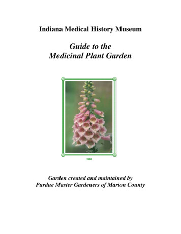
Kong et al. BMC Complementary and Alternative Medicine 2014, 1Page 3 of 75′-CCAAGGAGCCAGAACCTTCGAAACT-3′ (reverse);and GAPDH, 5′-ACCACAGTCCATGCCATCAC-3′ (forward) and 5′-TCCACCACCCTGTTGCTGTA- 3′(reverse). PCR was performed using the QuantiTect SYBRGreen PCR kit (Qiagen) in triplicates according to themanufacturer’s instructions. Relative levels of c-Fos, c-Jun,NFATc1, TRAP, and OSCAR were normalized to GAPDH.were transferred to polyvinylidene difluoride membranes(Milipore, Bedford, MA, USA). Non-specific interactions were blocked with 5% skim milk for 2 h andwere then probed with the appropriate primary antibodies. Membranes were incubated with the appropriate secondary antibodies attached to horseradishperoxidase, and immunoreactivity was detected withenhanced chemiluminescence reagents. Densitometricvalues were quantified for each band with the ImagePro-plus program version 4.0.Western blot analysisBMMs or osteoclasts were lysed in a buffer containing50 mM Tris–HCl, 150 mM NaCl, 5 mM EDTA, 1% Triton X-100, 1 mM sodium fluoride, 1 mM sodium vanadate, 1% deoxycholate, and protease inhibitors. Thelysates were centrifuged at 14,000 g for 20 min andsupernatants were collected. Protein concentrationsof supernatants were determined. Cellular proteins(30 μg) were resolved by 8–10% sodium dodecyl sulfatepolyacrylamide gel electrophoresis (SDS-PAGE) andStatistical analysisAll data are expressed as means standard deviation(SD). Statistical analysis was done using SPSS softwarepackage ver. 11.0 (SPSS, Chicago, IL); one-way ANOVAwas used for comparison among the different groups.Post hoc testing of differences between groups was performed by using Duncan’s test when the ANOVA wasFigure 1 Effect of AS extract on RANKL-induced osteoclast differentiation. (A) BMMs were cultured for 4 days with M-CSF (20 ng/ml) andRANKL (40 ng/ml) in the presence of varying concentrations of AS extract. Cells were fixed with 3.7% formalin, permeabilized with 0.1% TritonX-100, and stained with TRAP solution. (B) TRAP-positive cells were counted as osteoclasts. Asterisk indicates a statistically significant difference(p 0.05) between control and treated. (C) Cytotoxicity of AS extract on BMMs. BMMs were cultured for 3 days with varying concentrations of ASextract in the presence of M-CSF (20 ng/ml). XTT solution (50 μl) was added to each well after 3 days, and plates were incubated for 4 h. Theoptical density was measured at 450 nm. (D) RAW 264.7 cells were seeded at 3000 cells/plate in 48-well plates and cultured with the indicatedconcentrations of AS extract for 4 days. After 4 days, the cells were fixed and stained with Hematoxylin. Colonies with 50 or greater cells werecounted. Similar results were obtained in at least 3 independent experiments.
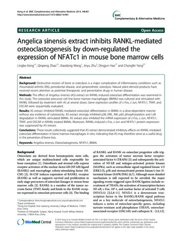
Kong et al. BMC Complementary and Alternative Medicine 2014, 1Page 4 of 7significant. All results were considered to be significantat the 5% critical level (P 0.05).ResultsInhibition of osteoclast differentiation by AS extractOsteoclasts were generated from mouse BMMs in thepresence of M-CSF (20 ng/ml) plus RANKL (40 ng/ml)to verify the effects of AS extract in osteoclastogenesis.The BMMs of the control group differentiated into mature TRAP-positive multinucleated osteoclasts while ASextract reduced the formation and numbers of TRAPpositive multinucleated cells in a dose-dependent manner (Figure 1A, B).The cytotoxic effect of AS extractAS extract generated a highly negative effect on osteoclastogenesis. We measured the effects of AS extract onbone marrow cells with the XTT assay to exclude thepossibility that the inhibition was due to cytotoxicity. ASextract demonstrated no cytotoxic effects at the samedoses which effectively inhibited osteoclast formation(Figure 1C). Also, AS extract did not affect in RAW264.7 cells colony formation (Figure 1D), suggesting thatosteoclastogenesis suppression by AS extract was notdue to toxic effects on BMMs.AS extract inhibits a variety of signals transduced byRANKLRANKL activates a variety of signal transducers that areinvolved in osteoclastogenesis, including p38, JNK, ERK,transcription factor NF-κB and I-κB, which are recognizedas the key factors of osteoclast differentiation [2,3]. Osteoclast precursors were pretreated with AS extract and stimulated with RANKL at various time points. Differentsignaling pathways were observed. Activation of ERK,JNK, p38, NF-κB p65 and degradation of I-κB by RANKLwere all significantly inhibited by AS extract (Figure 2).RANKL induced c-Fos, c-Jun, NFATc1, TRAP, and OSCARmRNA expression is reduced by AS extractOsteoclast differentiation is regulated by the inductionof various genes in response to RANKL and RANKbinding. The c-Fos, c-Jun and NFATc1 genes have an essential role in the osteoclast differentiation [2,13], andNFATc1 regulates OSCAR expression [18]. We examined the effects of AS extract on the RANKL-inducedregulation of c-Fos, c-Jun and NFATc1 expression, andto assess whether there were any effects on TRAP andOSCAR expression. Osteoclast precursors were pretreated with AS extract and further stimulated withRANKL at various time points. Results revealed that cFos, c-Jun and NFATc1 mRNA levels were increased in response to RANKL, but c-Fos, c-Jun and NFATc1 mRNAexpression was significantly inhibited by AS extract.Figure 2 Effect of AS extract on RANKL-induced MAPKs andNF-κB activation. AS extract inhibits the p38, ERK, JNK, p65phosphorylation and I-κB degradation in RANKL-stimulated BMMs. BMMswere pretreated with or without AS extract (50 mg/l) for 1 h prior toRANKL stimulation (100 ng/ml) at indicated time periods. Cells werelysed in lysis buffer, and lysates were analyzed by Western blotting withindicated antibodies. The intensities of protein bands were analyzedand normalized to actin. Similar results were obtained in at least 3ndependent iexperiments.Moreover TRAP and OSCAR mRNA expression was alsosignificantly inhibited by AS extract (Figure 3A). Thisraises the possibility that, AS extract may inhibit osteoclastdifferentiation through the inhibition of RANKL-inducedc-Fos, c-Jun and NFATc1 expression.
Kong et al. BMC Complementary and Alternative Medicine 2014, 1Figure 3 (See legend on next page.)Page 5 of 7
Kong et al. BMC Complementary and Alternative Medicine 2014, 1Page 6 of 7(See figure on previous page.)Figure 3 AS extract suppresses the mRNA expression of c-Fos, c-Jun, NFATc1, OSCAR and TRAP in BMMs treated with RANKL. (A)BMMs were pretreated with or without AS extract (50 mg/l) for 1 h and with RANKL (100 ng/ml) for the indicated time. Total RNA was isolatedusing TRIzol, and 1 μg of total RNA was used to transcribe cDNA. cDNA (1 μl) was used as a template for real time RT-PCR. The mRNA expressionof the indicated genes was analyzed by real time RT-PCR. (B) AS extract inhibits the expression of c-Jun, c-Fos and NFATc1 induced by RANKL.BMMs were pretreated with or without AS extract (50 mg/l) for 1 h and were treated with RANKL (100 ng/ml) for the indicated time. Cells werelysed in the lysis buffer, and lysates were analyzed by Western blotting with antibodies against c-Jun, c-Fos, NFATc1 and actin. The intensities ofthe protein bands were analyzed and normalized to actin. Similar results were obtained in at least 3 independent experiments.AS extract also inhibits c-Fos, c-Jun and NFATc1 proteinexpressionWestern blots were examined to verify the effects of ASextract on c-Fos, c-Jun and NFATc1 protein expression.c-Fos, c-Jun and NFATc1 protein levels were increasedin response to RANKL, but c-Fos, c-Jun and NFATc1expression was significantly inhibited by AS extract(Figure 3B). These results demonstrated that the inhibitoryeffects of AS extract involved the inhibition of majortranscription factors such as c-Fos, c-Jun and NFATc1.DiscussionBone resorption by osteoclasts is frequently caused byexcessive RANKL signaling which has been a valuabletarget for the treatment of pathological bone loss.RANKL activates a series of major intracellular signaltransducing pathways including NFATc1, NF-κB, phosphoinositide 3 kinase (PI3K)-Akt, JNK, ERK, and p38MAPK. AS is one of the most popular Chinese herbs inboth clinical field and research field. Su et al. [15] reported that a component of AS extract: ligustilide suppresses the LPS-induced production of NO, PGE2 andTNF-α by inhibiting both IKK/NF-κB and MAPK/AP-1signaling. Recently we [16] demonstrated that AS extractcould inhibit bone resorption by attenuating proinflammatory cytokines. However, the effect of AS extract onRANKL-induced osteoclast formation, especially RANKLmediated intracellular signal pathway still is an interestingquestion that remains to be investigated.The MAPK group of enzymes selectively phosphorylates serine and threonine residues in response to extracellular stimuli and transmits the stimuli from the cellsurface to the nucleus [7]. MAPK is primarily composedof JNK, ERK, and p38 in mammalian cells. RANKL andRANK receptor binding expressed in osteoclast precursors provides a link between distinct signaling molecules such as JNK, ERK, and p38 MAPK [1]. p38 MAPKsignaling is particularly crucial in the early stages ofosteoclast differentiation as it promotes the activity ofmicrophthalmia-associated transcription factor (MITF)and TRAP expression. Inhibition of p38 MAPK withSB203580 has a negative effect on osteoclast formation[19]. AS extract suppressed RANKL-induced the phosphorylation of p38, ERK and JNK activity in the presentstudy (Figure 2).The activation of signaling molecules induces transcription factors such as NF-κB, NFATc1, and AP-1 thatare essential for osteoclast differentiation [20]. NF-κB isan important signal mediator for inflammatory and immune reactions and is a major transcription factor forRANKL-activated osteoclastogenesis [21]. I-κB is attached to NF-κB preventing it from migrating into thenucleus, and phosphorylation with I-κB kinase (IKK)separates the two proteins. Subsequent ubiquitinationand proteosome degradation of I-κB allows the transferof NF-κB into the nucleus and transcription of the targetgene [22]. Although our previous study showed that onefactor of AS extract: ligustilide decreases NF-κB luciferase activity in a reporter assay [16], we also confirmedthat AS extract inhibits NF-κB/I-κB protein expressionin this experiments. These results are consistent withRANKL activation of NF-κB in osteoclastic precursorcells through IKK activation and susequent I-κB phosphorylation and degradation (Figure 2).NFATc1 is a master regulator of osteoclastogenesiswhich autoamplifies and conducts the expression of osteoclast specific genes such as activator protein-1 (AP-1),TRAP, calcitonin receptor, OSCAR, and cathepsin K[10,23-25]. The RANKL-induced NFATc1 expression ismediated by the activation of AP-1 consisting of c-Fos andc-Jun [26,27]. c-Fos was reported to be critical for transcriptional activation of NFATc1 in RANKL-induced osteoclastogenesis [7]. Putative c-Fos binding sites were mappedin the promoter region of NFATc1, and the NFATc1 expression was abolished in the osteoclast precursors lackingc-Fos [11].On the other hand, Ikeda et al. [26] reported thatRANKL could not induce expression of NFATc1 indominant-negative c-Jun transgenic mouse. It is possiblethat the induction of NFATc1 may be cooperatively upregulated by c-Fos and c-Jun, which make up each otherfor the induction [28]. The present study demonstrates thereduced both c-Fos and c-Jun expression in AS extracttreated BMM cells (Figure 3). Therefore, the reduced c-Fosand c-Jun expression is suggested to cause the impaired activation of NFATc1, followed by the inhibition of RANKLinduced osteoclast formation. Additionally mRNA levels ofmajor osteoclast marker TRAP and OSCAR was alsoinhibited by AS extract (Figure 3). These data suggest thatthe AP-1 transcription factor is the targets of AS extractinduced inhibition of osteoclastogenesis.
Kong et al. BMC Complementary and Alternative Medicine 2014, 1Page 7 of 7ConclusionsIn summary, the present study demonstrated that ASinhibited osteoclastogenesis from macrophages and boneresorption in vitro. AS also reduced the RANKL-inducedexpression of osteoclastic marker genes. In addition, ASattenuated RANKL-induced ERK, p38, JNK, NF-κB, AP-1and NFATc1 activation. Although additional experimentsare needed to confirm the efficacy of AS in treating diseaseconditions in vivo, our results indicate that AS has potential as a therapy for disorders associated with bone loss.kinase pathways are involved in osteoclast differentiation. Bone 2002,30:71–77.Takayanagi H, Kim S, Koga T, Nishina H, Isshiki M, Yoshida H: Inductionand activation of the transcription factor NFATc1 (NFAT2) integrateRANKL signaling in terminal differentiation of osteoclasts. Dev Cell 2002,3:889–901.Matsuo K, Owens JM, Tonko M, Elliott C, Chambers TJ, Wagner EF: Fos is atranscriptional target of c-Fos during osteoclast differentiation. Nat Genet2000, 24:184–187.Nakashima T, Takayanagi H: Osteoimmunology: cross talk between theimmune and bone systems. J Clin Immunol 2009, 29:555–567.Zhao Q, Wang X, Liu Y, He A, Jia R: NFATc1: function in osteoclasts.Int J Biochem Cell Biol 2010, 42:576–579.Grigoriadis AE, Wang ZQ, Cecchini MG, Hofstetter W, Felix R, Fleisch HA:c-Fos: a key regulator of osteoclast-macrophage lineage determinationand bone remodeling. Science 1994, 266:443–448.Su YW, Chiou WF, Chao SH: Ligustilide prevents LPS-induced iNOSexpression in RAW 264.7 macrophages by preventing ROS productionand down-regulating the MAPK, NF-κB and AP-1signaling pathways.Int Immuno Pharmacol 2011, 11:1166–1172.Yang C, Niu S, Lifeng Y: The aqueous extract of Angelica sinensis, apopular Chinese herb, inhibits wear debris-Induced Inflammatoryosteolysis in mice. J Surg Res 2012, 176:476–483.Wang H, Li W, Li J: The aqueous extract of a popular herbal nutrientsupplement, Angelica sinensis, protects mice against lethal endotoxemiaand sepsis. J Nutr 2006, 136:360.Kim K, Kim JH, Lee J, Jin HM, Lee SH, Fisher DE: Nuclear factor of activatedT cells c1 induces osteoclast-associated receptor gene expression duringtumor necrosis factor-related activation-induced cytokine mediatedosteoclastogenesis. J Biol Chem 2005, 280:35209–35216.Matsumoto M, Sudo T, Saito T, Osada H, Tsujimoto M: Involvement of p38mitogen-activated protein kinase signaling pathway in osteoclastogenesismediated by receptor activator of NF-kappa B ligand (RANKL). J Biol Chem2000, 275:31155–31161.Takayanagi H: Osteoimmunology. Nat Rev Immunol 2007, 7:292–304.Iotsova V, Caamano J, Loy J, Yang Y, Lewin A, Bravo R: Osteopetrosis inmice lacking NF-kappaB1 and NF-kappaB2. Nat Med 1997, 3:1285–1289.Hayden MS, Ghosh S: Signaling to NF-κB. Genes Dev 2004, 18:2124–2195.Crotti TN, Flannery M, Walsh NC, Fleming JD, Goldring SR, McHugh KP:NFATc1 regulation of the human beta 3 integrin promoter in osteoclastdifferentiation. Gene 2006, 372:92–102.Kim Y, Sato K, Asagiri M, Morita I, Soma K, Takayanagi H: Contribution ofnuclear factor of activated T cells c1 to the transcriptional control ofimmunoreceptor osteoclast-associated receptor but not triggeringreceptor expressed by myeloid cells-2 during osteoclastogenesis.J Biol Chem 2005, 280:32905–32913.Matsumoto M, Koqawa M, Wada S, Takayangi H, Tsujimoto M, Katayama S:Essential role of p38 mitogen-activated protein kinase in cathepsin Kgene expression during osteoclastogenesis through association ofNFATc1 and PU.1. J Biol Chem 2004, 279:45969–45979.Ikeda F, Nishimura R, Matsubara T, Tanaka S, Inoue J, Reddy SV: Criticalrolesof c-Jun signaling in regulation of NFAT family and RANKL-regulatedosteoclast differentiation. J Clin Invest 2004, 114:475–484.Wagner EF, Eferl R: Fos/AP-1 proteins in bone and the immune system.Immunol Rev 2005, 208:126–140.Mohamed S: Interleukin-10 inhibits RANKL-mediated expression ofNFATc1 in part via suppression of c-Fos and c-Jun in RAW264.7 cells andmouse bone marrow cells. Bone 2007, 41:592–602.10.11.12.13.14.Competing interestsThe authors declare that they have no competing interests.Authors’ contributionsDH, CY, LK: conception and design, analysis and interpretation of data; draftthe manuscript and revise it critically for important intellectual content; finalapproval of the version to be published: acquisition of data, analysis andinterpretation of data; QZ: acquisition of data, analysis and interpretation ofdata; DW: acquisition of data, draft the manuscript. JZ, DH: conception anddesign, revise the manuscript critically for important intellectual content, finalapproval of the version to be published, account for all aspects of the workin ensuring that questions related to the accuracy or integrity of any part ofthe work are appropriately investigated and resolved. All authors read andapproved the final manuscript. All authors read and approved the finalmanuscript.15.16.17.18.19.AcknowledgementsThe authors wish to thank Guo Hua (Honghui hospital) for overseeing theconduct of the study, Rui Ma, the Fourth Military Medical University (Xi’an,China) for statistical analysis assistance and Shu Zhu for overseeing datamanagement. We thank Lifeng Yu for regulatory and quality activities andfor his technical support. We thank Xiaorui Cao, PhD, for his assistance withmanuscript preparation and review.Received: 14 May 2014 Accepted: 10 December 2014Published: 12 December 2014References1. Han Bok K, Byeong Ki L: Inhibition of osteoclast differentiation and boneresorption by rotenone, through down-regulation of RANKL-inducedc-Fos and NFATc1 expression. Bone 2010, 46:724–731.2. Boyle WJ, Simonet WS, Lancey DL: Osteoclast differentiation andactivation. Nature 2003, 423:337–342.3. Arai F, Miyamoto T, Ohneda O, Inada T, Sudo T, Brasel K: Commitment anddifferentiation of osteoclast precursor cells by the sequential expressionof c-Fms and receptor activator of nuclear factor kappaB (RANK)receptors. J Exp Med 1999, 190:1741–1754.4. Suda T, Takahashi N, Udagawa N, Jimi E, Gillespie MT, Martin TJ: Modulationof osteoclast differentiation and function by the new members of thetumor necrosis factor receptor and ligand families. Endocr Rev 1999,20:345–357.5. Kobayashi N, Kadono Y, Naito A, Matsumoto K, Yamamoto T, Tanaka S:Segregation of TRAF6-mediated signaling pathways clarifies its role inosteoclastogenesis. EMBO J 2001, 20:1271–1280.6. Hotokezaka H, Sakai E, Kanaoka K, Saito K, Matsuo K, Kitaura H: U0126and PD98059, specific inhibitors of MEK, accelerate differentiationof RAW264.7 cells into osteoclast-like cells. J Biol Chem 2002,277:47366–47372.7. Matsuo K, Galson DL, Zhao C, Peng L, Laplace C, Wang KZ: Nuclear factorof activated T-cells (NFAT) rescues osteoclastogenesis in precursorslacking c-Fos. J Biol Chem 2004, 279:26475–26480.8. David JP, Sabapathy K, Hoffmann O, Idarraga MH, Wagner EF: JNK1 modulatesosteoclastogenesis through both c-Jun phosphorylation-dependent and –independent mechanisms. J Cell Sci 2002, 115:4317–4325.9. Lee SE, Woo KM, Kim SY, Kim HM, Kwack K, Lee ZH: Thephosphatidylinositol 3-kinase, p38, and extracellular 1186/1472-6882-14-481Cite this article as: Kong et al.: Angelica sinensis extract inhibits RANKLmediated osteoclastogenesis by down-regulated the expression of NFATc1in mouse bone marrow cells. BMC Complementary and Alternative Medicine2014 14:481.
University College of Medicine and conducted strictly followed by "the institutional guidelines for the care and use of laboratory animals at the Jiaotong University Col-lege of Medicine". Bone marrow cells were obtained by flushing the femurs and tibiae of 5-week-old ICR mice with α-minimum essential medium (α-MEM; Gibco