Transcription
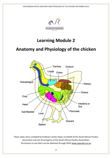
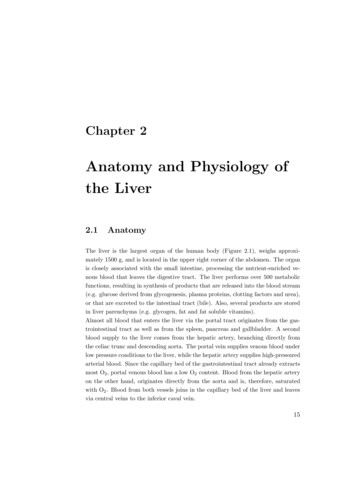
Chapter 2Anatomy and Physiology ofthe Liver2.1AnatomyThe liver is the largest organ of the human body (Figure 2.1), weighs approximately 1500 g, and is located in the upper right corner of the abdomen. The organis closely associated with the small intestine, processing the nutrient-enriched venous blood that leaves the digestive tract. The liver performs over 500 metabolicfunctions, resulting in synthesis of products that are released into the blood stream(e.g. glucose derived from glycogenesis, plasma proteins, clotting factors and urea),or that are excreted to the intestinal tract (bile). Also, several products are storedin liver parenchyma (e.g. glycogen, fat and fat soluble vitamins).Almost all blood that enters the liver via the portal tract originates from the gastrointestinal tract as well as from the spleen, pancreas and gallbladder. A secondblood supply to the liver comes from the hepatic artery, branching directly fromthe celiac trunc and descending aorta. The portal vein supplies venous blood underlow pressure conditions to the liver, while the hepatic artery supplies high-pressuredarterial blood. Since the capillary bed of the gastrointestinal tract already extractsmost O2 , portal venous blood has a low O2 content. Blood from the hepatic arteryon the other hand, originates directly from the aorta and is, therefore, saturatedwith O2 . Blood from both vessels joins in the capillary bed of the liver and leavesvia central veins to the inferior caval vein.15
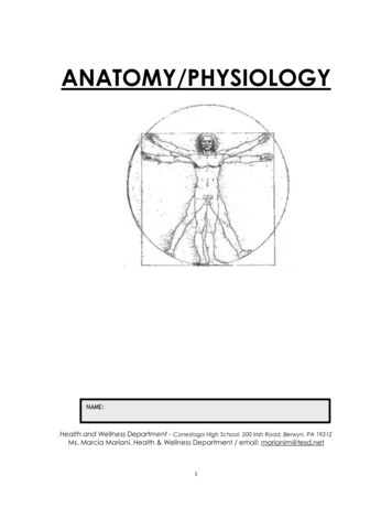
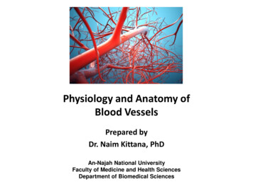
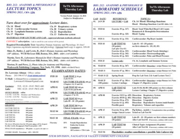
2. Anatomy and PhysiologyFigure 2.1: The liver.2.1.1Basic liver architectureThe major blood vessels, portal vein and hepatic artery, lymphatics, nerves andhepatic bile duct communicate with the liver at a common site, the hilus. Fromthe hilus, they branch and rebranch within the liver to form a system that travelstogether in a conduit structure, the portal canal (Figure 2.2). From this portalcanal, after numerous branching, the portal vein finally drains into the sinusoids,which is the capillary system of the liver. Here, in the sinusoids, blood from theportal vein joins with blood flow from end-arterial branches of the hepatic artery.Once passed through the sinusoids, blood enters the collecting branch of the centralvein, and finally leaves the liver via the hepatic vein. The hexagonal structure with,in most cases, three portal canals in its corners draining into one central vein, isdefined as a lobule (Figure 2.3). The lobule largely consists of hepatocytes (livercells) which are arranged as interconnected plates, usually one or two hepatocytesthick. The space between the plates forms the sinusoid. A more functional unit ofthe liver forms the acinus. In the acinus, the portal canal forms the center and thecentral veins the corners. The functional acinus can be divided into three zones: 1)the periportal zone, which is the circular zone directly around the portal canal, 2)the central zone, the circular area around the central vein, and 3) a midzonal area,which is the zone between the periportal and pericentral zone.2.1.2SinusoidsSinusoids (Figure 2.4) are the canals formed by the plates of hepatocytes. They areapproximately 8-10 µm in diameter and comparable with the diameter of normal16
2.1. AnatomyFigure 2.2: Network of branching and rebranching blood vessels in the liver.17
2. Anatomy and PhysiologyFigure 2.3: The liver lobule with portal canals (hepatic artery, portal vein and bileduct), sinusoids and collecting central veins.18
2.2. Physiologycapillaries. They are orientated in a radial direction in the lobule. Sinusoids arelined with endothelial cells and Kupffer cells, which have a phagocytic function.Plasma and proteins migrate through these lining cells via so-called fenestrations(100-150 nm) into the Space of Disse, where direct contact with the hepatocytesoccurs and uptake of nutrients and oxygen by the hepatocytes takes place. Onthe opposite side of the hepatocyte plates are the bile canaliculi situated (1 µmdiameter). Bile produced by the hepatocytes empties in these bile canaliculi andis transported back towards the portal canal into bile ductiles and bile ducts, andfinally to the main bile duct and gallbladder to become available for digestive processes in the intestine. The direction of bile flow is opposite to the direction of theblood flow through the sinusoids.Figure 2.4: Detailed view of the liver sinusoidal structure.2.22.2.1PhysiologyPressure distributionBlood pressure in afferent vessels and pressure distribution inside the liver, is essentially similar for most species. Pressure in the hepatic artery, originating fromthe descending aorta and the celiac trunc, is considered to be the same as aorticpressure. This includes a high pulsatile pressure between 120 and 80 mmHg witha frequency equal to the heart rate. Vessel compliance causes a gradual decreasein pulsation as the hepatic artery branches and rebranches inside the liver. Once19
2. Anatomy and Physiologyat the sinusoidal level, pulsation amplitude decreases to virtually zero and pressuredrops to approximately 2-5 mmHg. On the other hand, pressure in the portal vein,originating from capillaries of the digestive tract, has no pulsation and a pressureof 10-12 mmHg. In the sinusoids, both portal venous and hepatic arterial pressureis 3-5 mmHg. Consequently, the pressure drop inside the liver is much less in theportal venous system than in the arterial system. The pressure drop from the collecting central veins to the vena cava is then approximately 1-3 mmHg, fluctuatingslightly with respiration.2.2.2Flow distributionTotal human liver blood flow represents approximately 25% of the cardiac output,up to 1500 ml/min. Hepatic flow is subdivided in 25-30% for the hepatic artery(500 ml/min) and the major part for the portal vein (1000 ml/min). Assuming ahuman liver weighs 1500 g, total liver flow is 100 ml/min per 100 g liver. Comparingthis normalized flow rate to other species, it can be concluded that total liver bloodflow is 100-130 ml/min per 100 g liver, independent of the species. The ratio ofarterial:portal blood flow, however, is species-dependent. The hepatic artery originates directly from the descending aorta, and is therefore saturated with oxygen.It accounts for 65% of total oxygen supply to the liver. The hepatic artery alsoplays an important role in liver blood vessel wall and connective tissue perfusion.It also secures bile duct integrity. The blood from the portal vein is full of nutrientsderived from the intestine and allows the hepatocytes to perform their tasks. Bloodfrom the hepatic artery and the portal vein joins in the sinusoids. However, recentstudies by others as well as our own observations, have revealed that there are bothcommon and separate channels for arterial and portal blood. The hepatic arteryperfuses the liver vascular bed in a ’spotty’ pattern, while the portal vein perfusesthe liver uniformly. The liver is able to regulate mainly arterial flow by means ofso-called sphincters, situated at the in- and outlets of the sinusoids. One of themost important triggers for sphincter function is the need for constant oxygen supply. If the rate of oxygen delivery to the liver varies, the sphincters will react andthe ratio of arterial:portal blood flow alters.2.3Physiology of Liver PreservationLiver transplantation requires a period of preservation time, during which the liverafter explantation is stored and transported outside its natural environment. The20
2.3. Physiology of Liver Preservationlength of this preservation or better bridging period from donor to recipient dependson a large number of donor and recipient as well as logistical factors. Nowadays thistime zone is kept between 6-15 hours of preservation, while the liver is cut off fromits life sustaining mechanisms, i.e. blood flow and oxygen supply, and consequently,ischemic damage will occur. Three periods of ischemia are distinguished in a donorand transplantation procedure.1. The first period is defined as the time in between clamping of the liver’s afferent vasculature and start of the wash-out with ice-cold preservation solutionduring the procurement phase in the donor. In this period of ischemia, bloodflow to the liver is stopped and perfusion with the preservation solution viaa cannula takes place. During this period of ischemia the liver is still atbody temperature, for which reason this is called warm-ischemia. Duringthis warm-ischemic time, mainly damage to the hepatocytes occurs, and it istherefore very important to keep this period as short as possible. Especiallyin non-heart-beating donors (NHBDs) after cardiac arrest and awaiting thedonor operation and start of cold preservation, the warm-ischemic periodcan become too long, and result in a serious injury of hepatocytes with nonfunction after transplantation.2. The second period of ischemia starts at the moment when the ice-cold preservation solution enters the microvasculature of the liver to wash-out the blood.A good wash-out of blood cells is important to obtain optimal and uniformperfusion and cooling of the liver. Furthermore, remaining blood cells couldcause inflammatory reactions and trigger the immune response of the receiving patient against these donor cells. This cold ischemic period continuesduring the preservation period, in which the liver is transported to the recipient, and ends at the moment that the liver is taken out of its cold environmentjust prior to be placed in the ’warm’ recipient’s abdomen during the actualimplantation. This second period takes place at 0-4 C, including the washout with ice-cold solution and cold storage preservation period, and is definedas the period of cold ischemia. During this period of cold ischemia, not somuch hepatocytes, but more endothelial and biliary epithelial cells appear tobe injured.3. The last period of this bridging ischemia is the period needed to completethe vascular anastomoses during implantation. This procedure could take30-60 minutes, a period in which the liver lies in the abdominal cavity and isthus rewarmed by temperature of the body. Once all vessels are connected21
2. Anatomy and Physiologybetween donor organ and recipient vasculature, the liver is reperfused withblood and after the biliary anastomosis the transplantation is complete.To minimize the impact of cold ischemic injury, two principal requirements forsuccessful preservation have to be considered: the temperature effect using hypothermia and the effect of the preservation solution. Cooling the liver to hypothermic temperatures (0-4 C) lowers the rate ofmetabolism and the rate at which cellular components degrade. As a result,the need for nutrients and oxygen decreases and the organ can be preservedin a viable state for several hours (see further § 6.4). Although hypothermia is a key to successful preservation, it has negativeside effects as well. The major drawback of hypothermia is the induction ofcell swelling due to suppressed functioning of the cellular ion pumps. Thiscan be counteracted, however, by the use of an effective preservation solutioncontaining so-called impermeants, the use of intracellular-like solution andpossibly colloids. Another reason for using a specially developed preservation solution is to prevent ischemia-induced cellular acidosis, which requiresa powerful buffer. Finally, in the initial reperfusion phase during transplantation the sudden increase in available oxygen (’respiratory burst’) results inthe formation of toxic oxygen radicals. To counteract this last drawback, thepreservation solution should contain agents that are able to scavenge oxygenfree radicals (see further § 6.2).The combination of hypothermia and the use of an adequate preservation solution prevent hypothermic and preservation-induced injury, and make it possibleto keep the liver for a limited amount of time outside its physiologic environment.Cold storage allows preservation for 12-15 hours in the clinics and even beyond 48hours in the experimental setting. This time period is considered short to fulfillall requirements of an appropriate match between donor and recipient as well as ofcomplex logistic factors. Also, despite major achievements in organ preservation,and especially in older donors, prolonged static cold storage is still accompanied byan increased primary non-function as well as reduced long term functional outcomeafter liver transplantation. To date, preservation times are kept short and any prolongation without a compromise to the viability of the liver would be a significantimprovement. Thus, hypothermic machine perfusion preservation could substantially increase the quality of preservation and lengthen the preservation period.22
donor operation and start of cold preservation, the warm-ischemic period can become too long, and result in a serious injury of hepatocytes with non-function after transplantation. 2. The second period of ischemia starts at the moment when the ice-cold preser-vation solution enters the microvasculature of the