Transcription
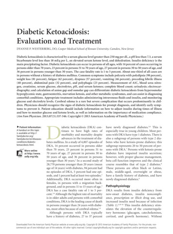
Evaluation and Managementof Intestinal ObstructionPATRICK G. JACKSON, MD, and MANISH RAIJI, MDGeorgetown University Hospital, Washington, District of ColumbiaAcute intestinal obstruction occurs when there is an interruption in the forward flow of intestinal contents. This interruption can occur at any point along the length of the gastrointestinaltract, and clinical symptoms often vary based on the level of obstruction. Intestinal obstruction is most commonly caused by intra-abdominal adhesions, malignancy, or intestinal herniation. The clinical presentation generally includes nausea and emesis, colicky abdominal pain,and a failure to pass flatus or bowel movements. The classic physical examination findings ofabdominal distension, tympany to percussion, and high-pitched bowel sounds suggest thediagnosis. Radiologic imaging can confirm the diagnosis, and can also serve as useful adjunctive investigations when the diagnosis is less certain. Although radiography is often the initialstudy, non-contrast computed tomography is recommended if the index of suspicion is high orif suspicion persists despite negative radiography. Management of uncomplicated obstructionsincludes fluid resuscitation with correction of metabolic derangements, intestinal decompression, and bowel rest. Evidence of vascular compromise or perforation, or failure to resolve withadequate bowel decompression is an indication for surgical intervention. (Am Fam Physician.2011;83(2):159-165. Copyright 2011 American Academy of Family Physicians.) Patient information:A handout on intestinalobstruction, written by theauthors of this article, isprovided on page 166.Intestinal obstruction accounts forapproximately 15 percent of all emergency department visits for acuteabdominal pain.1 Complications ofintestinal obstruction include bowel ischemia and perforation. Morbidity and mortality associated with intestinal obstructionhave declined since the advent of moresophisticated diagnostic tests, but the condition remains a challenging surgical diagnosis. Physicians who are treating patients withintestinal obstruction must weigh the risks ofsurgery with the consequences of inappropriate conservative management. A suggestedapproach to the patient with suspected smallbowel obstruction is shown in Figure 1.PathophysiologyThe fundamental concerns about intestinal obstruction are its effect on whole bodyfluid/electrolyte balances and the mechanical effect that increased pressure has onintestinal perfusion. Proximal to the pointof obstruction, the intestinal tract dilates asit fills with intestinal secretions and swallowed air.2 Failure of intestinal contents topass through the intestinal tract leads to acessation of flatus and bowel movements.Intestinal obstruction can be broadly differentiated into small bowel and large bowelobstruction.Fluid loss from emesis, bowel edema, andloss of absorptive capacity leads to dehydration. Emesis leads to loss of gastric potassium,hydrogen, and chloride ions, and significant dehydration stimulates renal proximaltubule reabsorption of bicarbonate and lossof chloride, perpetuating the metabolic alkalosis.3 In addition to derangements in fluidand electrolyte balance, intestinal stasis leadsto overgrowth of intestinal flora, which maylead to the development of feculent emesis.Additionally, overgrowth of intestinal florain the small bowel leads to bacterial translocation across the bowel wall.4Ongoing dilation of the intestine increasesluminal pressures. When luminal pressuresexceed venous pressures, loss of venousdrainage causes increasing edema andhyperemia of the bowel. This may eventually lead to compromised arterial flow tothe bowel, causing ischemia, necrosis, andDownloaded from the American Family Physician Web site at www.aafp.org/afp. Copyright 2011 American Academy of Family Physicians. For the private, noncommercial Volume 83, Number 2January 15,www.aafp.org/afp AmericanFamilyrequests.Physician 159use2011of one individualuser of the Web site. All other rights reserved.Contact copyrights@aafp.orgfor copyright questionsand/or permission
Intestinal Obstructionperforation. A closed-loop obstruction, inwhich a section of bowel is obstructed proximally and distally, may undergo this process rapidly, with few presenting symptoms.Intestinal volvulus, the prototypical closedloop obstruction, causes torsion of arterialinflow and venous drainage, and is a surgicalemergency.Causes and Risk FactorsThe most common causes of intestinalobstruction include adhesions, neoplasms,and herniation (Table 1). Adhesions resultingfrom prior abdominal surgery are the predominant cause of small bowel obstruction,accounting for approximately 60 percentof cases.5 Lower abdominal surgeries, including appendectomies, colorectal surgery,gynecologic procedures, and hernia repairs,confer a greater risk of adhesive smallbowel obstruction. Less common causes ofobstruction include intestinal intussusception, volvulus, intra-abdominal abscesses,gallstones, and foreign bodies.Management of Small Bowel ObstructionPatient presents with signs andsymptoms of small bowel obstructionClinically stable?NoYesExploratorylaparotomyRadiography orcomputed tomographyVascular compromiseor onNoExploratorylaparotomyNo oral intake,nasogastric intubation,intravenous rehydrationResolution within 24 to 48 hours?NoNo oral intake,nasogastric intubation,intravenous rehydrationResolution within 24 to 48 hours?YesYesExploratorylaparotomyNoUpper gastrointestinal/small bowel followthrough/enteroclysis?Advance dietYesResolution?NoExploratorylaparotomyFigure 1. Algorithm for evaluation and treatment of patients with suspected small bowelobstruction.160 American Family Physicianwww.aafp.org/afpVolume 83, Number 2 January 15, 2011
Intestinal ObstructionTable 1. Causes of IntestinalObstructionAdhesive disease (60 percent)Neoplasm (20 percent)Herniation (10 percent)Inflammatory bowel disease (5 percent)Intussusception ( 5 percent)Volvulus ( 5 percent)Other ( 5 percent)History and Physical ExaminationPatients should be asked about their historyof abdominal neoplasia, hernia or herniarepair, and inflammatory bowel disease,because these conditions increase the riskof obstruction. The hallmarks of intestinal obstruction include colicky abdominalpain, nausea and vomiting, abdominal distension, and a cessation of flatus and bowelmovements. It is important to differentiatebetween true mechanical obstruction andother causes of these symptoms (Table 2).Distal obstructions allow for a greater intestinal reservoir, with pain and distensionmore marked than emesis, whereas patientswith proximal obstructions may have minimal abdominal distension but markedemesis. The presence of hypotension andtachycardia is an indication of severe dehydration. Abdominal palpation may reveal adistended, tympanitic abdomen; however,this finding may not be present in patientswith early or proximal obstruction. Auscultation in patients with early obstructionreveals high-pitched bowel sounds, whereasthose with late obstruction may present withminimal bowel sounds as the intestinal tractbecomes hypotonic.Diagnostic Testing and ImagingLABORATORY TESTSLaboratory evaluation of patients with suspected obstruction should include a complete blood count and metabolic panel.Hypokalemic, hypochloremic metabolicalkalosis may be noted in patients withsevere emesis. Elevated blood urea nitrogenlevels are consistent with dehydration, andhemoglobin and hematocrit levels may beincreased. The white blood cell count maybe elevated if intestinal bacteria translocateinto the bloodstream, causing the systemicinflammatory response syndrome or sepsis.January 15, 2011 Volume 83, Number 2The development of metabolic acidosis, especially in a patient with an increasing serumlactate level, may signal bowel ischemia.RADIOGRAPHYThe initial evaluation of patients withclinical signs and symptoms of intestinalobstruction should include plain uprightabdominal radiography. Radiography canquickly determine if intestinal perforationhas occurred; free air can be seen above theliver in upright films or left lateral decubitus films. Radiography accurately diagnoses intestinal obstruction in approximately60 percent of cases,6 and its positive predictive value approaches 80 percent in patientswith high-grade intestinal obstruction.7However, plain abdominal films can appearnormal in early obstruction and in highjejunal or duodenal obstruction. Therefore,when clinical suspicion for obstruction ishigh or persists despite negative initial radiography, non-contrast computed tomography (CT) should be ordered.8In patients with small bowel obstruction, supine views show dilation of multipleloops of small bowel, with a paucity of air inthe large bowel (Figure 2). Those with largebowel obstruction may have dilation of theTable 2. Differential Diagnosis of Abdominal Pain,Distension, Nausea, and Cessation of Flatus andBowel MovementsAlternate diagnosisCluesAscitesAcute liver failure, history of hepatitis oralcoholismReview of medications; diagnosis ofexclusionHistory of peripheral vascular disease,hypercoagulable state, or postprandialabdominal angina; recent use ofvasopressorsFever, leukocytosis, acute abdomen, freeair on imagingRecent abdominal surgery with nopostoperative flatus or bowel movementAcutely dilated large intestine, history ofintestinal dysmotility, diabetes mellitus,sclerodermaMedications (e.g., tricyclicantidepressants, narcotics)Mesenteric ischemiaPerforated viscus/intraabdominal sepsisPostoperative paralytic ileusPseudo-obstruction(Ogilvie syndrome)www.aafp.org/afp American Family Physician 161
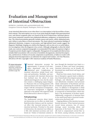
Intestinal Obstructioncolon, with decompressed small bowel in thesetting of a competent ileocecal valve. Uprightor lateral decubitus films may show ladderingair fluid levels (Figure 3). These findings, inconjunction with a lack of air and stool in thedistal colon and rectum, are highly suggestiveof mechanical intestinal obstruction.COMPUTED TOMOGRAPHYFigure 2. Supine view of the abdomen in a patient with intestinalobstruction. Dilated loops of small bowel are visible (arrows).Figure 3. Lateral decubitus view of the abdomen, showing air-fluidlevels consistent with intestinal obstruction (arrows).162 American Family Physicianwww.aafp.org/afpCT is appropriate for further evaluation ofpatients with suspected intestinal obstruction in whom clinical examination and radiography do not yield a definitive diagnosis.CT is sensitive for detection of high-gradeobstruction (up to 90 percent in some series),9and has the additional benefit of definingthe cause and level of obstruction in mostpatients.10-12 In addition, CT can identifyemergent causes of intestinal obstruction,such as volvulus or intestinal strangulation.CT findings in patients with intestinalobstruction include dilated loops of bowelproximal to the site of obstruction, with distally decompressed bowel. The presence of adiscrete transition point helps guide operative planning (Figure 4). Absence of contrastmaterial in the rectum is also an important sign of complete obstruction. For thisFigure 4. Axial computed tomography scanshowing dilated, contrast-filled loops ofbowel on the patient’s left (yellow arrows),with decompressed distal small bowel onthe patient’s right (red arrows). The cause ofobstruction, an incarcerated umbilical hernia,can also be seen (green arrow), with proximally dilated bowel entering the hernia anddecompressed bowel exiting the hernia.Volume 83, Number 2 January 15, 2011
Intestinal Obstructionreason, rectal administration of contrastmaterial should be avoided. A C-loop of distended bowel with radial mesenteric vesselswith medial conversion is highly suspiciousfor intestinal volvulus. Thickened intestinal walls and poor flow of contrast materialinto a section of bowel suggests ischemia,whereas pneumatosis intestinalis, free intraperitoneal air, and mesenteric fat strandingsuggest necrosis and perforation.Although CT is highly sensitive and specific for high-grade obstruction, its valuediminishes in patients with partial obstruction. In these patients, oral contrast material may be seen traversing the length of theintestine to the rectum, with no discrete areaof transition. Fluoroscopy may be of greatervalue in confirming the diagnosis.The American College of Radiology recommends non-contrast CT as the initialimaging modality of choice.13 However,because most causes of small bowel obstruction will have systemic manifestations or failto resolve—necessitating operative intervention—the additional diagnostic value of CTcompared with radiography is limited. Radiation exposure is also significant. Therefore,in most patients, CT should be ordered whenthe diagnosis is in doubt, when there is nosurgical history or hernias to explain the etiology, or when there is a high index of suspicion for complete or high-grade obstruction.within 24 hours of administration has a97 percent sensitivity for spontaneous resolution of intestinal obstruction.16,17There are several variations of contrastfluoroscopy. In the small-bowel followthrough study, the patient drinks contrastmaterial, then serial abdominal radiographsare taken to visualize the passage of contrastthrough the intestinal tract. Enteroclysisinvolves naso- or oro-duodenal intubation,followed by the instillation of contrast material directly into the small bowel. Althoughthis study has superior sensitivity comparedwith small-bowel follow-through,18 it is morelabor-intensive and is rarely performed.Rectal fluoroscopy can be helpful in determining the site of a suspected large bowelobstruction.CONTRAST FLUOROSCOPYMAGNETIC RESONANCE IMAGINGContrast studies, such as a small bowelfollow-through, can be helpful in the diagnosis of a partial intestinal obstruction inpatients with high clinical suspicion and inclinically stable patients in whom initial conservative management was not effective.14The use of water-soluble contrast material isnot only diagnostic, but may also be therapeutic in patients with partial small-bowelobstruction. A randomized controlled trialof 124 patients showed a 74 percent reduction in the need for surgical intervention inpatients receiving gastrografin fluoroscopywithin 24 hours of initial presentation.15Contrast fluoroscopy may also be useful indetermining the need for surgery; the presence of contrast material in the rectumMagnetic resonance imaging (MRI) may bemore sensitive than CT in the evaluation ofintestinal obstruction.20 MRI enteroclysis,which involves intubation of the duodenumand infusion of contrast material directlyinto the small bowel, can more reliablydetermine the location and cause of obstruction.21 However, because of the ease and costeffectiveness of abdominal CT, MRI remainsan investigational or adjunctive imagingmodality for intestinal obstruction.January 15, 2011 Volume 83, Number 2ULTRASONOGRAPHYIn patients with high-grade obstruction,ultrasound evaluation of the abdomen hashigh sensitivity for intestinal obstruction,approaching 85 percent.19 However, becauseof the wide availability of CT, it has largelyreplaced ultrasonography as the first-lineinvestigation in stable patients with suspectedintestinal obstruction. Ultrasonographyremains a valuable investigation for unstablepatients with an ambiguous diagnosis and inpatients for whom radiation exposure is contraindicated, such as pregnant women.TreatmentManagement of intestinal obstruction isdirected at correcting physiologic derangements caused by the obstruction, bowel rest,and removing the source of obstruction. Thewww.aafp.org/afp American Family Physician 163
Intestinal ObstructionSORT: KEY RECOMMENDATIONS FOR PRACTICEEvidenceratingReferencesCommentsAbdominal radiography is an effective initialexamination in patients with suspected intestinalobstruction.Computed tomography is warranted whenradiography indicates high-grade intestinalobstruction or is inconclusive.C6, 7C8-10Upper gastrointestinal fluoroscopy with smallbowel follow-through can determine the needfor surgical intervention in patients with partialobstruction.Antibiotics can protect against bacterialtranslocation and subsequent bacteremia inpatients with intestinal obstruction.Clinically stable patients can be treatedconservatively with bowel rest, intubationand decompression, and intravenous fluidresuscitation.Surgery is warranted in patients with intestinalobstruction that does not resolve within 48 hoursafter conservative therapy is initiated.C14, 15C22A22-26Radiography has greater sensitivity inhigh-grade obstruction than in partialobstruction.Computed tomography can reliably determinethe cause of obstruction, and whetherserious complications are present, in mostpatients with high-grade obstructions.Contrast material that passes into the cecumwithin four hours of oral administration ishighly predictive of successful nonoperativemanagement.Enteric bacteria have been found in culturesfrom serosal scrapings and mesenteric lymphnode biopsy in patients requiring surgery.Several randomized controlled trials haveshown that surgery can be avoided withconservative management.B25Clinical recommendationStudy found that conservative managementbeyond 48 hours does not diminish the needfor surgery, but increases surgical morbidity.A consistent, good-quality patient-oriented evidence; B inconsistent or limited-quality patient-oriented evidence; C consensus, diseaseoriented evidence, usual practice, expert opinion, or case series. For information about the SORT evidence rating system, go to http://www.aafp.org/afpsort.xml.former is addressed by intravenous fluidresuscitation with isotonic fluid. The use ofa bladder catheter to closely monitor urineoutput is the minimum requirement forgauging the adequacy of resuscitation; otherinvasive measures, such as arterial canalization or central venous pressure monitoring,can be used as the clinical situation warrants. Antibiotics are used to treat intestinalovergrowth of bacteria and translocationacross the bowel wall.22 The presence of feverand leukocytosis should prompt inclusion ofantibiotics in the initial treatment regimen.Antibiotics should have coverage againstgram-negative organisms and anaerobes,and the choice of a specific agent should bedetermined by local susceptibility and availability. Aggressive replacement of electrolytes is recommended after adequate renalfunction is confirmed.The decision to perform surgery for intestinal obstruction can be difficult. Peritonitis, clinical instability, or unexplainedleukocytosis or acidosis are concerning forabdominal sepsis, intestinal ischemia, orperforation; these findings mandate immediate surgical exploration. Patients with anobstruction that resolves after reduction of164 American Family Physicianwww.aafp.org/afpa hernia should be scheduled for electivehernia repair, whereas immediate surgeryis required in patients with an irreducibleor strangulated hernia. Stable patients witha history of abdominal malignancy or highsuspicion for malignancy should be thoroughly evaluated for optimal surgical planning. Abdominal malignancy can be treatedwith primary resection and reconstructionor palliative diversion, or placement of venting and feeding tubes.Treatment of stable patients with intestinal obstruction and a history of abdominalsurgery presents a challenge. Conservativemanagement of a high-grade obstructionshould be attempted initially, using intestinal intubation and decompression, aggressive intravenous rehydration, and antibiotics.The inclusion of oral magnesium hydroxide,simethicone, and probiotics decreased thelength of hospitalization in a randomizedcontrolled trial of 144 patients with partialsmall bowel obstructions (number neededto treat 7).23 Caution should be used whenclinical and radiologic evidence suggest complete obstruction, because the use of intestinalstimulation can exacerbate the obstructionand precipitate intestinal ischemia.Volume 83, Number 2 January 15, 2011
Intestinal ObstructionConservative management is successful in40 to 70 percent of clinically stable patients,with a higher success rate in those with partialobstruction.24-26 Although conservative management is associated with shorter initial hospitalization (4.9 versus 12 days), there is also ahigher rate of eventual recurrence (40.5 versus26.8 percent).27 With conservative management, resolution generally occurs within 24to 48 hours. Beyond this time frame, the riskof complications, including vascular compromise, increases. If intestinal obstruction is notresolved with conservative management, surgical evaluation is required.25Figures 2 through 4 provided by Cirrelda J. Cooper, MD.The AuthorsPATRICK G. JACKSON, MD, is chief of gastrointestinal surgery at Georgetown University Hospital, Washington, DC.MANISH RAIJI, MD, is a third year surgical resident atGeorgetown University Hospital.Address correspondence to Patrick G. Jackson, MD, 3800Reservoir Rd., 4th Floor, PHC, Washington, DC 20007.Reprints are not available from the authors.Author disclosure: Nothing to disclose.REFERENCES1. Irvin TT. Abdominal pain: a surgical audit of 1190 emergency admissions. Br J Surg. 1989;76(11):1121-1125.2. Wright HK, O’Brien JJ, Tilson MD. Water absorption inexperimental closed segment obstruction of the ileumin man. Am J Surg. 1971;121(1):96-99.3. Wangensteen OH. Understanding the bowel obstruction problem. Am J Surg. 1978;135(2):131-149.4. Rana SV, Bhardwaj SB. Small intestinal bacterial overgrowth. Scand J Gastroenterol. 2008;43(9):1030-1037.5. Shelton BK. Intestinal obstruction [published correctionappears in AACN Clin Issues. 2000;11(1):following tableof contents]. AACN Clin Issues. 1999;10(4):478-491.6. Maglinte DD, Heitkamp DE, Howard TJ, Kelvin FM, Lappas JC. Current concepts in imaging of small bowelobstruction. Radiol Clin North Am. 2003;41(2):263-283.7. Lappas JC, Reyes BL, Maglinte DD. Abdominal radiography findings in small-bowel obstruction: relevanceto triage for additional diagnostic imaging. AJR AmJ Roentgenol. 2001;176(1):167-174.8. Stoker J, van Randen A, Laméris W, Boermeester MA.Imaging patients with acute abdominal pain. Radiology.2009;253(1):31-46.9. Suri S, Gupta S, Sudhakar PJ, Venkataramu NK, Sood B,Wig JD. Comparative evaluation of plain films, ultrasound and CT in the diagnosis of intestinal obstruction.Acta Radiol. 1999;40(4):422-428.10. Furukawa A, Yamasaki M, Furuichi K, et al. Helical CT inJanuary 15, 2011 Volume 83, Number 2the diagnosis of small bowel obstruction. Radiographics.2001;21(2):341-355.11. Gazelle GS, Goldberg MA, Wittenberg J, Halpern EF,Pinkney L, Mueller PR. Efficacy of CT in distinguishingsmall-bowel obstruction from other causes of small-boweldilatation. AJR Am J Roentgenol. 1994;162(1):43-47.12. Frager DH, Baer JW, Rothpearl A, Bossart PA. Distinction between postoperative ileus and mechanical smallbowel obstruction: value of CT compared with clinicaland other radiographic findings. AJR Am J Roentgenol.1995;164(4):891-894.13. Ros PR, Huprich JE. ACR Appropriateness Criteria onsuspected small-bowel obstruction. J Am Coll Radiol.2006;3(11):838-841.14. Hayanga AJ, Bass-Wilkins K, Bulkley GB. Current management of small-bowel obstruction. Adv Surg. 2005;39:1-33.15. Choi HK, Chu KW, Law WL. Therapeutic value of gastrografin in adhesive small bowel obstruction afterunsuccessful conservative treatment: a prospective randomized trial. Ann Surg. 2002;236(1):1-6.16. Abbas S, Bissett IP, Parry BR. Oral water soluble contrastfor the management of adhesive small bowel obstruction. Cochrane Database Syst Rev. 2007;(3):CD004651.17. Anderson CA, Humphrey WT. Contrast radiographyin small bowel obstruction: a prospective, randomizedtrial. Mil Med. 1997;162(11):749-752.18. Dunn JT, Halls JM, Berne TV. Roentgenographic contrast studies in acute small-bowel obstruction. ArchSurg. 1984;119(11):1305-1308.19. Lim JH, Ko YT, Lee DH, Lee HW, Lim JW. Determiningthe site and causes of colonic obstruction with sonography. AJR Am J Roentgenol. 1994;163(5):1113-1117.20. Matsuoka H, Takahara T, Masaki T, Sugiyama M,Hachiya J, Atomi Y. Preoperative evaluation by magneticresonance imaging in patients with bowel obstruction.Am J Surg. 2002;183(6):614-617.21. Wiarda BM, Horsthuis K, Dobben AC, et al. Magneticresonance imaging of the small bowel with the trueFISP sequence: intra- and interobserver agreement ofenteroclysis and imaging without contrast material. ClinImaging. 2009;33(4):267-273.22. Sagar PM, MacFie J, Sedman P, May J, Mancey-Jones B,Johnstone D. Intestinal obstruction promotes gut translocation of bacteria. Dis Colon Rectum. 1995;38(6):640-644.23. Chen SC, Yen ZS, Lee CC, et al. Nonsurgical management of partial adhesive small-bowel obstruction withoral therapy: a randomized controlled trial. CMAJ.2005;173(10):1165-1169.24. Mosley JG, Shoaib A. Operative versus conservative management of adhesional intestinal obstruction. Br J Surg.2000;87(3):362-373.25. Fevang BT, Jensen D, Svanes K, Viste A. Early operation or conservative management of patients with smallbowel obstruction? Eur J Surg. 2002;168(8-9):475-481.26. Williams SB, Greenspon J, Young HA, Orkin BA. Smallbowel obstruction: conservative vs. surgical management. Dis Colon Rectum. 2005;48(6):1140-1146.27. Cox MR, Gunn IF, Eastman MC, Hunt RF, Heinz AW.The safety and duration of non-operative treatmentfor adhesive small bowel obstruction. Aust N Z J Surg.1993;63(5):367-371.www.aafp.org/afp American Family Physician 165
Jan 15, 2011 · Intestinal Obstruction. January 15, 2011 Volume 83, Number 2. www.aafp.org/afp. Ameri