Transcription
Periprocedural Evaluation & Management ofNurse Practitioner-ledParacentesis & Thoracentesis1SUE KIM-SAECHAO, RN, DNP, FNP-BCJHOANNA ANURAN-TORRES, RN, MSN, ACNP-BCUCLA INTERVENTIONAL RADIOLOGYFRIDAY, MARCH 20, 20153 8 TH A N N U A L C A N P C O N F E R E N C ENEWPORT BEACH, CA
Objectives21. Elaborate on the top 3 risks and complicationsassociated with paracentesis and thoracentesis2. List the pre-procedure laboratory testingutilized in evaluate the patient3. Evaluate the safety profile of drainages whileon anticoagulants
What is a Paracentesis or Thoracentesis?31. Paracentesis: therapeutic drainage of abdominal ascites(abnormal fluid from abdomen)2. Thoracentesis: therapeutic drainage of pleural effusion(abnormal fluid from between chest wall & lung)**Most typically performed in decompensated liver &metastatic cancer patients**
Who and where areparacentesis & thoracentesis’ being performed?4California: UCLA, UCSFInterventional radiology (IR), hepatology, liver transplantsurgery, pulmonology/critical care, internal medicine, familymedicine, etc.USA: Emory University1, Mayo ClinicGlobally: Wales2, United Kingdom
Benefits of an NP-ledparacentesis & thoracentesis program5Close proximity to staff physicians & in protocol-defined environments2. Removes pressure of patients needing to go the ER or being admittedfor routine drainages3. Same day appointments at different locations4. Few post procedure complications with proceduralists5. Same day discharge typically within an hour6. High patient satisfaction with quality NP care7. Established rappaport with patients & clinicians with follow-updrainages; communication support8. Foundation for other NP-led programs & educational opportunities forother services1.
Utility of NPs vs. MDsfor paracentesis’ & thoracentesis’6Academic medical centers:More consistent patient care with NPs vs. resident and fellow physiciansCost effective: physicians can spend more time focusing on less time consumingtasks, such as image interpretation, and staffing complex interventionalprocedures that NPs are unable to performConsistent point of facilitation of radiology servicesNon-academic medical centers:Cost effective, especially with time constraints in outpatient medicineContinuity and consistency of clinical care with time consuming procedure thatis relatively repetitive and can be easily mastered
NP-led vs. MD-performed drainagesSimilar rates of bleeding, leakage, peritonitis, andpneumothorax
Pre-procedure lab testing & preparation81. H&P: usually within 30d2. Check labs: CBC w/platelets, INR, PT/PTT, within 1-2weeks, if on anticoagulants or chemotherapy, no longerthan 30d3. Imaging: review relevant imaging4. Hold:a) Anticoagulants or NSAIDS, as directedby charge NP and by schedulersa) Hold diuretics, lactulose, and any othermeds interfering with procedureuntil procedure completed. Anti-hypertensives need to beevaluated5. NPO: not required for majority of cases, as sedationgenerally unnecessary
Strategies to Prevent Bleeding Complications91. Investigate for any anticoagulants patient is on: hold times varydepending on anticoagulantUp to 5 days with Plavix and only 1 hr with heparin gtt2. Severe thrombocytopenia (platelet count 20 103/μL) &coagulopathy with INR 2.0) are relative contraindications3. INR 2.0: receive fresh frozen plasma (FFP) prior to the procedureOne strategy is to infuse 1U FFP before the procedure & thenperform the procedure while the 2nd unit is infusing4. Platelet count 20 103/μL:Receive an infusion of platelets right before/during procedure5. Utilize imaging guidance to avoid vessels (color doppler)
General rule of safety with anticoagulants10Hold anticoagulants at least 2.5 half-lives. It takes 5 half-livesto clear the medication completelyEven more of a concern with the newer irreversibleanticoagulants, as there are no reversal agents
Safety profile of drainages with anticoagulants11Heparin: subQ qd-bid or IV; half-life 1-6h, IV half-life 0.51.5h. Reversal with protamine sulfateHold at least 12h before procedure w/subQ,1h with IVWarfarin (Coumadin): PO; half-life 20-60h. Reversal withvitamin K, prothrombin complex concentrates (PCC), FactorVIIa. Do not hold unless INR is too highCheck INR
Safety profile of drainages with anticoagulants12Anti-platelet agents:Aspirin: PO; half-life 2-4.5 hrs with 150 mg qd, 15-30h with 4g.Reversal with plateletsClopidogrel (Plavix): half-life 7-10 hrs. FFP, cryoprecipitate used,potential reversal with methylprednisolone and desmopressinDipyridamole (Persantine, Aggrenox): half-life 7-10 hrs. PlateletsprnPrasugrel (Effient): half-life 7-10 hrs. Platelets prnTicagrelor (Brillinta): half-life 7-10 hrs. Platelets, FFP, cryo prnHold ideally 5-10d before procedure
Safety profile of drainages with anticoagulants13Low molecular weight heparin (LMWH)1. Enoxaparin (Lovenox): subQ or IV; half-life 4.5-7h. Reversal withvitamin K, PCC or FFP. Hold 24h prior to procedure2. Fondaparinux (Arixtra): subQ; half-life 17-21h. Reversal withvitamin K, PCC or FFP. Hold at least 48h prior to procedure3. Dalteparin (Fragmin): subQ; half-life 5.7 /- 2h. Reversal withvitamin K, PCC or FFP. Hold at least 2.5 half lives4. Tinzaparin (Innohep): subQ; half-life 3-4h. Reversal with vitamin K,PCC or FFP. Hold at least 2.5 half lives
Safety profile with newer anticoagulants14Direct thrombin inhibitors1. Dabigatran (Pradaxa): PO qd-bid; half-life 12-17h. No reversal agent.Insufficient evidence of FFP, HD (60% removal of drug) may be used asmajority of dabigtran is unbound to proteinHold 24-48h or 3-5d with renal impairment2. Argatroban: IV; half-life 39-51 min. Monitor by PTT (INRinaccurate). No reversal agent. Hold at least 2.5 half lives3. Bivalirudin (Angiomax): IV, half-life 22 min. No reversal agentModified ultrafiltration (45-89% removal) and HD (25% removal) may facilitate theremoval of bivalirudin; Factor VII 90 mcg/kg IV; FFP or cryoprecipitate may alsohelp displace bivalirudin from thrombin. Hold at least 2.5 half lives
Safety profile with newer anticoagulants15Direct Factor Xa inhibitor1. Apixaban (Eliquis): PO bid; half-life 12-17h. No reversal agent. Holdat least 24-48h2. Rivoroxaban (Xarelto): PO qd-bid; half-life 5-9h, 9-13h in elderly.No reversal agent with acute bleed: use factor VII and prothrombincomplex concentrate. Hold 24h for healthy pts to 48h for renal/elderlyGlycoprotein IIb/IIIa inhibitors:1. Abciximab (Reopro): IV; half life 30 min. No reversal agent. Hold24h2. Eptifibatide (Integrilin): IV; half-life 2.5h. No reversal agent. Holdat least 2.5 half lives3. Tirofiban (Aggrastat): IV; half-life 2h. No reversal agent. HD,platelets for supportive management. Hold at least 2.5 half lives
Paracentesis16
Why does ascites develop?17Most common causes:CirrhosisHepatitis –infectious and autoimmuneEnd stage liver disease (ESLD)Malignancies and metastasisPortal vein thrombosis
Symptoms of ascites18Abdominal distentionAbdominal discomfort/painAbdominal compartment syndromeEarly satietyNauseaVomitingBack painRespiratory compromiseLower extremity edemaTesticular or vulvar edema
General management of ascites19Ascites:Dietary sodium restrictionDiureticsDietary sodium restriction & diuretics don’t often provide symptomaticrelief of refractory ascites in patients with advanced cancerParacentesis indications:Diagnostic: rule out spontaneous bacterial peritonitis (SBP), abdominal TB,malignancy with new onset ascitesTherapeutic: Ascites causing abdominal pain or abdominal compartmentsyndrome, early satiety, nausea, vomiting, or respiratory compromiseFirst drainage: often dual indication (diagnostic & therapeutic)
Contraindications to Paracentesis20Mild hematologic abnormalities do not increase the risk of bleeding.Bleeding may be increased if:Prothrombin time (PTT) 21 secondsInternational normalized ratio (INR) 1.6Platelet count 50,000 per cubic millimeterAbsolute contraindication: acute abdomen requiring surgery, i.e.) bowelperforationRelative contraindications are:PregnancyDistended urinary bladderAbdominal wall cellulitisDistended bowelIntra-abdominal adhesionsAbdominal masses at insertion site
Paracentesis Complication Risks21Bleeding 0-2.7%2. Local leakage of ascitic fluid 0.36%-2.35%3. Abdominal wall hematoma 0.9%1.Wound infection 0.83%Post-paracentesis Hypotension: can be 12 hrs post procedure*Hepatorenal syndrome, acute kidney injuryFailed attempt to collect peritoneal fluidBowel PerforationSpontaneous hemoperitonium: rare; dt mesenteric variceal bleeding afterlarge ascites 4LDilutional hyponatremiaDeath*With large volume paracentesis
Pre-procedure221. Review indication and perform time-out2. Consent status: screen prior to scheduling to determine if patient isconsentable or if it needs to be obtained from the DPOA3. Patient consent: patient must be A & O X 4 (must be able to state theirname, DOB, time, place, and be able to understand and elaboraterisks of procedure they are undergoing)4. Make sure patient has voided bladder5. Obtain consent: risks, benefits, and alternatives6. Baseline BP status evaluation: monitoring for hypotensivepatients or those on midodrine7. Patient must be cooperative or sedation may be arranged ifrequired
Procedure technique231.Position patient: generally supine or sitting for paras & sitting with support onbedside table for thoras2. Pre-procedural imaging performed to confirm fluid3. Insertion area marked with permanent marker; patient must maintainposition throughout procedure to prevent fluid shifting4. Procedure kit opened and sterile gloves donned5. Procedural area prepped sterilely, typically with chlorhexidine, allowing tofully dry and drapes applied6. Procedure kit prepped, including drawing up lidocaine and prepping catheter7. Ultrasound probe covered with sterile probe cover*8. Insertion site confirmed again with imaging*9. Local anesthetic given, typically 1% lidocaine or buffered with sodiumbicarbonate, with or without imaging guidance10. Catheter inserted with or w/o imaging guidance. Stylet removed once fluidnoted11. Drain fluid & obtain diagnostic samples, if indicated12. Remove catheter & apply bandage for hemostasis and to prevent infection andleaking*Optional based on provider preference
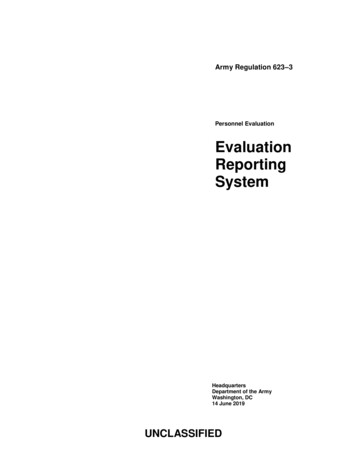
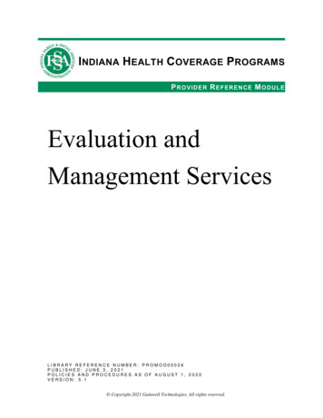
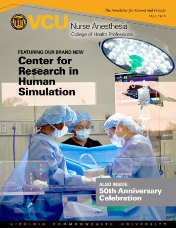
Paracentesis sites:avoid inferior epigastric arteries24
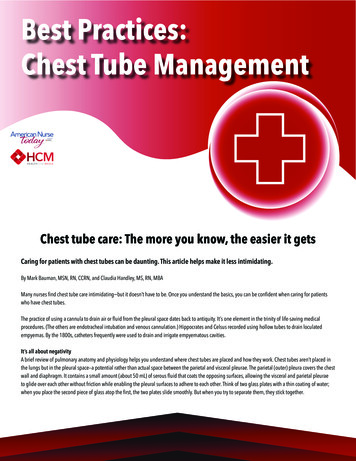
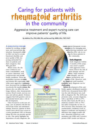
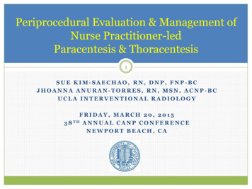
Ascites on Ultrasound25
Cleaning skin26
Sterile draping27
Prepping kit28
Local anesthesia administration29
Nicking for catheter insertion30
Catheter insertion31
Confirm catheter is in the fluid32
Remove stylet once catheter is inserted33
Connect to drainage tubing34
Drain into negative pressure evacuation containers35
Remove catheter when drainage completed36
Label & send specimens to lab for analysis37
Evidence Findings: Albumin Infusions38A meta-analysis from Kwok et al (2013) suggests that the use ofalbumin in cirrhotic patients undergoing paracentesis reducesparacentesis-induced circulatory dysfunction & reduces deathand renal impairmentLarge volume fluid removal: more than 5 litersIntravenous serum albumin (25% albumin, 6-8 gms/L) to prevent hypotension maybe indicated for some patients, i.e.) hypotensive, low albuminSmall volume initial paracentesis, followed by large volume with albumin andhemodynamic support, inpatient or outpatient, may be consideredKwok CS, Krupa L, Mahtani A, et al. Albumin Reduces Paracentesis-Induced Circulatory Dysfunction and ReducesDeath and Renal Impairment among Patients with Cirrhosis and Infection: A Systematic Review and MetaAnalysis. Biomed Res Int. 2013;2013:295153.
Laboratory tests: Ascitic fluid391. Cell count with differential: infection2. Albumin: calculate SAAG3. Protein: calculate SAAG4. Bacterial culture: infection5. Glucose level6. Gram stain: rarely provides clinically useful information for thedetection of SBP7. Triglyceride levels (elevated in chylous ascites)8. Bilirubin level (may be elevated in bowel perforation)9. Amylase level (elevation suggests pancreatic source)10. Lactate dehydrogenase (LDH) level11. Cytology: rule out malignancies
Ascitic fluid analysis: Cell Count40Cell count with differential: elevated counts suggest infection, such asspontaneous bacterial peritonitis (SBP). Anaerobic bacteria not associated withSBPPrimary SBP: infection of ascitic fluid without intra-abdominal infection withchronic liver disease due to translocation of enteric bacteria, such as e. coli,klebsiella pneumoniae, enterococcal species, and steptococcus pneumoniaeHigher risk for SBP: renal failure patients using peritoneal dialysis, SLE patients,and children with nephrosisAscitic fluid polymorphonclear leukocyte (PMN) count 250/uL with 50%PMNs in fluid is presumptive for SBP treat empirically, regardless of sxsSecondary SBP: infected ascitic fluid associated with an intra-abdominalinfection
Ascitic fluid analysis: SAAG41Serum ascites albumin gradient (SAAG): can be calculated to determine the causeof ascites. Draw serum albumin same day as fluid albumin & proteinSAAG (serum albumin) – (albumin level of ascitic fluid)Normal 1.7High gradient ascites ( 1.1 g/dL): dt portal HTN from liver or non-liver dzw/97% accuracyCauses: Budd-Chiari syndrome, CHF, or cirrhosisLow gradient ascites ( 1.1 g/dL): not dt portal pressureCauses: peritoneal carcinomatosis, TB, pancreatitis, nephrotic syndrome,serositisSAAG is preferred method for characterizing ascitesMore discriminant than older methods of classifying fluid astransudative vs. exudative
Thoracentesis42
Why does pleural fluid develop?43Normally, only small amounts of pleural fluid exist forlubrication ( 10mL)Liquid (pleural effusion) or air (pneumothorax) canaccumulate due to different conditions1. Transudative (CHF, 5-10% of cirrhosis pts develop hepatichydrothorax)2. Exudative (PNA, CA, PE, viral infx, recent chest or cardiacsurgery)Malignancy: most common cause of pleural fluidaccumulation as a complication of cancer
Symptoms of pleural effusion44Dry, nonproductive coughBreathing difficultyChest painFeverEdemaWeight loss
General management of pleural effusions45Pleural effusion:Observation & monitoringDiuretics, if indicatedAntibiotics, if clinically indicatedThoracentesis indications:Diagnostic: rule out infection, malignancy with new onset or unilateralpleural effusionTherapeutic: pleural effusions causing respiratory compromise orsignificantly increasing in volumeFirst drainage: often dual indication (diagnostic & therapeutic)
Thoracentesis Complication Risks46Bleeding/Hematoma2. Infection (2%)3. Limited coughing, dyspnea, or chest discomfort with postprocedural lung expansion1.PneumothoraxRe-expansion pulmonary edema after 1L: limit to 2 litersDry tap (no free fluid); may need chest tube if empyema presentVasovagal syncope/hypotensionDeath
Contraindications to Thoracentesis47Mild hematologic abnormalities do not increase the risk of bleeding.Bleeding may be increased if:1. Prothrombin time (PTT) 21 seconds2. International normalized ratio (INR) 1.63. Platelet count 50,000 per cubic millimeterAbsolute contraindication: acute chest requiring surgery, i.e.) traumaRelative contraindications are:Local cutaneous conditionSevere coagulopathyBronchial obstruction: mediastinal shift toward effusion - bronchoscopyrecommended
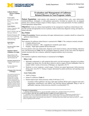
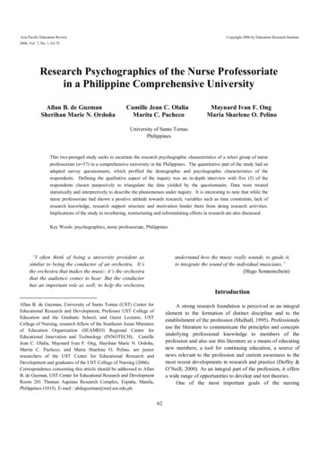
Pleural effusion on CXR48
Pleural fluid on CT49
Pleural effusion on ultrasound50
Technique for thoracentesis51
Prep the skin52
Drape the back53
Administer local lidocaine54
Nick the skin55
Close look at pleural space56
Align catheter device57Advance device over the superior aspect ofthe rib
Insert catheter device58
Advance catheter over stylet & remove stylet59
Collect diagnostic samples60
Drain into negative pressure evacuationcontainers61
Post-Thoracentesis CXR Indications621.Not required unless otherwise indicated by symptoms or signs ofcomplication1.Signs of pneumothorax post-procedureProgressive post-procedure chest pain and dyspneaVoice transmission absent superior to thoracentesis siteTactile fremitus absent superior to thoracentesis site
Pneumothorax63Pneumothorax (air between lung and chest wall):Rates up to 20-39% - without imaging guidanceRates up to 11%; 2% needing chest tubes - with imaging guidance1. Can be spontaneous2. Through hole between the ribs/lungs: trauma3. Iatrogenic: ‘ex vacuo’ related to lack of post-procedural lung re-expansionrelated to diseaseCT:Black airspacecompared tocontralateral chestCXR:No lung markingscompared tocontralateral chest
Pneumothorax management64I. Stable Patients with Small Pneumothoraces1. Observation for up to 3 to 6 hours2. Discharge home if a repeat chest radiograph excludes progression3. Follow up within 12-48 hours with repeat chest radiograph todocument resolution, if clinically indicatedII. Patients with Large Pneumothoraces1. Hospitalization2. Re-expansion of lung using a small-bore catheter or placing a 16-22 Fchest tube3. Suction if lung fails to re-expand
Transudative vs. Exudative Fluid65Transudates: thin, watery fluid oozing into cavity due to loss of osmoticpressure or back pressure from the circulation. Systemic diseaseCauses: CHF, hypoalbuminemic states such as cirrhosis and liver failure,portal vein thrombosis, nephrosis, PE, myxedema, etc.Exudate s: thicker, more viscous fluid usu. due to tissue damage,allowing albumin & water to seep out. Local diseaseCauses: malignancies, pneumonia, PE, peritoneal carcinomatosis,nephrotic syndrome, TB, autoimmune dz (RA, SLE), esophagealperforation, pancreatic or biliary disease, pericarditis, radiation(accidental or therapeutic), peritonitis, ischemic or obstructed bowel,medication SE’s, rare conditions
Other types of fluid66Blood (sanguinous):Related to trauma severing blood vessels, malignanciesEvaluate hematocrit (Hct): Hemothorax if 50% of the serum HctChyle (chylous):Related to trauma from surgery, MVA, malignancies,idiopathic, intestinal lymphgiectasia, subclavian venousthrombosis, tumors, filarasis, lymphangiomyomatosisWhite or milky in color due triglycerides fromdigestion (erosion of thoracic duct carrying lymphfluid from intestines to the heart)
Laboratory tests: Pleural fluid671.2.3.4.5.6.7.8.Cell count with differential: WBCs indicate infection and subtypes gives clues asto type of infection. RBCs indicate bleedingCultures & gram stains: may yield infectious organism prior to other cxs,including blood & sputum, including TBAmylase: 2X’s serum level or absolute value 160 indicative of pancreatitis,dissected or ruptured pancreatic pseudocyst, cancer, or esophageal ruptureGlucose: low if pleural fluid value 50% of normal serum value; differentialincludes RA, SLE, bacterial empyema, malignancy, TB, and esophageal rupturepH: normal is 7.60. 7.3 with normal arterial blood pH has same differential aslow pleural glucoseTriglyceride & cholesterol: 110 mg/dL and presence of chylomicrons indicateschylous effusion from rupture of thoracic duct most frequently resulting fromtrauma or malignancy, such as lymphomaAdenosine deaminase: high for empyema, RA. 70 U/L for TBCytology: rule out malignancy, such as lung cancer, pleural meothelioma, andmetastasis
Pleural effusion management68Pleural AnatomyPleural FluidBacteriologyPleural FluidChemistryNeed for DrainageMinimal effusion( 10 mm on lateraldecubitus view); freeflowingCx and GS unknownpH unknownNoSmall to moderateeffusion( 10 mm to one half ofhemithorax on lateraldecubitus view); freeflowingNegative Cx and GSpH 7.20NoLarge effusion( one half of hemithoraxon lateral decubitus view)or loculated fluid orthickened pleuraPositive Cx or GSpH 7.20YesAnyPuspH 7.0YesDweik (2010). Pleural disease. Cleveland Clinic
Conclusion: Key to Positive Outcomes691.2.3.4.5.6.Check labsReview H&PManage anticoagulants, if applicableUse imaging guidanceMonitor appropria
USA: Emory University1, Mayo Clinic Globally: Wales2, United Kingdom 4 . Benefits of an NP-led paracentesis & thoracentesis program 1. . Portal vein thrombosis 17 . Symptoms of ascites Abdominal distention Abdominal discomfort