Transcription
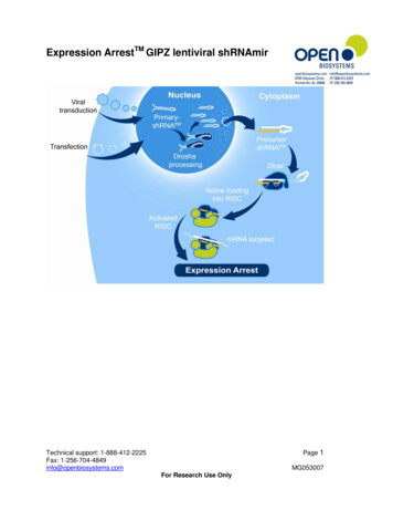
Expression ArrestTM GIPZ lentiviral shRNAmirPage 1Technical support: 1-888-412-2225Fax: 1-256-704-4849info@openbiosystems.comMG053007For Research Use Only
Expression ArrestTM GIPZ lentiviral shRNAmirCatalog # RHS4430, RHS4531, RHS4477, RMM4431, RMM4532, RMM4501CONTENTSTopicProduct descriptionShipping and storageDesign InformationVector InformationAntibiotic ResistanceVector Map- pGIPZProtocolsProtocol I-ReplicationProtocol II-Plasmid PrepProtocol III-Restriction DigestProtocol IV-TransfectionProtocol V-PackagingProtocol VI-TransductionRelated reagentsFAQsTroubleshootingReferencesLicensing informationPage22344567891013131718192123PRODUCT DESCRIPTIONThe GIPZ lentiviral shRNAmir library was developed by Open Biosystems in collaboration withDr. Greg Hannon (CSHL) and Dr. Steve Elledge (Harvard). This library combines the designadvantages of microRNA-adapted shRNA (shRNAmir) with the pGIPZ lentiviral vector to createa powerful RNAi trigger capable of producing RNAi in most cell types including primary and nondividing cells.SHIPPING AND STORAGEIndividual constructs are shipped as bacterial cultures of E. coli (Prime Plus) in LB-Lennox (lowsalt) broth with 8% glycerol, 100µg/ml carbenicillin and 25µg/ml zeocin. Individual constructsare shipped on wet ice. Collections are shipped in 96 well plate format on dry ice. Individualconstructs and collections should be stored at -80oC.Open Biosystems checks all cultures for growth prior to shipment.TO ALLOW ANY CO2 THAT MAY HAVE DISSOLVED INTO THE MEDIA FROM THE DRYICE IN SHIPPING TO DISSIPATE, PLEASE STORE CONSTRUCTS AT –80 C FOR ATLEAST 48 HOURS BEFORE THAWING.Important Safety Note:Follow NIH guidelines regarding lentiviral production and transduction; follow Biosafety Level 2(BL2) or BL2 laboratory criteria.Page 2Technical support: 1-888-412-2225Fax: 1-256-704-4849info@openbiosystems.comMG053007For Research Use Only
DESIGN INFORMATIONUnique MicroRNA-30 based hairpin designExpression Arrest short hairpin RNA constructs are expressed as human microRNA-30(miR30) primary transcripts (Figure 1). This design adds a Drosha processing site to the hairpinconstruct and has been shown to greatly increase knockdown efficiency (Boden et al. 2004).The hairpin stem consists of 22nt of dsRNA and a 19-nt loop from human miR30. Adding themiR30 loop and 125nt of miR30 flanking sequence on either side of the hairpin results in greaterthan 10-fold increase in Drosha and Dicer processing of the expressed hairpins when comparedwith conventional shRNA designs without microRNA (Silva et al. 2005. Increased Drosha andDicer processing translates into greater siRNA/miRNA production and greater potency forexpressed hairpins.Figure 1. Expression Arrest shRNA are expressed as miR30primary transcriptsUse of the miR30 design also allowed the use of 'rules-based' designs for target sequenceselection. One such rule is the destabilizing of the 5' end of the antisense strand which results instrand specific incorporation of miRNAs into RISC.The proprietary design algorithm targets sequences in coding regions and the 3’UTR with theadditional requirement that they contain greater than 3 mismatches to any other sequence in thehuman or mouse genomes.Each shRNA construct has been sequence verified before being cloned into the vector toensure a match to the target gene. To assure you the highest possibility of modulating the geneexpression level, each gene is represented by multiple shRNA constructs, each covering aunique region of the target gene.Page 3Technical support: 1-888-412-2225Fax: 1-256-704-4849info@openbiosystems.comMG053007For Research Use Only
VECTOR INFORMATIONVersatile vector designFeatures of the pGIPZ lentiviral vector (Figure 2-3, Table 1) that make it a versatile tool forRNAi studies include: Ability to perform transfections or transductions using the replication incompetentlentivirus TurboGFP and shRNAmir are part of a bicistronic transcript allowing the visual markingof shRNAmir expressing cells Amenable to in vitro and in vivo applications Puromycin drug resistance marker for selecting stable cell lines Molecular barcodes enable multiplexed screening in poolsFigure 2. pGIPZ lentiviral vectorTable 1. Features of the pGIPZ vectorVector ElementUtilityCMV PromoterRNA Polymerase II promoterCentral Polypurine tract helps translocation into the nucleus of noncPPTdividing cellsWREEnhances the stability and translation of transcriptsTurboGFPMarker to track shRNAmir expressionIRES-puroMammalian selectable markerAMPrAmpicillin bacterial selectable marker.5'LTR5' long terminal repeatpUC oriHigh copy replication and maintenance of plasmid in E.coliSIN-LTR3' Self inactivating long terminal repeatRRERev response elementZEOrBacterial selectable markerANTIBIOTIC RESISTANCEpGIPZ contains 3 antibiotic resistance markers (Table 2).Table 2. Antibiotic Resistances Conveyed by pGIPZAntibioticAmpicillin ml25µg/mlvariableUtilityBacterial selection marker (outside LTRs)Bacterial selection marker (inside LTRs)Mammalian selectable markerPage 4Technical support: 1-888-412-2225Fax: 1-256-704-4849info@openbiosystems.comMG053007For Research Use Only
VECTOR MAPFigure 3. Detailed Vector Map of pGIPZ lentiviral vectorPage 5Technical support: 1-888-412-2225Fax: 1-256-704-4849info@openbiosystems.comMG053007For Research Use Only
PROTOCOLSCulturing protocols and maintenance of pGIPZ The pGIPZ lentiviral shRNAmir library has passed through internal QC processes to ensure highquality and low recombination (Figures 4 and 5).Figure 4. Representative shRNAmir containing pGIPZ lentiviral clones grown for 16 hours at 30 C andthe plasmid isolated and normalized to a standard concentration. Clones were then digested with SacIIand run out on a gel. The expected band sizes are 1259bp, 2502bp, 7927bp. No recombinant productsare visible. 10kb molecular weight ladder (10kb, 7kb, 5kb, 4kb, 3kb, 2.5kb, 2kb, 1.5kb, 1kb)Figure 5. Gel image of a single plate from the GIPZ library cultured for 10 successive generations in anattempt to determine the tendency of the pGIPZ vector to recombine. Each generation was thawed,replicated and incubated overnight for 16 hours at 30 C then frozen, thawed and replicated. This processwas repeated for 10 growth cycles. After the 10th growth cycle, plasmid was isolated and normalized to astandard concentration. Clones were then digested with SacII and run on a gel. Expected band sizes1259bp, 2502bp, 7927bp. 10kb molecular weight ladder (10kb, 7kb, 5kb, 4kb, 3kb, 2.5kb, 2kb, 1.5kb,1kb)The pGIPZ vector appears stable without showing any recombination.Page 6Technical support: 1-888-412-2225Fax: 1-256-704-4849info@openbiosystems.comMG053007For Research Use Only
PROTOCOL I-REPLICATIONTable 3. Materials for plate replicationItemLB-Lennox Broth (low salt)Peptone, granulated, 2kg - DifcoYeast Extract, 500g, granulatedNaClGlycerolCarbenicillin or ampicillinZeocinPuromycin96 well microplatesAluminum sealsDisposable replicatorsDisposable groVWRVWRGenetixScinomixCatalog M-2200 or 56X5054SCI-5010-OSCulture conditions for replication of shRNA constructsFor archive replication, grow all pGIPZ clones at 30 C in LB-Lennox (low salt) media plus25µg/ml zeocin and 100µg/ml carbenicillin in order to provide maximum stability of the clones.Prepare media with glycerol and the appropriate antibiotics. Use the minimal volume ofinoculant (0.5-1µl in 1ml LB) or use replicating pins for 96 well microtiter plates. Incubate withshaking (or without shaking for microtiter plates) at 30 C for 16-20 hours. Freeze at –80 C forlong term storage. Avoid long periods of storage at room temperature or higher in order tocontrol background recombination products.2X-LB broth (low-salt) media preparationPeptone20g/lYeast Extract10g/lNaCl5g/lAppropriate antibiotic(s) at recommended concentration(s)*Glycerol8% for long term storageNote: LB media can be used instead of 2XLB*Glycerol can be omitted from the media if you are culturing for plasmid preparation. If makingcopies of the constructs for long term storage at –80 C, 8% glycerol is required.Replication of platesPrepare target plates by dispensing 160µl of LB media supplemented with 8% glycerol andappropriate antibiotic (25µg/ml zeocin and 100µg/ml carbenicillin).Prepare source plates:1. Remove foil seals while the source plates are still frozen. This minimizes crosscontamination.2. Thaw the source plates with the lid on. Wipe any condensation underneath the lid with apaper wipe soaked in ethanol.Page 7Technical support: 1-888-412-2225Fax: 1-256-704-4849info@openbiosystems.comMG053007For Research Use Only
Replicate:1. Gently place a disposable replicator in the thawed source plate and lightly move thereplicator around inside the well to mix the culture. Make sure to scrape the bottom ofthe plate of the well.2. Gently remove the replicator from the source plate and gently place in the target plateand mix in the same manner to transfer cells.3. Dispose of the replicator.4. Place the lids back on the source plates and target plates.5. Repeat steps 1-4 until all plates have been replicated.6. Return the source plates to the -80 C freezer.7. Place the inoculated target plates in a 37 C incubator for 14-20 hours.Note: Due to the tendency of all viral vectors to recombine, we recommend keeping theincubation times as short as possible and avoid subculturing. Return to your glycerol stock foreach plasmid preparation.PROTOCOL II- PLASMID PREPCulture conditions for individual plasmid preparationsMost plasmid mini-prep kits recommend a culture volume of 1–10ml for good yield.For shRNAmir constructs, 5ml of culture can be used for one plasmid mini-prep generallyproducing 5–10µg of plasmid DNA.1. Upon receiving your glycerol stock(s) containing the shRNAmir of interest store at –80 Cuntil ready to begin.2. To prepare plasmid DNA first thaw your glycerol stock culture and pulse vortex toresuspend any E. coli that may have settled to the bottom of the tube.3. Take a 10µl inoculum from the glycerol stock into 3-5ml of LB (low salt) with 100µg/mlcarbenicillin and 25µg/ml zeocin. Incubate at 37 C for 16 hours with vigorous shaking.Return the glycerol stock(s) to -80 C. If a larger culture volume is desired, use the 3-5mlovernight culture as a starter inoculum. Incubate at 37 C for 16 hours with vigorousshaking.4. Pellet the 3-5ml culture and begin preparation of plasmid DNA.5. Run 3-5µl of the plasmid DNA on a 1% agarose gel. pGIPZ with shRNAmir is11744bp.Note: Due to the tendency of all viral vectors to recombine, we recommend keeping theincubation times as short as possible and avoid subculturing. Return to your original glycerolstock or the colony glycerol stock for each plasmid preparation.Culture conditions for 96 well plasmid preparationInoculate 96 well bio-block containing 1ml per well of the above media with 1µl of the culture.Incubate at 37 C with shaking ( 170-200rpm). We have observed that incubation times from 16hours produces good plasmid yield. For plasmid preparation, follow the protocols recommendedby the plasmid isolation kit manufacturer.Note: The cells can be grown at 37 C for purposes of template preparation or sequencing. Forarchive replication, grow all pGIPZ clones at 30 C in LB-Lennox (low salt) media plus 25µg/mlzeocin and 100µg/ml carbenicillin in order to provide maximum stability of the clones.Page 8Technical support: 1-888-412-2225Fax: 1-256-704-4849info@openbiosystems.comMG053007For Research Use Only
PROTOCOL III-RESTRICTION DIGESTRestriction Digests of pGIPZ The following is a sample protocol for restriction enzyme digestion using KpnI, SacII, SalI, XhoIand/or NotI for diagnostic quality control of pGIPZ lentiviral vectors.1. Using filtered pipette tips and sterile conditions add the following components, in theorder stated, to a sterile PCR thin-wall tube.ComponentSterile, nuclease-free waterRestriction enzyme 10X bufferBSA (10X, 10mg/ml) if requiredDNA sample 80-240ng, in water or TE bufferRestriction enzyme 20UFinal volumeAmountXµl1µl1µlXµl0.25µl10µl2. Mix gently by pipetting.3. Incubate in a thermalcycler at 37 C for 2 hours to digest4. Load the gel with 10µl of each of the digested samples (KpnI, SacII, SalI, XhoI and/orNotI) on a 1% agarose gel. Run uncut sample alongside the digested samples. (Figure6)123456Figure 6: Restriction digests with pGIPZ. Lane 1– 10kb molecular weight ladder (10kb, 7kb, 5kb, 4kb,3kb, 2.5kb, 2kb, 1.5kb, 1kb). Lane 2 - Uncut pGIPZ vector. Lane 3 - KpnI digested pGIPZ produces 2bands at 1750bp and 9860bp. Lane 4- SacII digest produces 3 bands at 1178bp, 2502bp and 7930bp.Lane 5 -SalI produces 3 bands at 2188bp, 4298bp and 5124bp. Lane 6 – XhoI, NotI double digestproduces 2 bands at 1210bp and 10400bp.Page 9Technical support: 1-888-412-2225Fax: 1-256-704-4849info@openbiosystems.comMG053007For Research Use Only
PROTOCOL IV-TRANSFECTIONThe protocol below is optimized for transfection of the shRNA plasmid DNA into HEK293T cellsin a 24 well plate using serum free media. If a different culture dish is used, adjust the numberof cells, volumes and reagent quantities in proportion to the change in surface area (Table 4).It is preferable that transfections be carried out in medium that is serum free and antibiotic free.A reduction in transfection efficiency occurs in the presence of serum, however it is possible tocarry out successful transfections with serum present (see Transfection Optimization).Warm Arrest-In to ambient temperature (approximately 20 minutes at room temperature) priorto use. Always mix well by vortex or inversion prior to use.Maintain sterile working conditions with the DNA and Arrest-In mixtures as they will be added tothe cells.Table 4. Suggested amounts of DNA, medium and Arrest-In reagent for transfection of shRNA plasmidDNA into adherent cellsTissue CultureSurface areaTotal serumPlasmid DNAArrest-In (µg)**Dishper plate or well free media(µg)*(cm2)volume per well(ml)60 mm20242135 mm812106 well9.4121012 well3.80.51524 well1.90.250.52.596 well0.30.10.1 - 0.20.5 - 1*Recommended starting amount of DNA. May need to be optimized for the highest efficiency**Recommended starting amount of Arrest-In reagent. See Transfection Optimization.1. The day before transfection (day 0), plate the cells at a density of 5 x 10⁴ cells per well ofa 24 well plate.Full medium (i.e. with serum and antibiotics) will be used at this stage.2. On the day of transfection form the DNA/Arrest-In transfection complexes.The principle is to prepare the shRNA plasmid DNA and transfection reagentdilutions in an equal amount of serum free medium in two separate tubes. Thesetwo mixtures (i.e. the DNA and the Arrest-In) will be added to each other andincubated for 20 minutes prior to addition to the cells. This enables the DNA/ArrestIn complexes to form.a. For each well to be transfected, dilute 500ng shRNA plasmid DNA into 50µl (totalvolume) of serum free medium in a microfuge tube.b. For each well to be transfected, dilute 2.5µg (2.5µl) of Arrest-In into 50µl (totalvolume) serum free medium into a separate microfuge tube.Page 10Technical support: 1-888-412-2225Fax: 1-256-704-4849info@openbiosystems.comMG053007For Research Use Only
c. Add the diluted DNA (step a) to the diluted Arrest-In reagent (step b), mixrapidly then incubate for 20 minutes at room temperature.This will give a 1:5 DNA:Arrest-In ratio which is recommended for optimaltransfection into HEK293T cells.Your total volume will be 100µl at this stage.d. Set up all desired experiments and controls in a similar fashion as outlined inTable 5. It is also advisable to set up an Arrest-In only control.Table 5. Quantities of DNA for transfection experimentsType ofshRNA Plasmid Reporter* (ng)Carrier DNA**Serum freetransfectionDNA (ng)(ng)medium (finalexperimentvolume in µl)0050shRNA plasmid 500 – hairpin togene of interestDNA0500050Transfectionefficiency450-500 –50050Knockdownhairpin toefficiency ofreporterreporter050450-50050Control forknockdownefficiency500 – scramble0050Non-silencinghairpincontrol*It is not necessary to transfect a reporter into cells if you are using a construct which already has areporter for convenient estimation of transfection efficiency. Recommended reporters for other vectorsinclude GFP, luciferase, and/or β-gal (X-gal staining and/or ONPG assays).**Carrier DNA is required to increase the total DNA quantity for the formation of adequate DNA/Arrest-Incomplexes. Recommended carriers are pUC19 or pBluescript plasmids.3. Aspirate the growth medium from the cells. Add an additional 150µl of serum freemedium to each of the tubes containing transfection complexes and mix gently. Add the250µl DNA/Arrest-In complex mixture to the cells and incubate for 3-6 hours in a CO2incubator at 37oC.Your total volume will be 250µl at this stage.4. Following the 3-6 hour incubation, add an equal volume of growth medium (250µl)containing twice the amount of normal serum to the cells (i.e. to bring the overallconcentration of serum to what is typical for your cell line). Alternatively, the transfectionmedium can be aspirated and replaced with the standard culture medium (see Note).Return the cells to the CO2 incubator at 37oC.Note – Arrest-In has displayed low toxicity in the cell lines tested, thereforeremoval of transfection reagent is not required for many cell lines. In our handshigher transfection efficiencies have been achieved if the transfection medium isnot removed. However, if toxicity is a problem, aspirate the transfection mixturePage 11Technical support: 1-888-412-2225Fax: 1-256-704-4849info@openbiosystems.comMG053007For Research Use Only
after 3-6 hours and replace with fresh growth medium. Additionally, fresh growthmedium should be replenished as required for continued cell growth.5. After 48-96 hours of incubation, examine the cells microscopically for the presence ofreporter expression where applicable as this will be your first indication as to theefficiency of your transfection. Then assay cells for reduction in gene or reporter activityby quantitative/real-time RT-PCR, western blot or other appropriate functional assay;compare to untreated, reporter alone, non-silencing shRNA or other negative controls.Optimal length of incubation from the start of transfection to analysis isdependent on cell type, gene of interest, and the stability of the mRNA and/orprotein being analyzed. Quantitative/real-time RT-PCR generally gives the bestindication of expression knock-down. The use of western blots to determineknock-down is very dependent on quantity and quality of the protein, its half-life,and the sensitivity of the antibody and detection systems used.6. If selecting for stably transfected cells (optional), transfer the cells to medium containingpuromycin for selection. It is important to wait at least 48 hours before beginningselection.The working concentration of puromycin needed varies between cell lines. Werecommend you determine the optimal concentration of puromycin required to killyour host cell line prior to selection for stable shRNA transfectants. Typically, theworking concentration ranges from 1-10µg/ml. You should use the lowestconcentration that kills 100% of the cells in 3-5 days from the start of puromycinselection.Cells Grown In SuspensionTransfection of cells in suspension would follow all the above principles and the protocol wouldlargely remain the same, except that the DNA/Arrest-In mixture should be added to cells (post20 minute incubation for complex formation) to a total volume of 250µl serum free medium or toa total volume of 250µl of medium with serum (no antibiotics).Transfection Optimization using Arrest-InIt is essential to optimize transfection conditions to achieve the highest transfection efficienciesand lowest toxicity with your cel
Expression Arrest TM GIPZ lentiviral shRNAmir Catalog # RHS4430, RHS4531, RHS4477, RMM4431, RMM4532, RMM4501 CONTENTS Topic Page Product description 2 Shipping and storage 2 Design Information 3 Vector Information 4 Antibiotic Resistance 4 Vector Map- pGIPZ 5 Protoc