Transcription
612086Journal of Dental ResearchImmune Cells and Molecular NetworksResearch Reports: BiologicalImmune Cells and Molecular Networksin Experimentally Induced PulpitisJournal of Dental Research 1 –10 International & American Associationsfor Dental Research 2015Reprints and : 10.1177/0022034515612086jdr.sagepub.comE. Renard1*, A. Gaudin1,2*, G. Bienvenu1,2, J. Amiaud3, J.C. Farges4,M.C. Cuturi1, A. Moreau1†, and B. Alliot-Licht1,2†AbstractDental pulp is a dynamic tissue able to resist external irritation during tooth decay by using immunocompetent cells involved in innateand adaptive responses. To better understand the immune response of pulp toward gram-negative bacteria, we analyzed biologicalmediators and immunocompetent cells in rat incisor pulp experimentally inflamed by either lipopolysaccharide (LPS) or saline solution(phosphate-buffered saline [PBS]). Untreated teeth were used as control. Expression of pro- and anti-inflammatory cytokines, chemokineligands, growth factors, and enzymes were evaluated at the transcript level, and the recruitment of the different leukocytes in pulp wasmeasured by fluorescence-activated cell-sorting analysis after 3 h, 9 h, and 3 d post-PBS or post-LPS treatment. After 3 d, injured ratincisors showed pulp wound healing and production of reparative dentin in both LPS and PBS conditions, testifying to the reversiblepulpitis status of this model. IL6, IL1-β, TNF-α, CCL2, CXCL1, CXCL2, MMP9, and iNOS gene expression were significantly upregulatedafter 3 h of LPS stimulation as compared with PBS. The immunoregulatory cytokine IL10 was also upregulated after 3 h, suggesting thatLPS stimulates not only inflammation but also immunoregulation. Fluorescence-activated cell-sorting analysis revealed a significant, rapid,and transient increase in leukocyte levels 9 h after PBS and LPS stimulation. The quantity of dendritic cells was significantly upregulatedwith LPS versus PBS. Interestingly, we identified a myeloid-derived suppressor cell–enriched cell population in noninjured rodent incisordental pulp. The percentage of this population, known to regulate immune response, was higher 9 h after inflammation triggered withPBS and LPS as compared with the control. Taken together, these data offer a better understanding of the mechanisms involved in theregulation of dental pulp immunity that may be elicited by gram-negative bacteria.Keywords: dental pulp, innate immune response, lipopolysaccharides, chemokines, cytokines, leukocytesIntroductionDental pulp is a highly dynamic tissue equipped with a network of resident immunocompetent cells that may play a majorrole in the defense against pathogens and during tissue injury(Izumi et al. 1995; Kawashima et al. 2006; Gaudin et al. 2015).Dental pulp is able to mount inflammatory/innate and lateradaptive immune responses to fight bacteria and inactivatethose of their components that gain access to the pulp duringthe caries process (Hahn and Liewehr 2007a, 2007b). The cariogenic microflora contains many bacterial species. Grampositive bacteria greatly dominate the microflora in initial andmoderate dentin caries lesions, with the proportion of gramnegative bacteria growing with the progression of tooth decay(Hahn et al. 1989; Love and Jenkinson, 2002; Martin et al.2002; Chhour et al. 2005; Aas et al. 2008). Pathogen-associatedmolecular patterns (PAMPs) are released into the dental pulpthrough dentinal tubules. Odontoblasts, fibroblasts, leukocytes, and stem cells recognize PAMPs through pattern recognition receptors, in particular the Toll-like receptors (TLRs;Tokuda et al. 2001; Durand et al. 2006; Veerayutthwilai et al.2007; Mutoh et al. 2009; Farges et al. 2011; Liu et al. 2014; Heet al. 2015). Lipopolysaccharide (LPS) is an integral component of the membrane of gram-negative bacteria and is recognized by TLR4. The binding of LPS to TLR4 leads to thedevelopment of innate immune responses, either by theMyD88-dependent pathway (He et al. 2014) or by the Trifdependent pathway. This second pathway induces a type I IFNresponse (Elson et al. 2007). Type I IFN signaling is alsorequired for TLR4-mediated induction of IL10, a potent antiinflammatory molecule that regulates excessive production ofinflammatory cytokines during infection (Iyer et al. 2010).In the pulpitis process, a low-grade inflammation enablespulp tissue regeneration (tertiary dentinogenesis), whereasmore intense and severe inflammation leads to necrosis (Izumi1INSERM, Center for Research in Transplantation and Immunology,UMR 1064, Nantes, France2Faculty of Odontology, University of Nantes, Nantes, France3INSERM, UMR 957, Nantes, France4Laboratory of Tissue Biology and Therapeutic Engineering, UMR 5305,and Faculty of Odontology, Lyon, France*Authors contributing equally to this article.†Authors contributing equally as senior investigators.A supplemental appendix to this article is published electronically only g Author:B. Alliot-Licht, UMR 1064 INSERM, 30 Bd Jean Monnet, 44093 NantesCedex 01, France.Email: Brigitte.alliot-licht@univ–nantes.frDownloaded from jdr.sagepub.com at International Association for Dental Research on October 16, 2015 For personal use only. No other uses without permission. International & American Associations for Dental Research 2015
2Journal of Dental Research et al. 1995; Cooper et al. 2014). To characterize the immuneresponse triggered by the gram-negative bacterial PAMPs indental pulp, we investigated the expression of proinflammatoryand anti-inflammatory mediators in rat incisor dental pulp tissue upon LPS stimulation. The expression of dentin sialophosphoprotein (DSPP), an odontoblastic marker, was also assessedas an indicator of dental pulp regeneration. In addition, theimmune cells of the dental pulp were investigated by fluorescence-activated cell-sorting (FACS) analysis. This techniqueallows identification and quantification of small populations ofcells with a set of different markers. We previously showed thattotal leukocytes represent 1% of the cell population obtainedafter enzymatic digestion in healthy human pulp, and we established the presence of specific populations of leukocytesinvolved in immunoregulation (Gaudin et al. 2015). In thisstudy, we used a model of experimentally injured rat pulp toanalyze the levels of the key cellular actors in immune responsepresent in this tissue to throw light on the immune responseprocess following gram-negative bacteria aggression, with thefinal aim of identifying new targets for vital pulp therapies.Materials and MethodsExperimental Pulpitis ModelFemale Sprague-Dawley rats (Charles River Laboratories,Saint-Germain-Nuelles, France) weighing 200 to 250 g wereused throughout the study. The animal experiments wereapproved by the Animal Ethics Committee of the University ofNantes (CEEA No. 00286.01). Induction of pulpitis was performed under general anesthesia with an intramuscular mix ofxylazine (8 mg/kg) and ketamine (805 mg/kg) and local anesthesia with periapical injection of articaine as previouslydescribed (Kawanishi et al. 2004). The portion of the upperincisor exposed to the oral cavity was removed horizontallywith round tungsten steel burs to expose the pulp chamber atthe pulp horn, and the entrance of the pulp horn was enlargedwith K-files #40 to a length of 1 mm to create space to applyLPS or phosphate-buffered saline (PBS). Five microliters ofLPS from Escherichia coli O111:B4 (Sigma Chemical Co.,St. Louis, MO, USA) diluted in PBS at a final concentration of10 mg/mL or PBS alone was dropped on the injured pulp, andthe cavities were sealed with Cavit (ESPE, Seefeld, Germany).Animals were sacrificed 3 h, 9 h, and 3 d after injury, and theincisors were extracted.Histologic AnalysisTeeth were fixed in paraformaldehyde (PFA) at 4 C overnight,decalcified (4% ethylenediaminetetraacetic aciod [EDTA],0.2% PFA) at 50 C in a KOS Microwave (Milestone,Kalamazoo, MI, USA) for up to 10 d, and then embedded inparaffin and cut into 3-μm-thick sections for Masson trichromestaining (n 4). Images were created with bright-field microscopy (Nikon Eclipse E600). For the immunohistochemistry (n 2), 3-μm paraffin sections were stained with a polyclonal goatanti-Iba1 antibody (Abcam, Cambridge, UK) for macrophagedetection (Rehg et al. 2012). A standard ABC method was usedfor detection with a biotinylated donkey anti-goat secondaryantibody and peroxidase-conjugated streptavidin (Dako,Trappes, France) and a DAB substrate as a chromogen (Dako).Negative control without the primary antibody was performed.Counterstaining was carried out with Gill 2 hematoxylin.Stained slides were observed with a virtual microscope (NDPview; Hamamatsu, Japan).Real-time Polymerase Chain ReactionRNA extraction and reverse transcription were performed frompulp tissues of nontreated teeth and teeth treated with PBS orLPS (n 8). Total RNAs were isolated with RNeasy Mini Kit(Qiagen, Valencia, CA, USA) according to the manufacturer’sinstructions. For cDNA synthesis, 1 µg of total RNA was usedas a template for reverse transcription according to the manufacturer’s instructions (Invitrogen, Waltham, MA, USA).Primer sequences are compiled in Table 1. Hypoxanthine phosphoribosyltransferase (HPRT) gene was used as endogenouscontrol gene. The analysis was performed with a Viia7TM RealTime PCR System (Thermo Fisher Scientific, Waltham, MA,USA) with SYBR Green qPCR Master Mix (Life Technologies,Foster City, CA, USA). The mRNA expression level wasdefined as the fold change in mRNA levels in a given samplerelative to the level of a calibrator. The nontreated pulp wasused as calibrator and defined the 1 expression of each gene. ThemRNA expression level is calculated as 2-ΔΔCt, where ΔΔCt (CtTarget – CtHPRT) sample – (CtTarget – CtHPRT) calibrator.Flow CytometryPulp tissue was minced into small pieces and centrifuged. Thetissue was digested in collagenase D (Roche, Paris, France)and incubated at 37 C. After 45 min, digestion was stoppedthrough 10mM EDTA. Cells were then washed 2 times withPBS supplemented with 2% fetal calf serum and 2mM EDTA.The cell suspension was filtered through a 100-μm nylon filter.After resuspension in the same buffer, single-cell suspensions(20 million cells) were ready for antibody staining. For eachstaining, 1 million cells were incubated for 30 min on ice withisotype control or monoclonal conjugated antibodies (Table 2).After 2 washes, cells were fixed with PFA 2%. Cells wereacquired with FACS Canto II (BD Biosciences, San Jose, CA,USA), and results were analyzed with Flowjo Software(TreeStar, Inc., Ashland, NC, USA). All results in this studywere based on the same gating strategy; nonviable cells anddoublets were excluded (n 8). The spleen of healthy rat andisotypic monoclonal conjugated antibodies were used to determine the gate of positive cells in each experiment.Statistical AnalysisStatistical analysis was performed by a Student’s t test for FACSanalysis data and nonparametric Mann-Whitney U test forDownloaded from jdr.sagepub.com at International Association for Dental Research on October 16, 2015 For personal use only. No other uses without permission. International & American Associations for Dental Research 2015
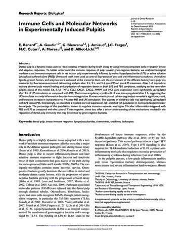
3Immune Cells and Molecular NetworksTable 1. Primer Sequences Used in the Current Study.PrimersForward 5′ 3′Reverse 5′ 3′Product Size, bpAccession 59250210178349173161176187199186111NM 012583.2NM 012589.2NM 012854.2NM 012675.3NM 012580.2NM 031512.2NM 031530.1NM 138880.2NM 053390.1NM 022611.1NM 130410.2NM 021578.2NM 030845.1NM 053647.1NM 031055.1NM 022221.1NM 133523.3NM 012611.3NM 012790.2bp, base pairs.Table 2. Monoclonal Conjugated Antibodies Used in the Current 8Ox62Ox6Iba1FluorochromeDilutionBiotinylated streptavidin PECy7FITCFITCAlexa 488PEAlexa 647Alexa 647FITCAlexa 1/1,000e1/500e1/2500e1/500e1/4,000equantitative polymerase chain reaction data through use ofGraphPad Prism software (La Jolla, CA, USA). Results arepresented as SEM. Differences associated with probabilityvalues of P 0.05 were considered statistically significant.ResultsHistology of Pulp Tissue andDSPP Gene ExpressionTo observe the effects of PAMPs on pulp tissue, histologic andimmunohistochemistry observations were carried out on theincisor teeth at different times posttreatment with LPS or PBS.Noninjured teeth were used to perform a control (Fig. 1A, B).In teeth treated with PBS, infiltrated cells were observed closeto the injured site as early as 9 h after cavity preparation (Fig.1D, E), and osteodentin matrix was observed 3 d postinjury(Fig. 1J, K). In rats treated with LPS, the area of infiltration byinflammatory cells and the osteodentin deposition were largerSpecificityCD45Ig κ chain antibodyCD11b/cTCRαβCD4CD8CD161 (NKRP1)GranulocyteCD103 (αE2integrin)MHC class IIIonized calcium binding adaptor molecule 1than in the PBS-treated teeth (Fig. 1G, H, M, N). However, theamount and the distribution of macrophages observed in theodontoblast and subodontoblastic zones were similar in allconditions (Fig. 1C, F, I, L, O). These data reveal that inflammation was limited to the horn of the teeth and disappearedafter 3 d. Compared with controls, the expression of the DSPPgene increased 3 h after PBS and LPS application and remainedsignificantly upregulated after 3 d. Furthermore, DSPP expression was significantly higher in the PBS-injured pulp as compared with LPS at day 3 (Fig. 1R).RNA Expression of Inflammatory MediatorGenes in LPS-induced PulpitisTo investigate the molecules involved in the inflammation, theexpression of inflammatory mediators was analyzed at thetranscript level in the injured pulp (Fig. 2). In contrast with thenontreated teeth and those with the PBS treatment, IL6, IL1-β,TNF-α, IL10, an anti-inflammatory cytokine, and CCL2,Downloaded from jdr.sagepub.com at International Association for Dental Research on October 16, 2015 For personal use only. No other uses without permission. International & American Associations for Dental Research 2015
4Journal of Dental Research Figure 1. Masson trichrome staining and immunohistochemical analyses of nontreated pulp (control) and experimentally rat pulpitis induced byphosphate-buffered saline (PBS) or lipopolysaccharide (LPS) treatment observed after 9 h and 3 d. On control pulp, the amputated site is shownby an arrow (A). In this area, the box delineates the region shown at higher magnification revealing the odontoblastic palisade (B). Inflammatoryinfiltrate was observed in the injured portion of the pulp 9 h after treatment with PBS (D: low magnification; E: higher magnification from boxed areain D). Nine hours after treatment with LPS, intense inflammatory infiltrate was observed in a major part of the pulp tissue (G: low magnification;H: high magnification). Three days after PBS treatment, wound-healing tissue with collagenous matrix was observed (J: low magnification; K: highDownloaded from jdr.sagepub.com at International Association for Dental Research on October 16, 2015 For personal use only. No other uses without permission. International & American Associations for Dental Research 2015
5Immune Cells and Molecular NetworksCXCL1, and CXCL2 chemokine ligands were highly upregulated 3 h after LPS treatment, after which the levels of all thesetranscripts rapidly returned to the baseline (untreated teeth).The expression of iNOS was also dramatically increased 3 hafter LPS treatment and decreased by 9 h, although it remainedupregulated after 3 d as compared with the untreated teeth. Asfar as metalloproteinase is concerned, the MMP3 transcriptlevel was significantly upregulated 9 h and 3 d after LPS treatment. In addition, as compared with PBS, MMP9 gene expression was significantly higher 3 and 9 h after LPS treatment.Furthermore, in LPS- and PBS-induced pulpitis, the levels ofheme oxygenase-1 (ΗΟ 1) transcripts were significantlyhigher 3 and 9 h after treatment as compared with control.Interestingly, 3 d after PBS application, HO-1 expressionappeared to be significantly higher than that of LPS. Finally,expression of IL12p35, IL12p40, IL23p19, and TGF-β wasalways at the same level as control in PBS- and LPS-injuredpulp during the experimentation (Fig. 2).Flow Cytometric Analysis of ImmunocompetentCells in Rat Incisor PulpWe then analyzed the percentage of leukocytes 9 h and 3 d afterLPS application on the pulp horn as compared with PBS andcontrol (untreated teeth). Dot plots of 1 representative experiment out of 8 were shown, and statistical analyses were performed either on the percentages of cells in total viable cells oron total viable cells and T cell population (Figs. 3, 4; AppendixFig.). FACS analysis revealed a significant increase of leukocytes (Ox1-Ox30 cells) 9 h after treatment with LPS and PBSwhen compared with control. The quantity of these cellsreturned to the baseline after 3 d (Fig. 3). Within the leukocytepopulation, a significant accumulation of myeloid cells (Ox42 )was in evidence 9 h after treatment with LPS and PBS versuscontrol. More precisely, percentages of granulocytes (His48 )were significantly upregulated 9 h after treatment with LPSversus the untreated pulp. In addition, dendritic cell (Ox62 )population was significantly increased under LPS treatment ascompared with PBS and control at the same time point. In contrast, the percentage of T cells (R7/3 ) and natural killer cells(R7/3- 3.2.3high) remained unchanged, whereas in this model ofpulpitis, the percentage of B cells (Ox12 ) was increased onlyafter 9 h of PBS exposure as compared with control. In addition, we identified, for the first time to our knowledge, amyeloid-derived suppressor cell (MDSC)–enriched cell population in dental pulp (R7/3- 3.2.3low subset). MDSCs are heterogeneous mixtures of myeloid cells at different stages ofdifferentiation, including precursors of granulocytes, macrophages, dendritic cells, and early myeloid progenitors (Dugastet al. 2008). In our study, MDSC-enriched cell populationswere significantly higher 9 h after LPS and PBS treatments ascompared with control, and they returned to the baseline after 3 d.DiscussionThis study gives details of an in-depth analysis of LPS-inducedinnate immune response in incisor rat dental pulp. Rodent incisors are continuously erupting teeth with high healing capability after pulp injury. They have been used to evaluate variationsin biological factors in acute pulpal inflammation (Kawanishiet al. 2004; Kawashima et al. 2005; Zheng et al. 2009). Wechose this model because it can provide the isolation of adequate cell amounts for the detection of subsets of immune cellsby FACS analysis, which would not be possible with molars.The formation of tertiary dentine and the increase in DSPPtranscripts confirm the reversible status of the induced pulpitiswith PBS or LPS treatment. As the quantity of osteodentind
Dental pulp is a highly dynamic tissue equipped with a net- . [PBS]). Untreated teeth were used as control. . and results were analyzed with Flowjo