Transcription
Cao et al. Molecular Pain 2012, ECULAR PAINRESEARCHOpen AccessDifferential lumbar spinal cord responses amongwild type, CD4 knockout, and CD40 knockoutmice in spinal nerve L5 transection-inducedneuropathic painLing Cao*, Holly Beaulac† and Adriana Eurich†AbstractBackground: Our previous studies have indicated that both lumbar spinal cord-infiltrating CD4 T cells andmicroglial CD40 contribute to the maintenance of mechanical hypersensitivity in a murine model of neuropathicpain spinal nerve L5 transection (L5Tx). To further delineate the CD4 and CD40-mediated mechanisms involved inthe development of L5Tx-induced neuropathic pain behaviors, we examined the lumbar spinal cord mononuclearcells of wild type (WT) BALB/c, BALB/c-CD4 knockout (KO), and BALB/c-CD40 KO mice via flow cytometry.Results: In WT mice, L5Tx induced significant but transient (at day 3 and/or day 7) increases of the total numbersof mononuclear cells, microglial cells (CD45loCD11b ), and infiltrating leukocytes (CD45hi) in the ipsilateral side ofthe spinal cord. In CD4 KO mice, significant elevation of microglia was detected only on day 7 post-L5Tx, while nosignificant increase in infiltrating leukocytes post-L5Tx was observed. CD40 KO mice did not exhibit any of thechanges observed in WT mice. Furthermore, neutralizing CD40 antibody treatment indicated an early involvementof CD40 signaling in the development of L5Tx-induced mechanical hypersensitivity.Conclusions: Altogether, data indicate that both CD4 and CD40 play a role in L5Tx-induced leukocyte infiltrationinto the lumbar spinal cord but have differential contributions to spinal cord microglial activation followingperipheral nerve injury.Keywords: Neuopathic pain, Nerve injury, CD4 T cells, CD40, MicrogliaBackgroundNeuropathic pain, defined as pain caused by a lesion ordisease of the somatosensory system [1], is still largelytreated suboptimally due to the limited understanding ofthe development and progression of neuropathic pain.Several research groups have demonstrated that centralnervous system (CNS) glial cell activation and neuroinflammation are critical in the induction and maintenance of neuropathic pain in part through centralsensitization [2-4].Our laboratory has been investigating the underlyingneuroimmune mechanisms in neuropathic pain using a* Correspondence: lcao@une.edu†Equal contributorsDepartment of Biomedical Sciences, College of Osteopathic Medicine,University of New England, Biddeford, ME, USAmurine model, spinal nerve L5 transection (L5Tx).Leukocyte infiltration into the affected nerve has beenreported in human patients suffering neuropathic pain[5]. We are particularly interested in the roles of spinalcord infiltrating leukocytes and potential interaction between the infiltrating leukocytes and spinal cord residentglial cells. Previously, we have shown that L5Tx induceda significant but transient increase of CD4 T lymphocyte infiltration into the lumbar spinal cord that peakedat day 7 post-L5Tx, and CD4 knockout (KO) mice displayed reduced mechanical hypersensitivity startingaround 7 days post-L5Tx [6]. As recently reviewed byGrace et al. [7], pre-clinical studies have provided evidence suggesting the involvement of multi-level interactions among infiltrating leukocytes, particularly CD4 Tlymphocytes, microglia, astrocytes, and neurons along 2012 Cao et al.; licensee BioMed Central Ltd. This is an Open Access article distributed under the terms of the CreativeCommons Attribution License (http://creativecommons.org/licenses/by/2.0), which permits unrestricted use, distribution, andreproduction in any medium, provided the original work is properly cited.
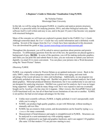
Cao et al. Molecular Pain 2012, h resultant proinflammatory responses in the development of neuropathic pain. Both localized break downof the blood-spinal cord barrier following nerve injury[8] and production of chemokines within the spinal cord[9-11] are thought to be critical in facilitating leukocyteinfiltration.To further investigate the role of infiltrating CD4 Tlymphocytes in the maintenance of L5Tx-induced behavioral hypersensitivity, we proposed the potential involvement of the interaction between infiltrating CD4 Tlymphocytes and microglia via microglia-expressingCD40. CD40, a 48 kD cell surface tumor necrosis factor(TNF) family receptor, has been found to be upregulatedin microglia upon activation both in vitro and in vivo[12-16]. CD40 has also been linked to the pathogenesisof various CNS diseases including multiple sclerosis andAlzheimer’s disease [17-19]. Our studies showed thatboth CD40 KO mice or bone marrow chimeric mice thathave little CD40 expression in the CNS displayedPage 2 of 8significantly reduced L5Tx-induced mechanical hypersensitivity during the maintenance phase starting at 3–5days post-L5Tx, suggesting the role of microglial CD40in the development of L5Tx-induced mechanical hypersensitivity [16].Our previous data indicate differential reductions ofL5Tx-induced mechanical hypersensitivity when comparing CD4 KO and CD40 KO mice, that is CD40 KOmice displayed greater and earlier reduction of mechanical hypersensitivity [6,16]. We suspect the coexistence of distinct CD4- and CD40- mediatedmechanisms in the development of neuropathic pain.Here, we further delineate these underlying mechanisms by examining lumbar spinal cord mononuclearcells collected from wide type (WT), CD4 KO andCD40 KO mice via flow cytometry. Follow-up studieswith neutralizing CD40 antibody were performed toidentify the specific timing of CD40 signalinginvolvement.Figure 1 Flow cytometric analysis of lumbar spinal cord mononuclear cells. Total mononuclear cells collected from each sample (pooledipsilateral or contralateral side of lumbar spinal cords from 3–4 mice) were labeled with mAbs against CD11b and CD45. All samples wereexamined with an Accuri C6 flow cytometer and analyzed with FlowJo. For each sample, total cell population was first identified (A), and thenmicroglia (CD45loCD11b ) (B) and infiltrating leukocytes (CD45hi) (C) were identified within the gated total cell population. The percentage ofeach of these populations within the total cells was recorded. The total number of microglia and infiltrating leukocytes per spinal cord werecalculated based on the recorded percentages, the total mononuclear cells collected, and the number of mice used in each sample.Representative plots shown here are from a “WT day 3 L5Tx ipsilateral” spinal cord mononuclear cell sample.
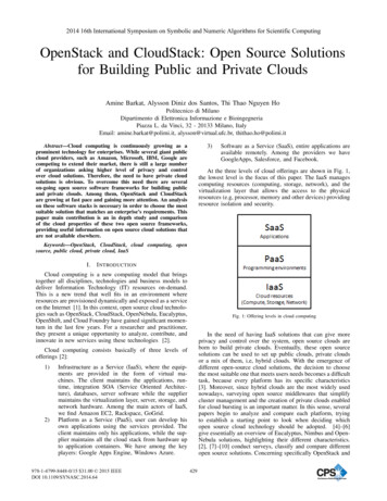
Cao et al. Molecular Pain 2012, ultsFlow cytometric analysis of lumbar spinal cordmononuclear cellsAdult male and female WT, CD4 KO and CD40 KOmice were subjected to either L5Tx or sham surgery. Atselected times, days 0 (naïve groups), 1, 3, 7, and 14post-surgery, mice (for each genotype, 3–4 mice pertreatment group) were euthanized and lumbar spinalcord samples (separated into ipsilateral and contralateralsides) were harvested and pooled to obtain mononuclearcells as described in the Materials and Methods (illustrated in Figure 1). Total mononuclear cells harvestedfrom lumbar spinal cord are known to contain both infiltrating leukocytes and resident microglia. In WT mice,L5Tx induced a significant but transient increase of thetotal number of lumbar spinal cord mononuclear cells inthe ipsilateral side of the spinal cord, which peaked between days 3 and 7 post-L5Tx (Figure 2A, two-wayANOVA, ptreatment 0.003, ptime 0.004, and ptreatment xtime 0.920). Although similar trends in the changes ofthe total lumbar spinal cord mononuclear cells wereobserved in both CD4 KO and CD40 KO mice, no statistically significant differences were detected in eithertype of KO mice (Figures 2B and C, two-way ANOVA,in B, ptreatment 0.304, ptime 0.392, and ptreatment x time 0.968; in C, ptreatment 0.131, ptime 0.114, and ptreatment x time 0.962). Further, total mononuclear cells wereanalyzed based on their microglial (CD45loCD11b ) andinfiltrating leukocyte (CD45hi) content (as illustrated inFigure 1). As expected, WT mice showed a significantincrease of microglial number in the ipsilateral side onboth days 3 and 7 post-L5Tx (Figure 3A, two-wayANOVA, ptreatment 0.007, ptime 0.001, and ptreatment xtime 0.460). Interestingly, significant increase ofPage 3 of 8microglial number was only observed at day 7 postL5Tx in CD4 KO mice (Figure 3B, two-way ANOVA,ptreatment 0.091, ptime 0.001, and ptreatment x time 0.077), and no significant microglial increase wasobserved in CD40 KO mice at any time after L5Tx(Figure 3C, two-way ANOVA, ptreatment 0.828, ptime 0.348, and ptreatment x time 0.955). Consistent with ourprevious report, significantly increased numbers of infiltrating leukocytes were detected in the ipsilateral side oflumbar spinal cord in WT mice at day 7 post-L5Tx(Figure 4A, two-way ANOVA, ptreatment 0.422,ptime 0.001, and ptreatment x time 0.839). However, ineither CD4 KO or CD40 KO mice, no significantincreases of infiltrating leukocytes were observed withinany treatment groups or sides relative to the surgery(Figure 4B and C, two-way ANOVA, in B, ptreatment 0.824, ptime 0.015, and ptreatment x time 0.823; in C,ptreatment 0.972, ptime 0.001, and ptreatment x time 0.988; for both B and C, no differences related to factor“time” were further identified within each surgery/sidegroup with the SNK post-hoc test). In addition, therewere no differences in all parameters measured amongnaïve mice at the basal levels across all three genotypes(Figures 2, 3 and 4, one-way ANOVA, p 0.05).Effects of anti-CD40 treatmentOur data from lumbar mononuclear cell analyses suggestthat spinal cord CD40 might play a more important rolecompared to CD4 in the development of peripheralnerve injury-induced behavioral hypersensitivity in partthrough the inhibition of microglial activation and theinfiltration of peripheral leukocytes into the spinal cord.We next examined the time when CD40 signaling is critical in the development of L5Tx-induced behavioralFigure 2 Numbers of total mononuclear cells in lumbar spinal cord following L5Tx in WT, CD4KO, and CD40KO mice. WT, CD4 KO, andCD40 KO mice were subjected to sham or L5Tx surgery. Lumbar spinal cord mononuclear cells from separate groups of mice of each genotypewere collected at indicated times post-surgery. The temporal changes of the numbers of total lumbar spinal cord mononuclear cells in WT(A, n 5–8), CD4 KO (B, n 4) and CD40 KO (C, n 4) mice are shown here (mean SEM). One-way ANOVA was performed to examine thebasal level genotypic differences among naïve mice and no significant differences were found. Two-way ANOVA for data sets in each graph wereperformed. * indicates significant differences between the indicated group and all other groups at the same time point. # indicates significantdifferences between the indicated group and the corresponding Day 0 group. An additional significant result from statistical comparison is alsoshown within the graph. “Tx” L5Tx, “Sh” sham operation, “ipsi” ipsilateral side, and “contra” contralateral side. For naïve mice, “ipsi” leftand “contra” right.
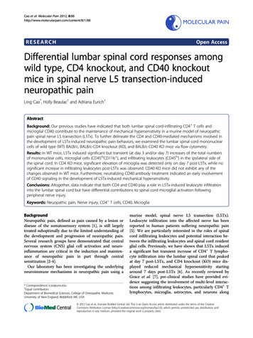
Cao et al. Molecular Pain 2012, e 4 of 8Figure 3 Numbers of microglia (CD45loCD11b ) in lumbar spinal cord following L5Tx in WT, CD4KO, and CD40KO mice. Totalmononuclear cells collected as described in Figure 2 were analyzed for their microglial content. The temporal changes of the numbers of lumbarspinal cord microglia in WT (A, n 5–8), CD4 KO (B, n 4) and CD40 KO (C, n 4) mice are shown here (mean SEM). One-way ANOVA wasperformed to examine the basal level genotypic differences among naïve mice and no significant differences were found. Two-way ANOVA fordata sets in each graph were performed. * indicates significant differences between the indicated group and all other groups at the same timepoint. # indicates significant differences between the indicated group and all other groups within the same treatment group (including thecorresponding Day 0 group). #1 indicates significant differences between the indicated group and days 0, 1 and 14 groups within the sametreatment group. An additional significant result from statistical comparison is also shown within the graph. “Tx” L5Tx, “Sh” sham operation,“ipsi” ipsilateral side, and “contra” contralateral side. For naïve mice, “ipsi” left and “contra” right.hypersensitivity. Neutralizing CD40 antibody was administered daily to WT mice either from day 1 to day 7or from day 6 to day 14 via intrathecal (i.t.) injection(L5Tx was performed on day 0). Saline-injected micewere used as controls. As expected, mice who receivedsaline (regardless of time period) displayed significantmechanical hypersensitivity after L5Tx that was maintained up to day 21 post-L5Tx (Figure 5) and is similarto what we have observed in non-treated WT mice following L5Tx surgery [6,16]. Further, when administeredfrom day 1 to day 7, both doses of anti-CD40, 1 μg/dayand 5 μg/day significantly reduced L5Tx-induced mechanical hypersensitivity (Figure 5A, two-way RMANOVA, ptreatment 0.0162, ptime 0.0001, and ptreatmentx time 0.0096). However, this effect on mechanicalhypersensitivity was gradually diminished following thelast treatment of anti-CD40 and by day 21 post-L5Txthere was no difference in mechanical sensitivity amongall treatment groups (Figure 5A). Interestingly, whenadministered from day 6 to day 14, anti-CD40 did notexert any effect on L5Tx-induced mechanical hypersensitivity (Figure 5B, two-way RM ANOVA, ptreatment 0.132, ptime 0.001, and ptreatment x time 0.782).DiscussionPreviously, we have shown that both infiltrating CD4 Tlymphocytes and spinal cord microglial CD40 areinvolved in the maintenance of L5Tx-induced mechanical hypersensitivity. To further identify the CD4- andCD40- mediated mechanisms, for the first time, weFigure 4 Numbers of infiltrating leukocytes (CD45hi) in lumbar spinal cord following L5Tx in WT, CD4KO, and CD40KO mice. Totalmononuclear cells collected as described in Figure 2 were analyzed for their content of infiltrating leukocytes. The temporal changes of thenumbers of lumbar spinal cord infiltrating leukocytes in WT (A, n 5–8), CD4 KO (B, n 4) and CD40 KO (C, n 4) mice are shown here (mean SEM). One-way ANOVA was performed to examine the basal level genotypic differences among naïve mice and no significant differences werefound. Two-way ANOVA for data sets in each graph were performed. # indicates significant differences between the indicated group and all othergroups within the same treatment group (including the corresponding Day 0 group). An additional significant result from statistical comparison isalso shown within the graph. “Tx” L5Tx, “Sh” sham operation, “ipsi” ipsilateral side, and “contra” contralateral side. For naïve mice, “ipsi” left and “contra” right.
Cao et al. Molecular Pain 2012, e 5 of 8Figure 5 Mechanical hypersensitivity of WT mice treated with anti-CD40. WT mice were subjected to L5Tx. Neutralizing CD40 antibody wasi.t. administered to WT mice daily either from day 1 to day 7 (A) or from day 6 to day 14 (B). Mechanical sensitivity of each mouse was testedbefore L5Tx and repeatedly after L5Tx using a series of von Frey filaments via the up-down method. All data are presented as mean SEM(n 6). Two-way RM ANOVA was performed for data sets within each graph. * indicates the significant differences between the indicated groupand the saline treated group within the same time point. In addition, within each treatment group, p 0.05 between any time point post-L5Txand both day 0 and day 1 time points (not indicated in the graph).evaluated lumbar spinal cord mononuclear cells fromWT, CD4 KO and CD40 KO mice via flow cytometry. Itis known that both microglia and infiltrating leukocytesare important components within lumbar spinal cordmononuclear cells, thus we focused our investigation onthese two populations.First, our data showed that the total numbers ofmononuclear cells collected from both CD4 KO andCD40 KO mice are comparable to that collected fromWT mice at all selected time points post-surgery. Furthermore, for all parameters evaluated, there are no significant genotypic differences detected in naïve mice.These observations indicate that depletion of either CD4or CD40 does not result in a reduction of total mononuclear cells within the lumbar spinal cord prior to anytreatment, and differences detected among mice withdifferent genotypes are most likely not due to thereduced availability of the specific cell populations thatwere analyzed.In CD4 KO mice (that are known to have no CD4 Tcells), unlike in WT mice, no significant leukocyte infiltration was observed at day 7 post-L5Tx. This is consistent with our previous finding that CD4 T cells are thedominant population detected at 7 days post-L5Tx inthe lumbar spinal cord. Unexpectedly, our data indicatethat a lack of CD4 T cells can affect L5Tx-induced earlyincrease of lumbar spinal cord microglial content. Increase in the total number of microglia is one of manysigns of microglial activation during the development ofneuropathic pain [2-4]. Although the peak time fordetecting infiltrating CD4 T cells is at 7 days post-L5Tx[16], it is possible that early-stage infiltrating CD4 Tcells play a critical role in microglial activation. In otherwords, lumbar spinal cord infiltrating CD4 T cells potentially contribute to the maintenance of L5Tx-inducedneuropathic pain in part through enhancing spinal cordmicroglial activation during the transition from the initiation phase to the maintenance phase of neuropathicpain behavioral development post-L5Tx.In CD40 KO mice, neither increased leukocyte infiltration nor increased microglial number were observed following L5Tx as that in WT mice, indicating that CD40is critical in both leukocyte infiltration and microglial activation post-L5Tx. Previously, we have demonstratedthe role of CD40 microglia in the maintenance ofL5Tx-induced neuropathic pain behavior [16]. It hasbeen shown that CD40-mediated, activated microglia arecapable of releasing several chemokines, including CCL2(also known as Monocyte chemoattractant protein-1(MCP-1)), CCL5 (also known as regulated and normalT cell expressed and secreted (RANTES)), and CXCL10(also known as interferon gamma-induced protein 10(IP-10)) [20]. Together, we propose that depletion ofCD40 significantly reduces the ability of spinal cordmicroglia to become fully activated, thus leading to adecreased production of various chemoattractant cytokines by activated microglia and subsequently reducedinfiltration of peripheral leukocytes. Further, it is knownthat CD40 stimulation can lead to increased expressionof certain adhesion molecules on both leukocytes andendothelial cells [21,22]. Elevated expression of adhesionmolecules has been observed in activated microglia[13,23,24]. Thus, depletion of CD40 may limit an injuryinduced increase of the total number of microglia at theinjury site by reducing the migration of microglia towards the injury site. Reduced expression of adhesionmolecules by endothelial cells could also contribute tothe reduced infiltration of peripheral leukocytes. Nevertheless, whether the CD40 depletion-related low level ofmicroglial activation is due to the lack of active CD40signaling during the development of neuropathic pain orCD40 depletion-induced developmental defects of
Cao et al. Molecular Pain 2012, roglia requires further investigation. In addition, wedid notice somewhat larger variations in all parameterswe measured within CD40 KO mice, particularlychanges between day 1 and day 7 post-surgery. Althoughi
cells of wild type (WT) BALB/c, BALB/c-CD4 knockout (KO), and BALB/c-CD40 KO mice via flow cytometry. Results: In WT mice, L5Tx induced significant but transient (at day 3 and/or day 7) increases of the total numbers of mononuclear cells, microglial cells (CD45loCD11b ), and infiltrating leuk