Transcription
Online Submissions: mdoi:10.3748/wjg.v17.i20.2543World J Gastroenterol 2011 May 28; 17(20): 2543-2551ISSN 1007-9327 (print) ISSN 2219-2840 (online) 2011 Baishideng. All rights reserved.TOPIC HIGHLIGHTNatalia A Osna, MD, PhD, Series EditorHepatic stellate cells and innate immunity in alcoholic liverdiseaseYang-Gun Suh, Won-Il JeongNatural killer cell; Kupffer cell; Endocannabinoid; Steatosis; Steatohepatitis; FibrosisYang-Gun Suh, Won-Il Jeong, Graduate School of MedicalScience and Engineering, Korea Advanced Institute of Scienceand Technology, Daejeon, 305-701, South KoreaAuthor contributions: Suh YG and Jeong WI contributedequally to the writing of the manuscript.Supported by A grant of the Korea Healthcare TechnologyR&D Project, Ministry for Health, Welfare and Family Affairs,South Korea (A090183)Correspondence to: Won-Il Jeong, DVM, PhD, Professorof Graduate School of Medical Science and Engineering, KoreaAdvanced Institute of Science and Technology, 373-1 Yuseonggu, Daejeon 305-701, South Korea. wijeong@kaist.ac.krTelephone: 82-42-3504239 Fax: 82-42-3504240Received: January 7, 2011 Revised: February 25, 2011Accepted: March 4, 2011Published online: May 28, 2011Peer reviewers: Ekihiro Seki, MD, PhD, Department of Medi-cine, University of California SanDiego, Leichag BiomedicalResearch Building Rm 349H, 9500 Gilman Drive MC#0702, LaJolla, CA 92093-0702, United States; Atsushi Masamune, MD,PhD,Division of Gastroenterology, Tohoku University Graduate School of Medicine,1-1 Seiryo-machi, Aoba-ku, Sendai980-8574, JapanSuh YG, Jeong WI. Hepatic stellate cells and innate immunity in alcoholic liver disease. World J Gastroenterol 2011;17(20): 2543-2551 Available from: URL: htm DOI: INTRODUCTIONConstant alcohol consumption is a major cause of chronicliver disease, and there has been a growing concernregarding the increased mortality rates worldwide. Alcoholic liver diseases (ALDs) range from mild to moresevere conditions, such as steatosis, steatohepatitis,fibrosis, cirrhosis, and hepatocellular carcinoma. Theliver is enriched with innate immune cells (e.g. naturalkiller cells and Kupffer cells) and hepatic stellate cells(HSCs), and interestingly, emerging evidence suggeststhat innate immunity contributes to the developmentof ALDs (e.g. steatohepatitis and liver fibrosis). Indeed,HSCs play a crucial role in alcoholic steatosis via production of endocannabinoid and retinol metabolites.This review describes the roles of the innate immunityand HSCs in the pathogenesis of ALDs, and suggeststherapeutic targets and strategies to assist in the reduction of ALD.Alcoholic liver disease (ALD) caused by chronic alcoholconsumption shows increased mortality rates worldwide[1,2]. As an adverse risk factor of alcohol abuse, ALDincludes a broad spectrum of liver diseases, ranging fromsteatosis (fatty liver), steatohepatitis, fibrosis, and cirrhosis to hepatocellular carcinoma[3,4]. Generally, steatosis isconsidered to be a mild or reversible condition, whereassteatohepatitis is a pathogenic condition, which has thepotential to progress into more severe diseases, such asliver fibrosis/cirrhosis, insulin resistance, and metabolicsyndrome in rodents and humans[5-7]. For the past decade,evidence has suggested that the innate immune cells ofliver and hepatic stellate cells (HSCs) play crucial roles inALD. For example, previous studies demonstrated thatalcoholic liver steatosis was induced by HSC-derived endocannabinoid and its hepatic CB1 receptor, and alcoholicliver fibrosis was accelerated due to abrogated antifibroticeffects of natural killer (NK) cells/interferon-γ (IFN-γ)against activated HSCs via the upregulation of transforming growth factor-β (TGF-β) and suppressor of cytokine 2011 Baishideng. All rights reserved.Key words: Alcoholic liver disease; Hepatic stellate cell;WJG www.wjgnet.com2543May 28, 2011 Volume 17 Issue 20
Suh YG et al . HSCs and innate immunity in liver diseasesignaling 1 (SOCS1)[8,9]. However, the molecular and cellular mechanisms underlying ALD remain controversial[4,6,10].Therefore, in the present review, we briefly describe the innate immunity of liver and HSCs, summarize the roles ofthese in ALD (with particular emphasis on alcoholic liversteatosis, steatohepatitis and liver fibrosis), and providebetter strategies for the prevention and treatment of ALD.studies have suggested that they contribute significantlyto liver injury, regeneration, and fibrosis[22-25].More interestingly, there are enigmatic cells in theliver that were previously called Ito cells or sinusoidalfat-storing cells, but are now standardized as HSCs[21].HSCs comprise up to 30% of NPC in the liver and arelocated in specialized spaces called Disse, between hepatocytes and sinusoidal endothelial cells. In addition,quiescent HSCs store retinol (vitamin A) lipid dropletsand regulate retinoid homeostasis in healthy livers. However, they become activated and transformed into myofibroblastic cells that have special features with retinol(vitamin A) loss and enhanced collagen expression whenliver injuries occur[19,21,26]. For several decades, activatedHSCs have been considered to be major cells that induceliver fibrosis via the production of ECM and inflammatory mediators (e.g. TGF-β) in humans and rodents[19-21].However, recent studies have suggested that the novelroles of HSCs are closely associated with other diseases,such as alcoholic liver steatosis and immune responses,by producing endocannabinoids and presenting antigenmolecules, respectively[8,27,28]. Moreover, HSCs can directlyinteract with immune cells, such as NK cells, NKT cellsand T cells, via the expression of retinoic acid early inducible-1 (RAE1), CD1d, and major histocompatibilitycomplex (MHC) Ⅰ and Ⅱ[22,28,29]. During HSC activation,they metabolize the retinols into retinaldehyde (retinal)via alcohol dehydrogenase (ADH), and the retinal is further metabolized into retinoic acid (RA) via retinaldehydedehydrogenase (Raldh)[3,29]. Surprisingly, activated HSCsexpress an NK cell activating ligand known as RAE1;however, RAE1 expression is absent in quiescent HSCs.This suggests that the activation processes of HSCs arenecessary for the expression of a NK cell activated ligand, RAE1. Furthermore, several TLRs have also beenidentified in HSCs[30]. Taken together, HSCs might beimportant not only in liver fibrosis, but also in other liverdiseases related to immune responses.INNATE IMMUNITY AND HSC IN LIVERThe innate immune system is the first line of defenseagainst pathogenic microbes and other dangerous insults, such as tissue injury, stress, and foreign bodies[11].It consists of three sub-barriers: physical (e.g. mucousmembrane and skin), chemical (e.g. secreted enzymesfor antimicrobial activity and stomach HCL), and cellular barriers (e.g. humoral factors, phagocytic cells,lymphocytic cells, etc), which immediately respond to thepathogens entering the body. Most body defense cellshave pattern recognition receptors (PRRs) that recognizethe overall molecular patterns of pathogens, known aspathogen associated molecular patterns. The examplesof PRRs are toll-like receptors (TLR), nucleotide-bindingoligomerization domain-like receptors, and the retinoicacid-induced gene I-like helicases[12].When extraneous molecules enter the human body,they have to be processed by the liver, either by metabolism or detoxification. Therefore, the liver is consideredas a barrier against pathogens, toxins, and nutrientsabsorbed from the gut via the portal circulation system.Consequently, the liver is enriched in innate immunesystem including humoral factors (e.g. complement andinterferon), phagocytic cells (e.g. Kupffer cells and neutrophils), and lymphocytes [e.g. NK cells, natural killerT (NKT) cells and T cell receptor γδ T cells][11,13-15]. In ahealthy liver, the principal phagocytic cells, the Kupffercells, representing 20% of the non-parenchymal cells(NPC), assist in the clearance of wastes via phagocytosis in the body[15,16]. However, when the liver is injured,Kupffer cells elicit immune and inflammatory responses(e.g. hepatitis, fibrosis, and regeneration) by producingseveral mediators, including tumor necrosis factor- α(TNF- α ), TGF- β , interleukin-6 (IL-6), and reactiveoxygen species (ROS)[17-19]. Among these, TGF-β playsa crucial role in the transdifferentiation of quiescentHSCs into fibrogenic activated HSCs, via the suppression of their degradation and the stimulation of theproduction of extracellular matrix (ECM), especially incollagen fibers[19-21]. In a healthy liver, liver lymphocytesconstitute about 25% of the NPC. Mouse liver lymphocytes contain 5%-10% NK cells and 30%-40% NKTcells, whereas rat and human liver lymphocytes consistof approximately 30%-50% NK cells and 5%-10% NKTcells[11,13,15,16]. These distributions of NK and NKT cellsare quite abundant compared with those in peripheralblood, which contains 2% of NKT cells and 13% of NKcells[13]. Previously, NK/NKT cells were regarded to assume a crucial role in mediating the immune responsesagainst tumor and microbial pathogens. However, recentWJG www.wjgnet.comALCOHOLIC LIVER STEATOSIS BYINNATE IMMUNITY AND HSCSAlcoholic liver steatosis has long been considered as amild condition; however, increasing evidence suggests thatit is a potentially pathologic state, which progresses into amore severe condition in the presence of other cofactors,such as the sustained consumption of alcohol, viral hepatitis, diabetes, and drug abuse[31,32]. It is believed that fat accumulation in the hepatocytes is a result of an imbalancedfat metabolism, such as decreased mitochondrial lipidoxidation and enhanced synthesis of triglycerides. Severalunderlying mechanisms of these processes indicate that itmight be related to an increased NADH NAD ratio[33,34],increased sterol regulatory element-binding protein-1(SREBP-1) activity[35,36], decreased peroxisome proliferator-activated receptor-α activity[37,38], and decreased AMPactivated protein kinase (AMPK) activity[8,36].Moreover, recent studies have suggested the involvement of innate immune cells, particularly Kupffer cells,2544May 28, 2011 Volume 17 Issue 20
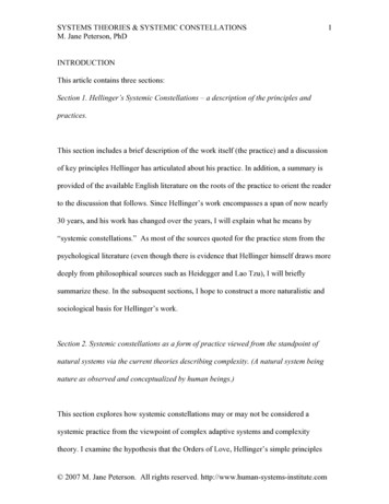
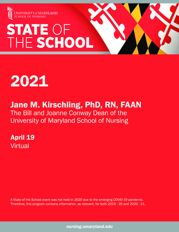
Suh YG et al . HSCs and innate immunity in liver diseasein alcoholic liver steatosis[39,40]. Generally, alcohol intakeincreases gut permeabilization, which allows an increaseduptake of endotoxin/lipopolysaccharide (LPS) in portalcirculation[18]. Kupffer cells are then activated in responseto LPS via TLR4 signaling cascade, leading to the production of several types of pro-inflammatory mediators suchas TNF-α, IL-1, IL-6, and ROS[3,4,39]. Of these mediators, the increased expression of TNF-α and enhancedactivity of its receptor (TNF-α R1) have been observedin alcoholic liver steatosis in mice[39-42]. In addition, it hasbeen reported that TNF-α has the potential to increasemRNA expression of SREBP-1c, a potent transcriptionfactor of fat synthesis, in the liver of mice and to stimulatethe maturation of SREBP-1 in human hepatocytes[43,44].Furthermore, a recent report demonstrated that alcoholmediated infiltration of macrophages decreased theamount of adiponectin (known as anti-steatosis peptidehormone) production of adipocytes, leading to alcoholicliver steatosis[45]. Therefore, Kupffer cells/macrophagesmight contribute to the development of alcoholic liversteatosis via the upregulation of the SREBP1 activity inhepatocytes and the downregulation of the productionof adiponectin in adipocytes. In contrast, IL-6 producedby Kupffer cells/macrophages is a positive regulator inprotecting against alcoholic liver steatosis via activation ofsignal transducer and activator of transcription (STAT)3,consequently inhibiting of SREBP1 gene expression inhepatocytes[46-48].Endocannabinoids, endogenous cannabinoids, are lipidmediators that interact with cannabinoid receptors (CB1and CB2) to produce effects similar to those of marijuana[49]. There are the two main endocannabinoids, arachidonoyl ethanolamide (anandamide) and 2-arachidonoylglycerol (2-AG). Recently, an intriguing report suggested thatalcoholic liver steatosis is mediated mainly through HSCderived endocannabinoid and its hepatocytic receptor[8].The study suggested that chronic alcohol consumptionstimulated HSC to produce 2-AG, and the interaction withthe CB1 receptor upregulated the expression of lipogenicgenes SREPB1c and fatty acid synthase but downregulatedthe activities of AMPK and carnitine palmitoyltransferase1. Consequently fat is accumulated in the hepatocyte. Morerecently, a related study reported that the increased expression of CB1 receptors on hepatocytes because of alcoholconsumption was mediated by RA acting via a RA receptor(RAR)-γ[27]. This study also showed that 2-AG treatmentin mouse hepatocytes increased the production of RA byRaldh1, the catalytic enzyme of retinaldehyde into RA. RAthen binds with RAR-γ, increasing the expression of CB1receptor mRNA and protein, and consequently exacerbating the alcohol-mediated fat accumulation via enhancedendocannabinoid and lipogenic signaling pathways[27].Reports stating that alcohol consumption simultaneouslyelevated the expression of RAR and the production ofretinol metabolites, including RA, in mouse and rat liver,supported these findings[50-52]. Moreover, hepatocytes andHSCs are major sources of retinoids, including retinol andRA, in the body[26,53]. In contrast to the CB1 receptors, theassociation of CB2 receptors with the development ofWJG www.wjgnet.comEtOHHSC2-AG CB1 R AMPK CPT1 Fatty acidβ-oxidation- -SREBP-1 FASLipid/fataccumulation 2-AG CB1 RRaldh1RARARγCB1 R promoterHepatocyteFigure 1 Regulatory mechanisms of the hepatic lipogenesis and CB1 receptor expression via hepatic stellate cell-derived endocannabinoids/CB1receptors and retinoic acid/retinoic acid receptor-γ in hepatocytes, respectively. CB1 R: CB1 receptor; AMPK: AMP-activated protein kinase; HSC:Hepatic stellate cell; 2-AG: 2-arachidonoylglycerol; SREBP-1: Sterol regulatoryelement-binding protein-1; FAS: Fatty acid synthase; RA: Retinoic acid; RAR:Retinoic acid receptor.hepatic steatosis has not yet been studied in depth. Onestudy showed that the expression of CB2 receptors wasincreased in the livers of patients with non-alcoholic fattyliver disease[54]. In an animal model, however, feeding ofhigh-fat diet for 15 wk induced severe fatty liver in wildtype mice, but not in hepatic CB2 knockout mice[55]. Theinvolvement of endocannabinoid, RA, and their receptorshas been integrated in Figure 1.Interestingly, in contrast with previous reports thatendocannabinoids activated HSCs to induce liver fibrosisand alcoholic liver steatosis[8,56], Siegmund et al reportedthat HSCs’ sensitivity to anandamide (AEA)-induced celldeath was because of low expression of fatty acid amidehydrolase and that 2-AG also induced apoptotic deathof HSCs via ROS induction[57-59]. These data indicatedthat endocannabinoids might play negative roles in liverfibrosis. Therefore, the functions of endocannabinoids toHSCs are still unclear and need to be studied further.ALCOHOLIC STEATOHEPATITIS BYINNATE IMMUNITY AND HSCSAlcoholic steatohepatitis has a mixed status with fat accumulation and inflammation in the liver, which has the potential to progress into more severe pathologic states suchas alcoholic liver fibrosis, cirrhosis, and hepatocellularcarcinoma. In response to alcohol uptake, many hepaticcells participate in the pathogenesis of alcoholic steatohepatitis. However, as described above, mainly Kupffercells and HSCs initiate and maintain hepatic inflammationand steatosis[4,8,60-63]. Considering their specific location atthe interface between the portal and systemic circulation,Kupffer cells are the central players in orchestrating theimmune response against endotoxin (LPS) via TLR4 signaling pathways[62,64]. TLR4 initiates two main pathways,and when TLR4 binds LPS, TIR domain-containing adaptor protein and myeloid differentiation factor 88 (MyD88)are recruited, resulting in the early-phase activation of nu-2545May 28, 2011 Volume 17 Issue 20
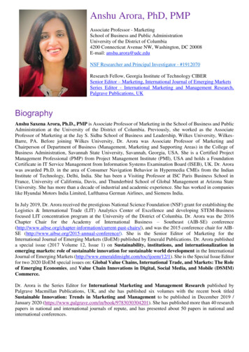
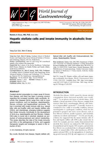
Suh YG et al . HSCs and innate immunity in liver diseaseclear factor-κB (NF-κB). The activation of NF-κB leadsto the production of pro-inflammatory cytokines, including TNF-α, IL-6, and monocyte chemotatic protein-1(MCP-1). Meanwhile, TIR-domain containing adaptor inducing IFN-β (TRIF) and TRIF-related adaptor moleculeactivate interferon regulatory factor 3 (IRF3), leading tothe production of type I IFN and late activation of NF[62,65]. Recent studies reported that alcohol-mediatedκBliver injury and inflammation were primarily induced byin a TLR4-dependent, but MyD88-independent, mannerin NPCs (Kupffer cells and macrophages), whereas IRF3activation in parenchymal cells (hepatocytes) renderedprotective effects to ALD[66,67]. In addition, the importanceof gut-derived endotoxin/LPS in ALD was suggested byexperiments where animals were treated with either antibiotics or lactobacilli to remove or reduce the gut microfloraprovided protection from the features of ALD[68]. Amongpro-inflammatory cytokines, TNF-α primarily contributesto the development of ALD, and its levels are increased inpatients with alcoholic steatohepatitis[39] and in the liver ofalcohol-fed animals[40,69]. Moreover, Kupffer cells secreteother important cytokines, including IL-8, IL-12, andIFNs, which contribute to the intrahepatic recruitmentand activation of granulocytes that are characteristicallyfound in severe ALD, and influence immune system polarization[70]. Interestingly, TLR4 is expressed not only oninnate immune cells, such as Kupffer cells and recruitedmacrophages, but also on hepatocytes, sinusoidal endothelial cells, and HSCs in the liver[30].In addition to LPS, oxidative stress-mediated cellularresponses also play an important role in activations of innate immune cells and HSCs. Furthermore, Kupffer cellsrepresent a major source of ROS in response to chronicalcohol exposure[71,72]. One important ROS is the superoxide ion, which is mainly generated by the enzyme complexNADPH oxidase. Underlining the important role of ROSin mediating ethanol damage, treatment with antioxidantsand deletion of the p47phox subunit of NADPH oxidasein ethanol-fed animals reduced oxidative stress, activation of NF-κB, and TNF-α release in Kupffer cells, thuspreventing liver injury[71,73]. Moreover, NADPH oxidaseinduces TLR2 and TLR4 expression in human monocytic cells[74], and direct interaction of NADPH oxidaseisozyme 4 with TLR4 is involved in LPS-mediated ROSgeneration and NF-κB activation in neutrophils[75].Besides Kupffer cells, HSCs also contribute to alcoholic steatohepatitis by producing endocannabinoids andreleasing proinflammatory cytokines and chemokines,such as TNF-α, IL-6, MCP-1, and macrophage inflammatory protein-2[63,76-78]. Moreover, Kupffer cells activatedby alcohol stimulate the proliferation and activation ofHSCs via IL-6 and ROS-dependent mechanisms in a coculturing system[17,79]. Furthermore, retinol metabolitesof HSCs activate latent TGF-β, leading to suppressionof apoptosis of HSCs[80-82]. Recently, an intriguing reviewprovided novel roles for HSCs in liver immunology, whereHSCs, depending on their activation status, can produceseveral mediators, including TGF-β, IL-6, and RA, whichare important components in naïve T cell differentiationWJG www.wjgnet.comIFN-gApoptosiscell cycle arrestSTAT1(-)NK( DHRetinolKillingFigure 2 Mechanism of natural killer cell cytotoxicity against activatedhepatic stellate cells. STAT: Signal transducer and activator of transcription;IFN: Interferon; NK: Natural killer; HSC: Hepatic stellate cell; MHC: Major histocompatibility complex; RAE1: Retinoic acid early inducible-1; RA: Retinoic acid;ADH: Alcohol dehydrogenase.into regulatory T cells (Treg cells) or IL-17 prod
Supported by A grant of the Korea Healthcare Technology R&D Project, Ministry for Health, Welfare and Family Affairs, South Korea (A090183) . Jolla, CA 92093-0702, United States; Atsushi Masamune, MD, PhD,Division of Gastroenterology, Tohoku University Gradu- . Natalia A Osna, MD, PhD,