Transcription
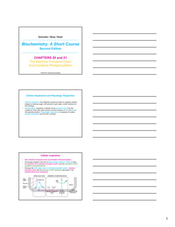
Tymoczko Berg StryerBiochemistry: A Short CourseSecond EditionCHAPTERS 20 and 21The Electron-Transport ChainAnd Oxidative Phosphorylation 2013 W. H. Freeman and CompanyCellular Respiration and Physiologic Respiration Cellular respiration: the metabolic process by which an organism obtainsenergy by reacting oxygen with glucose to give water, carbon dioxide andATP (energy).In physiology, respiration is defined as the transport of O2 from theoutside air to the cells within tissues, and the transport of CO2 and H2O inthe opposite direction. Physiologic respiration is necessary to sustaincellular respiration and thus life in animals.Cellular respiration CAC, Electron-transport chain and Oxidative Phosphorylation.The energy trapped in the form of high-energy electrons (CAC) is usedfor reduction of O2 (electron-transport chain ) and the synthesis of ATP(oxidative phosphorylation).Whereas the CAC takes place in the mitochondrial matrix, electrontransport chain and the process of ATP synthesis take place in themitochondrial inner membrane.1
ATP Synthesis and Proton Gradients ATP need in humans: a sedentary male of 70 kg (154 lbs) requires 85 kgof ATP/day. Humans possess only about 250 g of ATP, less than 1% of thedaily required amount each ATP molecule is recycled from ADPapproximately 300 times/dayATP recycling - primarily through oxidative phosphorylation-the culminationof cellular respirationCellular respiration: high-transfer-potential electrons flow through a seriesof large protein complexes embedded in the inner mitochondrial membrane,called the respiratory chain, to reduce O2 to H2O.The electron flow through these complexes is a series of highlyexergonic oxidation–reduction reactions that power the pumping ofprotons from the inside of the mitochondria to the outside, establishing aproton gradient, called the proton-motive force.The final phase of oxidative phosphorylation is carried out by an ATPsynthesizing assembly that is driven by the flow of protons back intothe mitochondrial matrix shows that proton gradients are aninterconvertible currency of free energy in biological systems.An overview of oxidative phosphorylationOxidation and ATP synthesis are coupled by transmembrane proton fluxes. The respiratory chain (yellow structure) transfers electrons fromNADH and FADH2 to O2 and simultaneously generates a protongradient. ATP synthase (red structure) converts the energy of the protongradient into ATPRespiration is an ATP-generating process in which an inorganic compound(such as molecular oxygen) serves as the ultimate electron acceptor. Theelectron donor can be either an organic compound or an inorganic one.Mitochondria Are Bounded by a Double Membrane The outer mitochondrial membrane is permeable to most smallions and molecules because of the channel protein mitochondrialporin. The inner membrane, which is folded into ridges called cristae, isimpermeable to most molecules. The inner membrane is the site ofelectron transport and ATP synthesis. The citric acid cycle and fatty acid oxidation occur in the matrix.2
Oxidative Phosphorylation Depends on Electron Transfer In the electron-transport chain, electrons from NADH and FADH2 are usedto reduce O2 to H2O, in a highly exergonic reaction. The reduction of molecular oxygen by NADH and FADH2 is accomplishedthrough a number of electron-transfer reactions, which take place in a set ofmembrane proteins known as the electron-transport chain. Electrons from NADH and FADH2 flow through the components of theelectron chain.Figure 20.6 Components ofthe electron-transport chainThe Electron‐Transfer Potential of an Electron IsMeasured As Redox Potential In oxidative phosphorylation, the electron-transfer potential of NADH orFADH2 is converted into the phosphoryl-transfer potential of ATP. The measure of phosphoryl-transfer potential is given by G ′ for thehydrolysis of the phosphoryl compound. The corresponding measure for the electron–transfer potential is E ′,the reduction potential (also called the redox potential or oxidation–reductionpotential). The reduction potential of a redox couple X/ X is measured experimentally bymeasuring the electromotive force generated by a sample halfcell connected to a standard reference half-cellFigure 20.4 The measurement of redoxpotential. Apparatus for the measurementof the standard oxidation–reductionpotential of a redox couple. Electrons flowthrough the wire connecting the cells,whereas ions flow through the agar bridge.The Redox Potential The sample half-cell consists of an electrode immersed in a solution of 1 Moxidant (X) and 1 M reductant (X ). The standard reference half-cell consists of an electrode immersed in a 1 MH solution that is in equilibrium with H2 gas at 1 atmosphere (1 atm). The electrodes are connected to a voltmeter, and an agar bridge allows the flowof ions between the half-cells. Electrons then flow from one half-cell to the otherthrough the wire connecting the two cells. The reduction potential of the H :H2 couple is defined to be 0 volts. A negative reduction potential meansthat X has lower affinity for electrons thandoes H2 ; it is a STRONGER REDUCINGagent, and it has LOWER ELECTRONAFFINITY. The following reaction occurs:X H X½H Thus, a strong reducing agent (such asNADH) will donate electrons and has anegative reduction potential, whereas astrong oxidizing agent (such as O2) willaccept electrons and has a positivereduction potential.3
Standard Redox Potential ΔE ′ and standardfree‐energy change ΔG ′The standard free-energy change G ′ is related to the change in reduction potential E ′ by G ′ nF E ′in which n is the number of electrons transferred, F is a proportionality constantcalled the faraday (96.48 kJ mol 1 V 1, or 23.06 kcal mol 1 V 1), E ′ is in volts, and G ′ is in kilojoules or kilocalories per mole.For the reduction of molecular oxygen to water:This value is substantially negative indicating a highly spontaneous reactionThe Electron‐Transport Chain Is a Series ofCoupled Oxidation–Reduction Reactions Electron flow from NADH to O2 isaccomplished by a series of intermediateelectron carriers, that are coupled asmembers of sequential redox reactions. Electrons flow down the electron-transportchain until they finally reduce O2. The members of the electron-transportchain are arranged so that the electronsalways flow to components with a higherelectron affinity. A more negative reduction potentialmeans that X has lower affinity forelectrons; more POSITIVE reductionpotential means that X has HIGHERaffinity for electronsFigure 20.10 The components of the electrontransport chain are arranged incomplexes. The complexes shown in yellowboxes are proton pumps. Cyt c stands forcytochrome c.4
Components of the electron‐transport chain Electrons flow down an energygradient from NADH to O2through 4 large proteincomplexes embedded in theinner mitochondriamembrane Fe and cytochrome c arecomponent of all of thecomplexes. These complexes pumpprotons out of themitochondria, generating aproton 8/Fe is a component of all of the complexes in theelectron transport chainFigure 20.8 A heme component ofcytochrome c oxidase.Fe is a component of all of the complexes inthe electron transport chain For iron (Fe) to participate in several places in the electron-transport chain itmust have several different reduction potentials. How can this happen? The oxidation–reduction potential of iron ions can be altered by theirenvironment. The iron ion is not free, it is embedded in differentproteins, enabling iron to have various reduction potentials and to play a roleat several different locations in the chain. In both iron–sulfur proteins andcytochromes, iron shuttles between its oxidized ferric (Fe3 ) and its reducedferrous state (Fe2 ).Figure 20.7 Iron–sulfur clusters. (A) A single Fe ion bound by 4 cysteineresidues. (B) 2Fe-2S cluster with iron ions bridged by sulfide ions. (C) 4Fe-4Scluster. Each of these clusters can undergo oxidation–reduction reactions.5
Coenzyme Q (CoQ) Another key feature of the electron-transport chain is the prominence ofcoenzyme Q (CoQ) as an electron carrier. Coenzyme Q, also known as ubiquinone because it isa ubiquitous quinone in biological systems, is a hydrophobic moleculewhich can diffuse rapidly within the inner mitochondrial membrane,where it shuttles protons and electrons. The most-common form in mammals contains 10 isopreneunits (CoQ10).Coenzyme Q (CoQ) Quinones can exist in three oxidation states (Figure 20.9). In the fully oxidizedstate (Q), coenzyme Q has two keto groups. The addition of one electron yields a semiquinone radical anion (Q ),whereas the addition of one electron and one proton results in thesemiquinone form (QH ). The addition of a second electron and proton generatesubiquinol (QH2), the fully reduced form of coenzyme Q. Thus, for quinones, electron-transfer reactions are coupled to protonbinding and release, a property that is key to transmembrane protontransport.RespirasomeThe large protein complexes that transfer electrons inthe respiratory chain are associated in asupramolecular complex termed the respirasome. Asin the CAC, the complex facilitates the rapid transfer ofsubstrate and prevent the release of reactionintermediates.The respirasome is composed of 4 large proteincomplexes:1. NADH-Q oxidoreductase2. Q–cytochrome c oxidoreductase3. Cytochrome c oxidase Electron flow within these transmembranecomplexes leads to the transport of protonsacross the inner mitochondrial membrane.4. Succinate-Q reductase, contains the succinatedehydrogenase that generates FADH2 in the CAC.Electrons from this FADH2 enter the electrontransport chain at Q-cytochrome c oxidoreductase. Succinate-Q reductase, in contrast with theother complexes, does not pump protons.6
Components of the electron‐transport chainThe electron‐transport chainHigh H Low H Figure 20.15 The electron-transport chain. High-energy electrons in the form ofNADH and FADH2 are generated by the citric acid cycle. These electrons flowthrough the respiratory chain, which powers proton pumping and results in thereduction of O2.Electron Transport Chain And OxidativePhosphorylation In the electron transport chain, electrons flow of from NADH to O2,an exergonic process:NADH ½O2 H H2O NAD G ′ 220.1 kJ/ mol During the electron flow, protons are pumped from the mitochondrialmatrix to the outside of the inner mitochondrial membrane, creatinga proton gradient. The energy of the proton gradient powers thesynthesis of ATP (oxidative phosphorylation)ADP Pi H ATP G ′ 30.5 kJ/ mol7
A Proton Gradient Powers the Synthesis of ATP A molecular assembly ATP synthase, in the inner mitochondrial membrane,carries out the synthesis of ATP. In 1961, Peter Mitchell suggested the chemiosmotic hypothesis. He proposed that electron transport and ATP synthesis are coupled by aproton gradient across the inner mitochondrial membrane. The H concentration becomes lower in the matrix membrane potential The pH gradient and membrane potential constitute a proton-motiveforce that is used to drive ATP synthesisProton-motive force ( p) chemical gradient ( H) charge gradient ( Ψ)ATP Synthase Is Composed of a Proton‐ConductingUnit and a Catalytic UnitATP synthase is a large, complex enzyme located in the inner mitochondrialmembrane, composed of an F0 and F1 subunit :1. F0 subunit, is a hydrophobic segment that spans the inner mitochondrialmembrane and contains the proton channel of the complex. This channel consists of a ring comprising from 8 -15 c subunits, depending onthe source of the enzyme, that are embedded in the membrane. A single a subunit binds to the outside of the ring.2. F1 subunit: 85-Å-diameter ball, protrudes into the mitochondrial matrix. Contains the catalytic activity of the synthase. Consists of 5 types of polypeptide chains (α3, β3, γ, δ, and ε).Figure 21.3 The structure of ATPsynthase. A schematic structure of ATPsynthase is shown. The F0 subunit isembedded in the inner mitochondrialmembrane, whereas the F1 subunit residesin the matrix.ATP Synthase Mechanism: Rotational Catalysishttp://www.youtube.com/watch?v Shs3lFU OFMATP synthase catalyzes the formation of ATP from ADP and orthophosphate.There are three active sites on the enzyme, each performing one of threedifferent functions at any instant. The proton-motive force causes the threeactive sites to sequentially change functions as protons flow through themembrane-embedded component of the enzyme. The rotation ofthe γ subunit interconverts the three β subunits.Figure 21.4 ATP synthasenucleotide-binding sitesare not equivalent.The γ subunit passesthrough the center ofthe α3β3hexamer andmakes the nucleotidebinding sites in the βsubunits distinct from oneanother.8
An overview of oxidative phosphorylationFigure 21.10 Anoverview ofoxidativephosphorylation.The electrontransport chaingenerates a protongradient, which isused to synthesizeATP.If a resting human being requires 85 kg of ATP/day for bodily functions, then3.3 1025 protons must flow through ATP synthase/day, or3.3 1021 protons/second.Shuttles Allow Movement Across MitochondrialMembranesThe inner mitochondrial membrane must be impermeable to most molecules,yet much exchange has to take place between the cytoplasm and themitochondria. This exchange is mediated by an array of membrane-spanningtransporter proteins9
Electrons from Cytoplasmic NADH Enter Mitochondriaby ShuttlesNADH cannot simply pass into mitochondria for oxidation by the respiratorychain, because the inner mitochondrial membrane is impermeable to NADH andNAD electrons from NADH, rather than NADH itself, are carried across themitochondrial membrane.Figure 21.11 The glycerol 3phosphate shuttle. Electronsfrom NADH can enter themitochondrial electron-transportchain by reducing DHAP toglycerol 3-phosphate.Electron transfer to an FADprosthetic group in amembrane-bound glycerol 3phosphate dehydrogenasereoxidizes glycerol 3phosphate. Subsequent electrontransfer to Q to form QH2 allowsthese electrons to enter theelectron-transport chain.The Entry of ADP into Mitochondria IsCoupled to the Exit of ATPADP enters the mitochondrial matrix only if ATP exits, and vice versa. This processis carried out by the translocase, an antiporter:Figure 21.13 The mechanism of mitochondrial ATP-ADP translocase. Thetranslocase catalyzes the coupled entry of ADP into the matrix and the exit of ATPfrom it. The binding of ADP (1) from the cytoplasm favors eversion of the transporter(2) to release ADP into the matrix (3). Subsequent binding of ATP from the matrix tothe everted form (4) favors eversion back to the original conformation (5), releasingATP into the cytoplasm (6)Mitochondrial Transporters Allow MetaboliteExchange Between the Cytoplasm and MitochondriaThe inner mitochondrial membrane must be impermeable to most molecules, yetmuch exchange has to take place between the cytoplasm and the mitochondria.This exchange is mediated by an array of membrane-spanning transporter proteins.Figure 21.14 Mitochondrial transporters. Transporters (also called carriers) aretransmembrane proteins that carry specific ions and charged metabolites across theinner mitochondrial membrane.10
Cellular Respiration Is Regulated by the Need for ATPThe stoichiometries of proton pumping and ATP synthesis, need not beinteger numbers or even have fixed values. The best current estimates for the number of H pumped out of thematrix by NADH-Q oxidoreductase, Qcytochrome c oxidoreductase, and cytochrome c oxidase per electronpair are 4, 4, and 2, respectively (10 H ) The synthesis of 1 molecule of ATP is driven by the flow of about 3 H through ATP synthase. An additional H is consumed in transportingATP from the matrix to the cytoplasm Hence, about 2.5 moleculesof cytoplasmic ATP are generated as a result of the flow of a pair ofelectrons from NADH to O2. For electrons that enter at the level of Q- cytochrome c oxidoreductase,such as those from the oxidation of succinate or cytoplasmicNADH, the yield is about 1.5 molecules of ATP per electron pair.The Complete Oxidation of Glucose Yields About 30Molecules of ATPThe Rate of Oxidative Phosphorylation Is Determinedby the Need for ATPUnder most physiological conditions, electron transport is tightly coupled tophosphorylation. Electrons do not usually flow through the electron-transport chainto O2 unless ADP is simultaneously phosphorylated to ATP.Experiments on isolatedmitochondria demonstrate theimportance of ADPlevel (Figure 21.15). The rate ofoxygen consumption by mitochondriaincreases markedly when ADP isadded and then returns to its initialvalue when the added ADP has beenconverted into ATP.When ADP concentration rises, as would be the case in active muscle that iscontinuously consuming ATP, the rate of oxidative phosphorylation increases tomeet the ATP needs of the cell. The regulation of the rate of oxidativephosphorylation by the ADP level is called respiratory control or acceptorcontrol11
The energy charge regulates the use of fuels.Electrons do not flow from fuel molecules to O2 unless ATP needs to besynthesized. The levels of ATP and ADP also affect the rate of the CAC-Figure 21.16 The energy charge regulates the use of fuels. The synthesis ofATP from ADP and Pi controls the flow of electrons from NADH and FADH2 tooxygen. The availability of NAD and FAD in turn control the rate of the citric acidcycle (CAC).Regulated Uncoupling Leads to the Generation of Heat Some organisms possess the ability to uncouple oxidative phosphorylation fromATP synthesis to generate heat. In animals, brown fat (brown adipose tissue) isspecialized tissue for this process of nonshivering thermogenesis. Brown adiposetissue is very rich in mitochondria, which contains a large amount of uncouplingprotein 1 (UCP-1), also called thermogenin. UCP-1 forms a pathway for the flowof protons from the cytoplasm to the matrix. In essence, UCP-1 generates heatby short-circuiting the mitochondrial proton battery.New studies haveestablished thatadults, womenespecially, havebrown adiposetissue on theneck and upperchest regionsFigure 21.17 The action of an uncoupling protein. Uncoupling protein 1 (UCP-1)generates heat by permitting the influx of protons into the mitochondria without thesynthesis of ATP. The energy is used for generating heat.Oxidative Phosphorylation Can Be Inhibited atMany StagesMany potent and lethal poisons exert their effects by inhibiting oxidativephosphorylation at one of a number of different locations. Inhibition of the electron-transport chain: rotenone, which is used asa fish and insect poison; amytal, a barbiturate sedative; antimycin A, anantibiotic isolated from Streptomyces that is used as a fishpoison; cyanide (CN ); azide (N3 ); and carbon monoxide (CO). Inhibition of ATP synthase : Oligomycin, an antibiotic used as anantifungal agent, and dicyclohexylcarbodiimide (DCCD), used in peptidesynthesis in the laboratory,) Uncoupling electron transport from ATP synthesis: 2,4dinitrophenol (DNP) and certain other acidic aromatic compounds Inhibition of ATP export: atractyloside (a plant glycoside) or bongkrekicacid (an antibiotic from a mold)12
Inhibition of the electron‐transport chain:Figure 21.20 Sites of action of someinhibitors of electron transport.The proton gradient is an interconvertible form of free energyProton gradients power a variety of energy-requiring processes such as:the active transport of calcium ions by mitochondria, the entry of someamino acids and sugars into bacteria, the rotation of bacterial flagella, andthe transfer of electrons from NADP to NADPH. As we have already seen,proton gradients can also be used to generate heat13
4 Standard Redox Potential ΔE ′ and standard free‐energy change ΔG ′ The standard free-energy change G ′ is related to the change in reduction potential E ′ by G ′ nF E ′ in whichn is the number of electrons transferred,F is a proportionality constant called thefaraday (96.48 kJ
