Transcription
Department of Rehabilitation ServicesPhysical TherapyStandard of Care: CardiacInpatient Physical Therapy Management of the Surgical and Non-Surgical Patient with CardiacDiseaseCase Type / Diagnosis: This standard of care applies to patients with cardiac disease including,but not limited to: coronary artery disease (CAD), myocardial infarction (MI), valvular disease,cardiomyopathy (CMP), heart failure (HF), arrhythmias, pulmonary hypertension, pulmonaryembolisms/deep vein thromboses, and congenital heart disease. It also applies to patients statuspost (s/p) cardiac surgical and non-surgical procedures including, but not limited to: coronaryartery bypass graft (CABG), valve replacement or repair, percutaneous coronary intervention(PCI), percutaneous transluminal coronary angioplasty (PTCA), aortic aneurysm repairs,radiofrequency ablation (RFA), and transcatheter valve repairs. This standard of care does notspecifically address patients who are s/p mechanical circulatory support device (MCSD) or s/porthotopic cardiac transplant (OHT). For standards associated with Physical Therapymanagement of these patient populations, please refer to the respective standards of care.TABLE OF CONTENTSCardiac Pathologies: Coronary Artery Disease and Myocardial Infarction Valvular Disease CHF/Cardiomyopathyo Cardiac Amyloidosis Arrhythmias Pulmonary Hypertension PE/DVT Pericardial Effusion/Tamponade Congenital Heart Defects Aortic dissection Cardiogenic ShockCardiac Tests & Procedures Common Cardiac Diagnostic Testso TTE/TEEo Stress Test (MIBI)o Cardiac MRIo Cardiac PET Scano Cath/PCIStandard of Care: Cardiac1Copyright 2019 The Brigham and Women's Hospital, Inc., Department of Rehabilitation Services. All rights reserved
Arrhythmia managemento Cardioversiono AblationCardiac Surgeryo CABGo Valve Repair/Replacemento Aortic Arch Repairo Mazeo ASD/VSDTAVRCardiac Support Devices Chest Tubes PPM/ICDso Semi-permanent pacemakerso Temporary pacemakers Pulmonary Artery Catheters IABP Impella ECMOCardiac Physical ExamPhysical Therapy InterventionAppendix 1: 6MWT 2MWT RPEAppendix 2: Cardiac and Critical Care MedicationsAppendix 3:Pulmonary Artery Catheter valuesCardiac Pathologies:Coronary Artery Disease and Myocardial InfarctionDefinitions: Coronary Artery Disease (CAD), also known as atherosclerotic heart disease, is aprogressive disease resulting in lipid deposits in coronary arteries resulting in coronaryartery stenosis and ischemia. Acute Coronary Syndrome: an umbrella term used to describe events and symptomsrelated to cardiac ischemiao Angina: typically presenting as chest pain, pressure, or discomfort. It can alsopresent as jaw, back, neck, or left arm pain or stiffness. Can be mistaken forindigestion. Defined as stable, unstable, or variant angina. Symptoms can bemasked by diabetes (silent MI) and can also present in atypical ways in womenStandard of Care: Cardiac2Copyright 2019 The Brigham and Women's Hospital, Inc., Department of Rehabilitation Services. All rights reserved
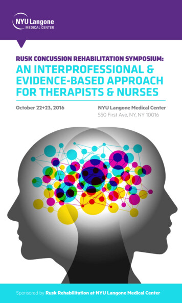
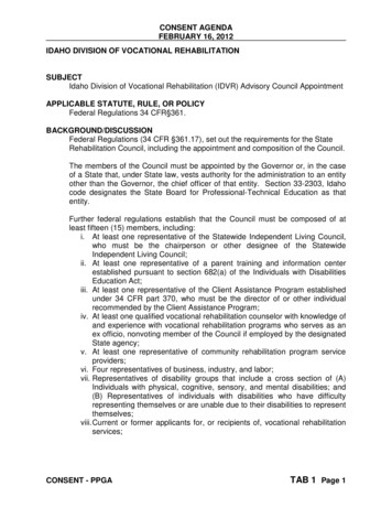
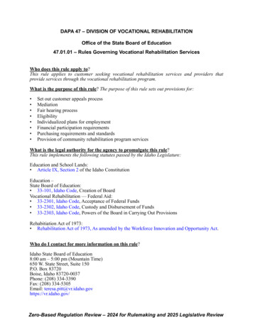
Stable angina (angina pectoris): presents during activity/exercise atpredictable heart rates and blood pressures (known as rate pressureproduct) and is relieved by coronary vasodilators or rest. Typicallymanaged by medication that reduces the cardiac workload (i.e.vasodilators), stress management, and activity pacing. Unstable angina: can occur at rest, spontaneously, or with reducedworkloads, and may not be easily managed by rest or medication.Typically indicates a blockage with intervention required, eitherpercutaneous or surgical Variant angina (Prinzmetal’s Variant Angina): typically occurs in theyounger population due to coronary artery vasospasm and most oftenoccurs at rest or overnight.1o Myocardial infarction (MI): typically classified as ST segment elevation (STEMI)or non-ST segment elevation (NSTEMI), determined by 12-lead ECG, related toelevation of the ST segment above the isoelectric line STEMI: An ST-elevation myocardialinfarction (STEMI) is a result of thecomplete blockage of a coronary artery,therefore typically has a higher risk of deathor disability due to increased myocardialcell damage/death. Tissue damage tends toextend through the full depth of the cardiacwall local to the area of ischemia NSTEMI: A non-ST elevation myocardialinfarction (NSTEMI) is defined as when apartial blockage of a coronary artery,though severity within this group can varypending the level of occlusion.2A.B.C.Normal Sinus RhythmSTEMINSTEMIChart Review: Treatment: can include pharmacologic therapy, percutaneous intervention (i.e. stenting), orcoronary artery bypass grafting (see CABG below) MI rule out (R/O): Patients admitted with acute coronary syndrome (ACS), chest pain(angina), or suspected MI are not appropriate for PT until they have either been ruled out fora MI event, or until they are medically/surgically managed. During a R/O for MI, three sets(one every 8 hours) of cardiac enzymes (creatinine kinase [CK-MB isoform], troponin [TnI]) are drawn and electrocardiograms (ECGs) are performed.1,3o Creatinine Kinase (CK-MB isoform): Creatinine phosphokinase is an enzymereleased after cell injury or death of cardiac muscle. CK-MB measurement can assistin the diagnosis of an MI, estimate the size of infarction and evaluate the occurrenceof re-perfusion. An early peak and rapid clearance from the blood can indicate reperfusion. Values may also be elevated due to other reasons. Communication withthe medical team should occur when there is question of the appropriateness of PT.Standard of Care: Cardiac3Copyright 2019 The Brigham and Women's Hospital, Inc., Department of Rehabilitation Services. All rights reserved
o Brigham and Women’s Hospital has started using a troponin assay which is moresensitive and specific for myocardial tissue necrosis. The numerical value associatedwith myocardial necrosis has changed. Rule-in: troponin 52 ng/L, OR Δ 5 ng/L Rule-out: troponin 10 ng/L for women and 12 ng/L for men, AND Δ 3ng/L If after q1h troponin neither rule-in or rule-out, obtain 3h troponino ECGs: Electrocardiography changes are almost always present in the event of an MI.Early tracings typically show peaked or “hyperacute” T waves. As the MI progresses,this is followed by ST-segment depression or elevation, Q-wave development andlastly, T-wave inversions. Presentation can vary, with this progression occurring overa few hours to several days, with Q-waves only developing in 30-50% of acute MIs. It is important to note that ST-segment depression or elevation can also becaused by coronary artery spasm, electrolyte abnormalities, left ventricularhypertrophy, interventricular conduction delays (BBB), atrial fibrillation orflutter, Digoxin and pacemakers.PT Examination: Determine medical stability:o If the patient rules in for a MI, care must be taken to determine when a patient isstable to participate in a PT examination or intervention. In general, it can beexpected that the patient may resume progressive monitored activity once cardiacbiomarkers have peaked and down trended for two sets and/or once the patient ishemodynamically stable at rest.o On occasion, once a patient has undergone intervention to treat the myocardialischemia (i.e.: heart catheterization with stenting), no further biomarkers will bedrawn, removing the ability to watch for down trending levels. In this event,communication with the team is vital to determine whether it is medically appropriateand safe to proceed with physical therapy evaluation and intervention.o Of note, in some instances, cardiac enzymes may be elevated e.g., cardiac stressrelated to volume overload (heart failure) or tachy-arrhythmias, or rise frompreviously lower values e.g., after a cardiac catheterization, and may not indicate anew MI event. Clarify appropriate activity orders with the responding clinician. Assessment of enduranceo Following ACSM Guidelines for Exercise Testing and Prescription4, it is appropriateto evaluate a patient’s exercise tolerance soon after an MI occurs. Submaximalexercise tests (such as a 6MWT, see Appendix 1) are recommended before hospitaldischarge at 4-6 days after acute MI. Submaximal exercise testing can be used todetermine an appropriate exercise prescription and guide the medical team on theeffectiveness of their interventions, based on a patient’s hemodynamic response.o See endurance/exercise testing.Standard of Care: Cardiac4Copyright 2019 The Brigham and Women's Hospital, Inc., Department of Rehabilitation Services. All rights reserved
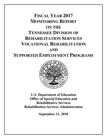
PT Intervention: Exercise Testing and Prescription Refer to PT Intervention/Aerobic Exercise Training for information regarding exercisetesting and prescription in the inpatient setting for a patient with cardiac dysfunction. A homewalking program should be established based on results of submax exercise test prior todischarge. Post MI, a referral to a cardiac rehab program is highly recommended. Patients should beeducated on purpose of cardiac rehab, locations near their home (found on T-drive) andprovided with a referral prior to discharge.Valvular DiseaseDefinitions: Any of the four valves of the heart can become diseased or dysfunctional for variousreasons, but commonly seen reasons include congenital abnormalities, progressivecalcification/atherosclerotic changes, rheumatic heart disease, infective endocarditis,connective tissue disorders, and changes associated with heart failure and ventriculardilation. Valve disease is typically diagnosed via echocardiogram which shows alterationsin a person’s blood flow.Common terms of dysfunction/disease of heart valves: Insufficiency (or regurgitation): valves do not close properly, allowing blood to flow inthe reverse direction during ventricular systole or diastole. Consequences of insufficiencyinclude chamber hypertrophy and retrograde chamber dilation.o Example: mitral insufficiency (mitral regurgitation): the mitral valve does notclose properly during systole, allowing leakage of blood backwards from the leftventricle into the left atrium; it is the most common type of valvular heart disease. Mitral Valve Prolapse: the cusps of the mitral valve become enlarged andfloppy, bulging backward into the left atrium Stenosis: narrowing of the opening of the valve, often due to progressive calcification orfrom rheumatic heart disease. This narrowing reduces forward blood flow, leading toprogressive dilation and hypertrophy of the chamber preceding the valve and potentially areduction in cardiac outputo Aortic valve stenosis is common and is classified in severity by a grading systembased on velocity of blood flow, pressure gradients from the left ventricle to theaorta, and the aortic valve area. Cardiac output is preserved and the patient isoften asymptomatic until the stenosis is severeAortic StenosisAreaMean gradientJet velocity2Mild 1.5cm 25 mmHg 3.0 m/sModerate1.0 – 1.5cm235-40 mmHg3.0 – 4.0 m/sSevere 1.0cm2 40 mmHg 4.0 m/so Higher pressures on the left side of the heart lead to greater chance of valvulardysfunction in the mitral and aortic valves, increasing the workload on the heart.The heart can compensate for a time with chamber hypertrophy, dilation, andStandard of Care: Cardiac5Copyright 2019 The Brigham and Women's Hospital, Inc., Department of Rehabilitation Services. All rights reserved
systemic BP adjustments however ultimately symptoms of heart failure candevelop.3Chart Review: Medical History:o Etiology of valvular dysfunctiono Associated comorbiditieso History of management Chart Review:o Reason for admission: typically related to progression of symptoms of heartfailure, admitted for medical management. Can be admitted for elective cardiacsurgery (see below).o Type of valve dysfunction and severity. See grading system aboveo Recent imaging including echo or right heart catheterization to evaluate forprogressiono Management plan, including changes in medication and/or progression to valverepair or replacement. Diuretics for volume management or other plans for pharmacologicmanagement. Valvuloplasty (for aortic stenosis) – use of femorally inserted ballooncatheter to separate calcified leaflets or to stretch the annulus. This is apalliative measure for patients not eligible for valve replacement Cardiac surgery Percutaneous valve replacement Aortic valves can be replaced transfemorally, particularly in older,frail patients, due to risks associated with cardiac surgery. SeeTAVR below. On occasion, patients undergo mitral valvereplacements with a similar techniquePT Examination: Prior level of function including most recent limitations. It is important, as with patientswith heart failure, to establish a baseline level of activity tolerance, both when a patienthas been feeling well and when they are feeling poorly, most likely due to symptomsrelated to volume overload. See CHF section for further information Lung and heart sounds, listening for murmurs, pulmonary congestion, both pre and postactivity to assess for response to exertion Refer to endurance/exercise testingPT Intervention: Refer to PT Intervention/Aerobic Exercise Training for information regarding exercisetesting and prescriptionStandard of Care: Cardiac6Copyright 2019 The Brigham and Women's Hospital, Inc., Department of Rehabilitation Services. All rights reserved
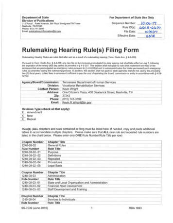
Heart Failure/CardiomyopathyDefinition/Pathology: The American Heart Association defines heart failure (HF) as a chronic, progressivecondition in which the heart muscle is unable to pump sufficient blood to meet the body'smetabolic demand. Heart failure is caused by conditions that damage or weaken the heart,including coronary artery disease, myocardial infarction, hypertension, valvular disease,cardiomyopathy, congenital heart defects, heart arrhythmias, myocarditis or other chronicconditions such as diabetes, HIV, hyperthyroidism, hypothyroidism, or a buildup of iron(hemochromatosis) or protein (amyloidosis).5 There are four types of heart failure, categorized by either a structural perspective (Leftsided vs. Right-sided) or functional perspective (Systolic and Diastolic):5-7o Left-sided heart failure is caused by failure of the left ventricle to adequatelypump blood to periphery, which can lead to a backup of fluid on the lungs andshortness of breath. Left ventricular failure and the resultant pulmonarycongestion is referred to as Congestive Heart Failure (CHF)o Right-sided heart failure is caused by failure of right ventricle to pumpdeoxygenated peripheral blood to the lungs, which can lead to back of fluid intothe abdomen, legs and veins. Right-sided heart failure can occur because ofprogressive left-sided heart failure, pulmonary hypertension, valvular disease orfrom pulmonary disease.o Systolic heart failure is caused by impaired contractile function of the ventricleresulting in reduced stroke volume, cardiac output and ejection fractiono Diastolic heart failure is caused by impaired ventricular relaxation duringdiastole leading to impaired filling and causes a reduced stroke volume andcardiac output but the ejection fraction remains the same. Impaired ventricularrelaxation is often related to ventricular stiffness which is associated with olderage and amplified by HTN, diabetes mellitus and kidney disease. When treating a patient with heart failure, it is important to understand the type and causeof heart failure to best design your education and treatment plan. Heart failure is typically classified based on the severity of the patient’s symptoms andthe effect the symptoms have on physical activity. The table below shows the mostcommonly used classification system, the New York Heart Association FunctionalClassification.8 This classification places patients in stages I-IV based on their physicalactivity limitations.NYHAClassLevel of Clinical ImpairmentINo limitation of physical activity. Ordinary physical activity does not cause unduebreathlessness, fatigue or palpitations.IISlight limitation of physical activity. Comfortable at rest, but ordinary physical activityresults in undue breathlessness, fatigue or palpitations.Standard of Care: Cardiac7Copyright 2019 The Brigham and Women's Hospital, Inc., Department of Rehabilitation Services. All rights reserved
IIIMarked limitation of physical activity. Comfortable at rest, but less than ordinary activityresults in undue breathlessness, fatigue or palpitations.IVUnable to carry on any physical activity without discomfort. Symptoms at rest can bepresent. If any physical activity is undertaken, discomfort is increased.Of note, Cardiac amyloidosis (CA) is a unique and rare form of heart failure that causesthickening of the heart walls and leads, most commonly, to diastolic heart failure. Due to the wayamyloid affects the heart and other body systems, patients present differently than those withmore common forms of heart failure and therefore, our physical therapy approach should bedifferent. Patients with CA often have an impaired hemodynamic response to activity orexercise, including exercise-induced syncope.9 If you are treating a patient with this diagnosis, besure to consult a team lead or cardiopulmonary specialist when developing your plan of care. Theremainder of this section will focus on the more common presentation of heart failure.Chart Review: Medical Historyo Cause of heart failure and categorization based structure or function (e.g. ischemic vs. non-ischemic, congenital, systolic vs. diastolic, preserved vs.reduced ejection fraction)o Onset and duration of HF symptoms and NYHA Stage Hospital Courseo Reason for admission. Patients are often admitted due to symptoms related toincreased volume status, e.g. - progressive dyspnea on exertion, increased lowerextremity edema and decline in activity toleranceo Take note of any relevant lab values, imaging or tests. Below are commontests/measures done by the medical team in a patient with CHF: Chest X-ray: Determines the level of pulmonary congestion, commonfindings include interstitial edema, atelectasis and pleural effusions.10 Echocardiogram Lab Values: Many lab values and electrolytes are affected by fluidretention that occurs secondary to the inadequate cardiac output in CHF.Fluid overload can lead to impaired renal function, hepatic function andaltered electrolytes.10 Below are common changes in lab values found in CHF:o BNP, protein in urine (Albumin), BUN/Cr, PaCO2, liverenzymeso PaCO2, SpO2, Sodiumo / Potassium- may be increased or decreased Severe hypokalemia ( 2.5 mEq/L) or hyperkalemia( 5 mEq/L) should be a precaution to PTintervention due to the increased risk for cardiacStandard of Care: Cardiac8Copyright 2019 The Brigham and Women's Hospital, Inc., Department of Rehabilitation Services. All rights reserved
arrhythmias. Be sure to collaborate with medicalteam prior to intervention.o Troponin can be elevated with heart failure, often due toincreased demand on the heart rather than acute myocardialinfarction. Be sure to trend troponin and look at clinicalpicture to determine appropriateness for PT intervention. Patients with heart failure are at risk for gout due to the fluidfluctuations, use of diuretics and potential for renal dysfunction.11Therefore, blood work may reveal hyperuricemia and the patientmay present with joint pain and inflammation.Cardiac Catheterization: may be done to determine the cause of heartfailure and potential for intervention (e.g. - valve repair/replacement,CABG). Additionally, cardiac catheterizations can provide the medicalteam with accurate information regarding ejection fraction, cardiac output,stroke volume and filling pressures to assist in prognosis and to optimizemedical management.12Medications*13o The focus of drug therapy in heart failure is on optimizing preload, improvingcardiac pumping performance and reducing afterload. Below are commonmedications seen in patients with heart failure: Pre-load red
Post MI, a referral to a cardiac rehab program is highly recommended. Patients should be educated on purpose of cardiac rehab, locations near their home (found on T-drive) and provided with a referral prior to discharge. Valvular Disease Definitions: Any of the four valves of t