Transcription
ng.comSymbiosisResearch ArticleJournal of Exercise, Sports & OrthopedicsOpen AccessDynamic Gene Expression Profile Changes in Synovial FluidFollowing Meniscal Injury; Osteoarthritis (OA) Markers FoundDanica D. Vance1*, Liyong Wang2, Evadnie Rampersaud2, Bryson P. Lesniak1, Jeffery M. Vance2,Margaret A. Pericak-Vance2 and Lee D. Kaplan1UHealth Sports Performance and Wellness Institute, University of Miami, Miller School of Medicine, Miami, FloridaJohn P. Hussman Institute for Human Genomics, University of Miami, Miami Miller School of Medicine, Miami, Florid12Received: 02 June, 2014; Accepted: 04 July, 2014; Published: 18 July, 2014*Corresponding author: Danica D. Vance, UHealth Sports Performance and Wellness Institute, University of Miami, Miller School of Medicine, Miami,Florida, USA, 33136,Tel: 305-781-1766; E-mail: ddv305@gmail.comIntroductionAbstractPurpose: We hypothesize that molecular changes leadingto Osteoarthritis (OA) occur soon after a meniscal tear and longbefore the manifestation of OA. We sought to characterize the geneexpression profile in synovial fluid at different time points followinga meniscal tear with a focused examination on inflammatory andarthritis related genetic markers.Methods: Synovial fluid was collected from 8 male patients(24 48yrs old) with evidence of meniscal injury on MagneticResonance Imaging (MRI). Illumina humanht-12 microarray andRNA-Seq were used to characterize gene expression profile in cellpellets and supernatant, respectively. Unsupervised clusteringanalysis was performed to identify patterns of gene expressionprofile among samples. Metacore was utilized to perform pathwayanalysis. T-tests with False Discovery Rate (FDR) corrections wereused to compare subset of genes among individuals.Results: Individuals with short injury duration ( 2 months)had distinct expression signatures than the rest, in both cell pelletand supernatant analyses. In the cell pellets, among genes previouslylinked to inflammatory or arthritic conditions, IL 1B, IL1RN, ILF2,NFKB1, IL10RB, IL18BP, ILF3, IL13RA, BMP2K and IL10RB weresignificantly upregulated (adjusted p 0.05) in individuals withlong duration compared to individuals with shorter injury durationin the microarray analysis. In the cell-free supernatant, 764 RNAspecies were identified and 65 RNA species were down-regulatedand 78 RNA species were up-regulated (FDR 0.05) in individualswith short compared to individuals with long injury duration. Themost differentially expressed gene in the supernatant is SLC2A9 (P 1.2x10⁻¹³; FDR 9.6x10⁻¹¹). In the cell pellet, the top 5% expressedgenes are enriched for inflammatory and cytoskeleton remodelingpathways. Among them LAIR 1, TMSB4X, CCR6, IL18 and IL10; all havebeen implicated in arthritic conditions.Conclusions: Molecular changes contributing to OA developmentoccur earlier than previously described and potentially evolve orchange over time.Clinical Relevance: Our data suggest the potential for earlyinterventions to halt detrimental molecular changes followingmeniscal injury, thereby reducing the susceptibility of patients to OAfollowing a meniscal tear.Keywords: Meniscus; Meniscal tear; Gene expression; Synovialfluid; Microarray; OsteoarthritisSymbiosis GroupThe meniscus is a critical component of the knee joint,contributing to shock absorption, load distribution and jointstability [1]. A meniscal tear is one of the most common injuriesseen in orthopedics [2]. In addition to the physical symptomsand limitations caused by the acute injury, researchers havelinked meniscal injury to the progression and development ofosteoarthritis (OA) [1,3,4]. Following meniscal damage, changesin load distribution contribute to joint space narrowing andincreased mechanical stresses on articular cartilage, whichin turn lead development of OA [1]. Osteoarthritic changesin articular cartilage have been seen as early as 2 years postmeniscal damage [4].In addition to the mechanical changes occurring in the kneejoint following meniscal tears, differences at the molecular levelin both meniscal tissue and synovium have been reported [57]. RNA extracted from injured meniscal tissue was found tohave significantly elevated levels of several arthritis-relatedgenetic markers in patients under the age of 40 [5]. Similarly,Scanzello et al. [6] also found significant differences in geneexpression patterns in synovium tissue after a meniscal injurywhen comparing patients with evidence of inflammation to thosewithout inflammation [6,7].Yet to be extensively investigated are the dynamic molecularchanges that occur in the synovial fluid following a meniscaltear. Because the meniscus tissue has limited blood supply,the synovial fluid is an important source of nutrients and wasteproducts. Previous studies have successfully used synovial fluidto examine protein complexes and specific cytokines that mightcontribute to acute knee pain [8-10]. This is one of the first studiesto look at gene expression globally in synovial fluid following ameniscal tear, one of the major risks for development of OA in theknee. Indeed, radiographic OA changes in cartilage can be seen asearly as 2 years post-injury. However, changes at the molecularlevel could occur earlier. Examining gene expression of synovialfluid following a meniscal tear will provide further insights on theinflammatory and cellular changes that occur in the knee joint*Corresponding author email: ddv305@gmail.com
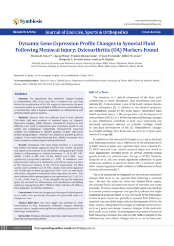
Dynamic Gene Expression Profile Changes in Synovial Fluid Following Meniscal Injury;Osteoarthritis (OA) Markers Foundafter injury. Determining these changes and when they beginwould be critical to successful intervention following meniscalinjury to prevent or delay the development of OA. Furthermore,if a link can be established between these molecular changes andclinical outcomes including future degenerative changes in theknee joint, these data could potentially provide a non-invasivemethod to help identify those individuals who are at high riskof developing OA. Early stage identification of OA patientsmakes room for potential therapeutic interventions when theinterventions are probably the most effective. We hypothesizethat molecular changes leading to OA occur soon after a meniscaltear and long before the manifestation of OA. Our purposewas to characterize the gene expression profile of synovialfluid following a meniscal tear to determine if known OA andinflammatory genes can be detected early post-injury.MethodsSynovial fluid collectionThe Institutional Review Board (IRB) (Protocol #: 20100506)of the participating institution approved this study. Patientswere enrolled through the sports medicine clinic. Potential studyparticipants were individuals scheduled to undergo a partialarthroscopic meniscectomy (Figure 1). Subjects were less than50 years of age, had no previous knee pathology or surgery of theinjured knee including any ligamentous or cartilage injury, andhad MRI evidence of a meniscal tear. Additional exclusion criteriaincluded patients with Kellgren- Lawrence Grade III or GradeIV osteoarthritis of the knees observed during the operation,Body Mass Index (BMI) greater then 30, age 50 years old orthose diagnosed with one of the following; systemic rheumatoidarthritis, neuromuscular disease or diabetes. These exclusioncriteria were met to limit the presence of osteoarthritis withinthe knee joint. Synovial fluid from the affected knee joint wascollected by needle aspiration from eight male patients (24 48years old) at the time of surgery, prior to initial incision. Collectedfluid was immediately transferred to RNAse free 1.5ml endorphintubes and kept on ice while transferring to the lab for processing.In addition, medical information regarding injury phenotype(A)Copyright: 2014 Vancewas collected directly from patient and the patient’s clinic notes(Table 1).Synovial Fluid processingAll sample processing and molecular analysis were carriedout at Hussman Institute for Human Genomics (HIHG), Universityof miami. In preparation for RNA extraction, collected synovialfluid samples were centrifuge at 1500g at 4 C for 5 minutes oruntil pellet formation. Next, the supernatant was removed and1ml of qiazol lysis reagent was added to the pellet followed byvigorous vortexing. Supernatant and cell pellet were stored at-80 C until RNA extraction.Rna extractionTotal RNA was extracted from cell pellet using theqiazol-chloroform-isopropanol protocol as instructed by themanufacture (Qiagen). Cell-free RNA was extracted using rneasykit (Qiagen) with modified protocol to recover small RNA species.RNA was quantified by Qubit RNA assay (Invitrogen LifeTechnologies) and qualified by Agilent 2100 RNA 6000 Pico Chip(Agilent Technologies).Microarray and RNA-Seq AnalysisFor the cell-pellet RNA, genome-wide gene expressionprofiling was done using the Illumina Whole Genome HighThroughput (HT) 12 Assay. For the cell-free RNA, nebnext Small RNA Library Prep Set for Illumina was used for librarypreparation. Next-generation sequencing of these librarieswere carried out on the Illumina hiseq 2500 instrument. Bothmicroarray and RNA-Seq were performed at Center for GenomicTechnology at the Hussman Institute for Human Genomicsfollowing manufacturer’s instructions.Statistical data analysisAll statistical analyses were performed in the Center ofGenetic Epidemiology and Statistical Genetics at the HIHG. Forthe microarray data, the raw expression data (Log2 values)were transformed using variance-stabilizing transformation(VST) [11] with the LUMI Bioconductor R package [12], whichtakes into account the large number of technical replicates on(B)Figure 1: Arthroscopic views of meniscal tear. (A) Complex degenerative tear (B) Flipped bucket handle tear.Citation: Vance DD (2014) Dynamic Gene Expression Profile Changes in Synovial Fluid Following Meniscal Injury; Osteoarthritis(OA) Markers Found. J Exerc Sports Orthop 1(3): 1-7. DOI: http://dx.doi.org/10.15226/2374-6904/1/3/00115Page 2 of 7
Dynamic Gene Expression Profile Changes in Synovial Fluid Following Meniscal Injury;Osteoarthritis (OA) Markers FoundTable 1: Data 456784838394 monthsNo4 monthsnoParrot-BeakRight MM, LMnone1 monthRight MM2 monthsLeft MM1 monthyesmild3 months7 monthsno44Type of MeniscalTearmoderate2 monthsyesLeft LMEffusionLeft LMyes4130InjuryDurationInjury Location( LM LateralMeniscus,MM MedialMeniscus)Right MMRight MMLeft MMthe Illumina arrays. Normalization was conducted using theRobust Spline Normalization (RSN) algorithm, which combinesfeatures of quantile and loess normalization. Quality control wasperformed using the lumiQ command. For the RNA-Seq data,the STAR program was used to align the trimmed sequencingreads against human reference genome to localize RNA species.Given the wide range of number of reads generated for eachsample, normalization is an essential step. The EdgeR programnormalizes the reads by estimating effective library size based onthe depth of reads. The normalized gene expression unit is CountPer Million (CPM).In order to correlate the RNA expression profile with clinicalcharacteristics, unsupervised cluster analysis was conductedusing LUMI software for microarray data and the EdgeR programfor RNA-Seq data. For gene expression analysis, we firstcharacterized what genes are expressed in synovial fluid followingmeniscal injury, examining those genes with the greatest averageexpression (top 5%) across all 8 samples. Pathway analysiswas conducted using MetaCore (GeneGo, Inc.) software. Then,we carried out differential gene expression analysis comparingsamples with short duration time ( 2 months) to samples withlonger duration time ( 3 months). For the microarray data,T-tests with FDR corrections [13] were performed to comparethe gene expression profiles of a subset of OA-related genes (n 203 genes, 340 transcripts) within samples of varied injuryduration. Genes with p value 0.05 were considered significant.For the RNA-Seq data, EdgeR was used to evaluate differentialgene expression among the 764 RNA species identified in the cellfree synovial fluid.ResultsFor this pilot study, we were able to ascertain eight malepatients with available funds. Average age of study participantwas 36.5 8.3 years old (range; 24-48 yrs). For all patients, tiveMM: Horizontal,LM radialDouble Flap ofPosterior HornComplex,Degenerative ofposterior hornFlipped buckethandleComplex,DegenerativeCopyright: 2014 VanceOA Grades (Kellgren andLawrence ) (P Patella,T Trochlear, MFC medialfemoral condyle, LFC Lateralfemoral Condyle)P Grade 1 lesionno changes observedP Grade 2 lesionMFC Grade 1-2 lesionP Grade 2 lesion, T Grade 2lesionP Grade 2 lesion, MFC Grade2 lesionP Grade 1 changesP Grade 1 lesionassessment of injury was made in clinic and then confirmed byMRI by an independent musculoskeletal radiologist. All 8 patientsunderwent an arthroscopic partial meniscectomy for a meniscaltear (Figure 1). The type of meniscal tear, degree and location ofarthosis were systematically observed by the surgeon during theoperation (Table 1). Additional injury phenotype was collectedfrom the patient and initial clinic note (Table 1). Specifically,duration of injury was calculated by the self-reported initialinjury time and the time of sample collection, i.e. time of surgery.The median value for time between initial injury and surgery was4 months (range: 1 month to 7 months).The top 5% expressed genes in cell pellets across all samplesare enriched for pathways involved in oxidative phosphorylation,immune response and cytoskeleton remodeling. (SupplementaryFigure 1) Included in the top 5% expressed genes were severalgenes with previously reported association to inflammation and/or Rheumatic or Osteoarthritis; such as LAIR 1, TMSB4X, CCR6,IL18 and IL10 [2, 14-17].Unsupervised cluster analysis of gene expression profile isillustrated in Figure 2 (heat map of microarray data in cell pellet)and Figure 3 (principal component analysis of RNA-Seq data incell-free supernatant). With the gene expression profile in cellpellets, we observed a clustering of three samples (samples 6 8)with distinct gene expression profiles from the rest (Figure 2).When we compared different injury phenotypes including; age,etiology of injury, injury duration, location of injury (medial vs.Lateral meniscus) and presence of effusion (Table 1), we foundthat injury duration best stratified the expression patterns.Specifically, samples 6, 7, and 8 had the shortest injury duration( 2 months). Sample 5 also had a 2 months of injury durationand was clustered right next to the tight cluster formed bysamples 6 8. With the gene expression profile in cell-freesupernatant, we observed a clustering of samples 6 8, as seen inthe microarray analysis. Samples with the longest injury durationCitation: Vance DD (2014) Dynamic Gene Expression Profile Changes in Synovial Fluid Following Meniscal Injury; Osteoarthritis(OA) Markers Found. J Exerc Sports Orthop 1(3): 1-7. DOI: http://dx.doi.org/10.15226/2374-6904/1/3/00115Page 3 of 7
Dynamic Gene Expression Profile Changes in Synovial Fluid Following Meniscal Injury;Osteoarthritis (OA) Markers Found7865241Copyright: 2014 Vance3Figure 2: Unsupervised cluster analysis of gene expression in cell pellet.Heatmap showing expressed genes across individuals. Horizontal axis displays individual samples, vertical axis displays each expressed genes byz-scores (scaled value of normalized intensity scores). Red decreased intensity; green increased intensity.Figure 3: Unsupervised cluster analysis of gene expression in cell-free supernatant.Citation: Vance DD (2014) Dynamic Gene Expression Profile Changes in Synovial Fluid Following Meniscal Injury; Osteoarthritis(OA) Markers Found. J Exerc Sports Orthop 1(3): 1-7. DOI: http://dx.doi.org/10.15226/2374-6904/1/3/00115Page 4 of 7
Dynamic Gene Expression Profile Changes in Synovial Fluid Following Meniscal Injury;Osteoarthritis (OA) Markers Found( 4 months), i.e. Sample 1 3, were clustered on the other end offirst dimension (Figure 3).To examine the gene expression differences between sampleswith short versus long injury duration we examined the foldchange in a subset of 203 genes (340 transcripts) with reportedinvolvement in either inflammatory or osteoarthritis pathways[13, 18-27], that were expressed in the cell pellet of synovialfluid. The complete list of genes can be found in (supplementaryTable 1). The following genes met FDR Corrected significance (p 0.05) (Table 2): Interleukin-1 beta (IL1B), Interleukin 1 receptorantagonist (IL1RN), Interleukin enhancing binding factor 2 (ILF2),Nuclear Factor of Kappa light polypeptide gene enhancer in B-cells1(NFKB1), Interleukin 10 receptor beta (IL10RB), Interleukin 10receptor alpha (IL10RA) Interleukin 18 binding protein (IL18),Interleukin enhancing binding factor 3 (ILF3), Interleukin 13receptor, alpha 1 (IL13RA), Bone morphogenetic protein 2inducible kinase (BMP2K).In the cell-free supernatant, 764 RNA species were identifiedto be present and all of them were evaluated for differentialexpression between samples with shorter ( 2 months) andlonger ( 3 months) injury duration. 65 RNA species were downregulated and 78 RNA species were up-regulated (FDR 0.05) inindividuals with short injury duration. The most differentiallyexpressed gene in the supernatant is SLC2A9 (P 1.2x10⁻¹³; FDR 9.6x10⁻¹¹) (Table 3).DiscussionWhile examining, highly expressed genes in the synovial fluidfrom patients with meniscal injury having no OA symptoms, weobserved that the expressions of many genes, due to the earlyinjury are shown to be associated with OA and inflammation.These reported genes include LAIR1, TMSB4X, CCR6, IL18, IL10[2, 14- 17]. LAIRI is the highest expressed gene among all thesamples. LAIRI is an immune inhibitory receptor for collagenfound on the most immune cells (pubmed gene ID: 3903) [28].Its inhibitor LAIR2 is found in the synovial fluid of patientshaving Rheumatoid Arthritis (RA) or OA. Patients with RA showhigher expression level of LAIR2. Similarly, IL-10, a prominentchondroprotective anti-inflammatory marker released by thesynovium and cartilage, was also highly expressed across allsamples. IL-10 has been shown to be present in synovial fluid ofOA patients [29].The second highest average expressed gene across all sampleswas TMSB4X, which encodes a protein (TB4) that activates theexpression of MMPS in fibroblasts (pubmed ID: 7114). Recently,TB4 level in synovial fluid of patients with RA was found whencompared to OA patients. It was also significantly associated withthe levels of MMP-9, MMP-13, IL-6 and IL-8 [15], suggesting TB4may play an early role in starting the inflammatory and cartilagedegradation cascade.Table 2: Differentially expressed OA-related genes in cell pellets between samples with short and long injury duration.Gene Symbollog FCAverage Expression (Z sted P Value0.00098.15-1.45P Value8.467.44-2.19IL1BCopyright: 2014 Vance0.0240.00639.150.0320.0064Table 3: Top differentially expressed RNAs in supernatant between samples with short and long injury duration.0.032Gene SymbleType of RNAlogFCAverage Expression (logCPM)P ValueFDRSLC2A9protein 2protein coding11.786.83C1orf114protein coding-6.50MBTPS1protein coding-5.98FBXO45N4BP2INHBBAL590452.1protein codingprotein codingprotein codingprotein 6E-103.65E-104.15E-10Citation: Vance DD (2014) Dynamic Gene Expression Profile Changes in Synovial Fl
UHealth Sports Performance and Wellness Institute, University of Miami, Miller School of Medicine, Miami, Florida. 2. John P. Hussman Institute for Human Genomics, University of Miami, Miami Miller School of Medicine, Miami, Florid. Research Article . Journal of Exercise, Spo