Transcription
U.S. ARMY MEDICAL DEPARTMENT CENTER AND SCHOOLFORT SAM HOUSTON, TEXAS 78234-6100NURSING CARE RELATED TO THESENSORY AND NEUROLOGICALSYSTEMSSUBCOURSE MD0919EDITION 100
DEVELOPMENTThis subcourse is approved for resident and correspondence course instruction. Itreflects the current thought of the Academy of Health Sciences and conforms to printedDepartment of the Army doctrine as closely as currently possible. Development andprogress render such doctrine continuously subject to change.ADMINISTRATIONFor comments or questions regarding enrollment, student records, or shipments,contact the Nonresident Instruction Section at DSN 471-5877, commercial (210) 2215877, toll-free 1-800-344-2380; fax: 210-221-4012 or DSN 471-4012, e-mailaccp@amedd.army.mil, or write to:COMMANDERAMEDDC&SATTN MCCS HSN2105 11TH STREET SUITE 4192FORT SAM HOUSTON TX 78234-5064Approved students whose enrollments remain in good standing may apply to theNonresident Instruction Section for subsequent courses by telephone, letter, or e-mail.Be sure your social security number is on all correspondence sent to the Academy ofHealth Sciences.CLARIFICATION OF TRAINING LITERATURE TERMINOLOGYWhen used in this publication, words such as "he," "him," "his," and "men" are intendedto include both the masculine and feminine genders, unless specifically stated otherwiseor when obvious in context.USE OF PROPRIETARY NAMESThe initial letters of the names of some products are capitalized in this subcourse. Suchnames are proprietary names, that is, brandnames or trademarks. Proprietary nameshave been used in this subcourse only to make it a more effective learning aid. The useof any name, proprietary or otherwise, should not be interpreted as an endorsement,deprecation, or criticism of a product. Nor should such use be considered to interpretthe validity of proprietary rights in a name, whether it is registered or not.
TABLE OF CONTENTSLesson1ParagraphsINTRODUCTIONNURSING CARE RELATED TO THE SENSORY SYSTEMSection I. Introduction to the Special SensesSection II. AssessmentSection III. Treatments and ProceduresSection IV. Disorders of the EyeSection V. Disorders of the EarExercises2NURSING CARE RELATED TO THE NEUROLOGICAL SYSTEMSection I. Anatomy and PhysiologySection II. Neurological AssessmentSection III. Diagnostic ProceduresSection IV. Care of the Unconscious PatientSection V. Infectious Nervous System DisordersSection VI. Degenerative Nervous System DisordersSection VII. Cranial Nerve DisordersSection VIII. Head and Spine InjuriesSection IX. Disorders of the 2--2-45
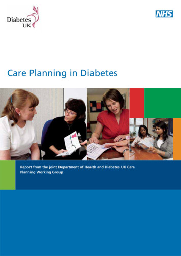
CORRESPONDENCE COURSE OF THEU.S. ARMY MEDICAL DEPARTMENT CENTER AND SCHOOLSUBCOURSE MD0919NURSING CARE RELATED TO THE SENSORY AND NEUROLOGICAL SYSTEMSINTRODUCTIONA patient is not a passive object on which we perform nursing care procedures.A patient should never be thought of, or discussed, as merely a condition or injury. It isnot acceptable to think of, or refer to, your patient as "the gallbladder" or "the fracturedhip" or "the bleeding ulcer." A patient is much more than the condition that brought himto the hospital. A patient is a human being, possessing physical and emotional needsand wants.The relationship between the patient and the nursing staff is extremely dynamicand personal. The patient places his trust in the nursing staff and they, in return, mustutilize all their knowledge and skills to ensure the patient's well-being and assist in hisreturn to good health and independence. This is accomplished by developing atherapeutic relationship between the patient, his family, and the health careprofessionals.The nursing paraprofessional relates very closely with the patient and his familyby virtue of his participation in providing nursing care and his presence at the bedside.It is often the nursing paraprofessional who makes the initial observation that somethingis not as it should be. Nursing paraprofessionals who are responsible, educated, andobservant are assets to the therapeutic environment necessary for patient recovery. Tohelp foster this therapeutic environment, the nursing paraprofessional must do thefollowing:Assess the patient's ability to express himself.Assess the patient's mechanisms for coping.Assess the patient's level of understanding about his condition and the teachingprovided by the health care professionals.Assist the patient to establish a trusting relationship with the health careproviders.Utilize the nursing process to provide the best possible care.Continue to pursue his own education in order to sustain and enhanceprofessional knowledge and skills.MD0919ii
The purpose of this subcourse is to enhance your knowledge of medical surgicalnursing care related to the sensory and neurological systems and the role of thenursing paraprofessional in providing that care.Subcourse Components:This subcourse consists of two lessons. The lessons are as follows:Lesson 1, Nursing Care Related to the Sensory System.Lesson 2, Nursing Care Related to the Neurological System.Credit Awarded:To receive credit hours, you must be officially enrolled and complete anexamination furnished by the Nonresident Instruction Section at Fort Sam Houston,Texas. Upon successful completion of the examination for this subcourse, you will beawarded 12 credit hours.You can enroll by going to the web site http://atrrs.army.mil and enrolling under"Self Development" (School Code 555).A listing of correspondence courses and subcourses available through theNonresident Instruction Section is found in Chapter 4 of DA Pamphlet 350-59, ArmyCorrespondence Course Program Catalog. The DA PAM is available at the followingwebsite: 919iii
LESSON ASSIGNMENLESSON 1Nursing Care Related to the Sensory System.TEXT ASSIGNMENTParagraphs 1-1 through 1-28.LESSON OBJECTIVESAfter completing this lesson, you should be able to:1-1.Identify the special senses.1-2.Name the stimulus for each of the specialsenses.1-3.Identify the receptor organ for each of thespecial senses.1-4.Name the two sensory functions of the ear.1-5.Explain the process by which airborne soundwaves are converted to nerve impulses.1-6.Name the two types of gravitational forces thatprovide sensory input for equilibrium.1-7.Define direct pupil reaction.1-8.Define consensual pupil reaction.1-9.Identify the steps in instilling eyedrops orointments.1-10. Identify the steps in irrigating the eye.1-11. Identify the steps in instilling ear drops.1-12. Identify the steps in irrigating the ear.1-13. Define myopia.1-14. Define hyperopia.1-15. Define presbyopia.MD09191-1
1-16. Define conjunctivitis.1-17. Identify the nursing implications in caring for apatient undergoing ophthalmic surgery.1-18. Identify the nursing implications in caring for apatient with vision loss.1-19. Identify 4 types of hearing loss.1-20. Identify steps in removal of a foreign body fromthe ear.SUGGESTIONMD0919After studying the assignment, complete the exercisesat the end of this lesson. These exercises will help youto achieve the lesson objectives.1-2
LESSON 1NURSING CARE RELATED TO THE SENSORY SYSTEMSection I. INTRODUCTION TO THE SPECIAL SENSES1-1.THE SPECIAL SENSESa. The human body is continuously bombarded by all kinds of stimuli. Some ofthese stimuli are received by sensory receptors distributed throughout the entire body.Other stimuli are received by highly complex receptor organs. These are referred to asthe special senses.b. From each special sense organ, information is sent to the brain throughspecific cranial nerves. When the information reaches the specific area of the brain'scerebral cortex, it is perceived at the conscious level as sight, sound, smell, taste, andbalance. These special senses allow us to detect changes in our environment,providing information necessary for homeostasis.c. The Special Senses are:(1)Sight.(a) The receptor organ is the eye.(b) The stimulus is light rays.(2)Hearing.(a) The receptor organ is the ear (cochlea).(b) The stimulus is sound waves.(3)Smell.(a) The receptor organ is the nose (olfactory hair cells).(b) The stimulus is airborne molecules.(4)Taste.(a) The receptor organs are the taste buds in the mouth.(b) The stimulus is fluid-borne molecules.MD09191-3
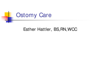
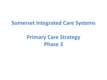
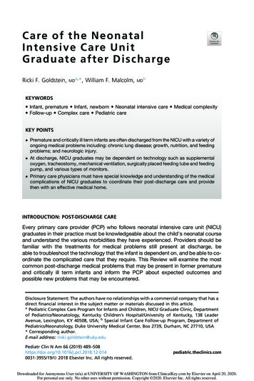
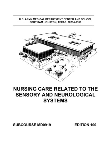
(5)Balance (equilibrium).(a) The receptor organ is the ear (membranous labyrinth).(b)1-2.The stimulus is gravitational forces.SMELLLocated in the upper recesses of the nasal chambers is a special layer of tissuecalled olfactory epithelium. Within the olfactory epithelium are special hair cells(chemoreceptors) that react to airborne molecules. Information received by the haircells is transmitted from the olfactory nerves to the olfactory bulbs and along theolfactory tract to the brain, where it is interpreted as the sensation of smell.1-3.TASTELocated on the tongue and back of the mouth are sensory receptors called tastebuds. Special hair cells in the taste buds are chemoreceptors that react to molecules ofmaterial taken into the mouth (food, liquids, and so forth.). Information received by thehair cells is transmitted to the brain, where it is interpreted as the sense of taste.1-4.VISIONThe eye (figure 1-1) is the special sense organ responsible for vision. Rays oflight (reflected from an object) pass through the cornea, aqueous humor, pupil, lens,and vitreous humor to stimulate the receptor tissue (rods and cones) in the retina. Theresulting nerve impulses travel to the brain, where they are interpreted as sight.1-5.HEARINGThe human ear (figure 1-2) serves two major sensory functions--hearing andequilibrium.a. Sound stimuli travel as airborne waves, which are collected by the externalear. The airborne waves pass through the external auditory meatus (ear canal) to thetympanic membrane, which separates the external and middle ear.b. The physical vibration of the airborne waves is converted to mechanicalvibration by the tympanic membrane and the ossicles. The ossicles (malleus, incus,and stapes) articulate with both the tympanic membrane and the oval window, whichopens into the vestibule of the inner ear.c. When the ossicles are set into mechanical vibration, the stapes acts as aplunger against the oval window, imparting pressure pulses to the fluid (perilymph) ofthe inner ear.MD09191-4
Figure 1-1. The eye.Figure 1-2. The ear.MD09191-5
d. Fluid vibrations of the perilymph are converted to nerve impulses when thehair cell receptors within the cochlea are stimulated by the fluid vibrations. The nerveimpulses are carried to the brain where they are interpreted as sound.1-6.EQUILIBRIUMa. Equilibrium is the state of balance of the body. Through a variety of sensoryinputs and postural reflexes, the body can be maintained in a desired posture. All of thevarious sensory inputs related to the maintenance of equilibrium and posture areintegrated within the brain as "body sense." The internal ear provides one of the inputsystems for general body sense.b. A primary sensory input for equilibrium consists of gravitational forces.Gravitational forces are of two types: static, when the body is standing still, and kinetic,when the body is in motion. Kinetic motion may be in a straight line (linear), or in anangular direction (curvilinear).c. The fluid-filled membranous labyrinth of the inner ear has two sac-likestructures called the sacculus and the utriculus. On the wall of each sac is a collectionof special hair cells, which serve as receptors for static and linear kinetic gravitationalforces.d. Associated with the utriculus are three tubular structures called thesemicircular canals. Two of the semicircular canals are vertically oriented and the thirdis essentially horizontal. All three semicircular canals are oriented at right angles toeach other.e. Each semicircular canal ends with an enlarged area where it opens into theutriculus. This area is called the ampulla. On the wall of each ampulla, at a right angleto the axis of the canal, is a little ridge of hair cells. The hair cells bend in directionalresponse to the kinetic gravitational forces initiated by movement of the head.f. All the information from the hair cells of the sacculus, utriculus, and ampullaeis transmitted to the brain by the vestibular nerve. The vestibular and auditory nervesare contained within the same fibrous sheath from the inner ear to the brain. Within thebrain, the two nerves split into different pathways.Section II. ASSESSMENT1-7.EXAMINATION OF THE EYEAn examination of the eye includes an external examination, examination byophthalmoscope, and an assessment of the functions of the eye.a. External Examination. The anterior segment of the eyes and theirappendages can be examined by visual inspection.MD09191-6
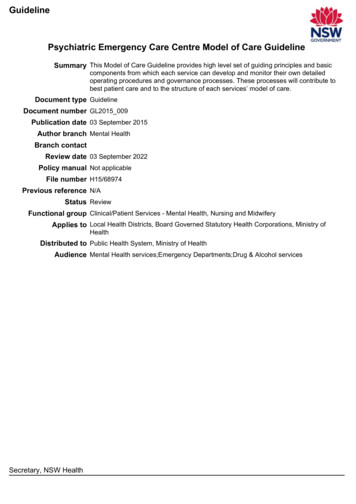
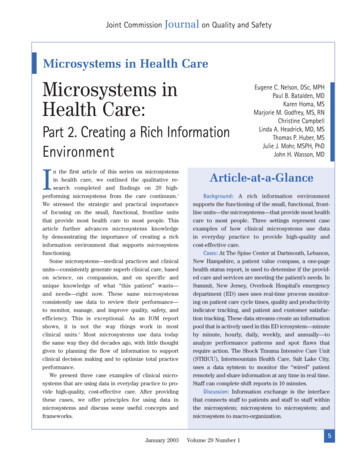
(1) Note the general appearance of the eyelids, eyelashes, and lacrimalapparatus. Observe for:(a) Redness around the eye.(b) Discharge or crusting.(c)Growths on eyes or eyelids.(d) Excessive tearing.(2) Position and mobility can be observed by having the patient rotate theeyes, looking up, down, and to each side.b. Pupillary Response. Normal pupils are rounded, centrally placed, andgenerally equal in size. (About 25 percent of normal individuals have pupils slightlyunequal in size.)(1) Reaction to light. Seat the patient in an area with even lighting andinstruct him to fix his gaze on a distant object. Cover one eye and shine a flashlight infront of the exposed eye. The pupil should contract (constrict) because of the light.This response is called a direct reaction. The covered pupil should also contract. Thisresponse is called a consensual reaction.(2) Near point reaction. When the gaze is changed from a distant object toan object close at hand, the pupils should contract.c. Ophthalmoscopic Examination. By looking through the various lenses ofan ophthalmoscope, the trained examiner can view and assess the internal structures ofthe eye. This examination is routinely performed by the physician.d. Functional Examinations.(1) Focusing power (power of accommodation) is tested by placing a line ofprint close to the eye, then slowly moving it back to the point at which the patient is ableto read it. The nearest point at which it is readable is the near point of accommodation.(2) Visual field refers to all that can be seen with both eyes fixed straightahead. To perform a gross examination of visual field, the confrontation method is used(figure. 1-3). Have the patient and the examiner face each other at a distance of about2-3 feet, each focusing his gaze at the other's nose. The examiner should then extendhis arm to the side, point his finger, and slowly move his arm back in, along a planehalf-way between himself and the patient. The examiner's finger should appear in thepatient's visual field at the same time the examiner sees it (assuming the examiner'svisual field is grossly normal).MD09191-7
Figure 1-3. Confrontation method.(3) Color sense is tested by using specially designed color plates todistinguish reds, greens, and blues.(4) Visual acuity testing is done with the Snellen chart or one of itsmodifications. Each eye is tested separately, both with and without glasses, if worn.(a) Since the distance at which rays of light from an object arepractically parallel and no accommodation of the lens is necessary to focus the object,the test is performed at a distance of 6 meters (20 feet),(b) The Snellen chart contains rows of letters of varying sizes,arranged to that the normal eye can see them at distances of 6, 9, 12, 15, 21, 30, and60 meters. (20, 30, 40, 50, 70, 100, and 200 feet.) If a patient is seated 6 meters (20feet) from the chart and can read the line of letters for 6 meters, his vision is expressedby the fraction 6/6 (or 20/20).(c) Vision is expressed by a fraction, the numerator denoting thedistance at which the test was performed (normally, 6 meters or 20 feet), and thedenominator denoting the smallest line of letters which could be read at that distance. Ifa patient is seated 6 meters from the chart and the smallest line of letters he is able toread is the one that should be read at a distance of 30 meters, then his vision isexpressed at 6/30 (or 20/100).(d) If the largest letters on the chart cannot be read at a distance of 6meters, the patient is moved toward the chart until he can read the largest letters.Vision is then expressed as a fraction, with the numerator denoting the distance atwhich the largest line could be read, and the denominator denoting the number of thelargest line.MD09191-8
(e) If the patient cannot read the largest line at a distance of one meter,the examiner tests the patient's ability to see hand motion in front of his face. If thepatient cannot see the examiner's hand at a distance of one or two meters, he is testedfor light perception. A light is flashed from different directions and the patient is askedfrom which direction the light appears and when it goes on and, it goes off. If the patientcan do this, the examination is recorded as "light perception present". If no lightperception is present, a person is technically blind.(5) These functional examinations are routinely performed by the physicianor eye specialist. However, it is recommended that nursing personnel beknowledgeable of these examinations. To do so will facilitate identification of visualabnormalities. A gross examination using "field expedient techniques" can beperformed when the proper equipment and personnelare not available. For example:(a) Color sense can be observed by having the patient identify thecolor of objects around him.(b) Gross acuity can be tested by having the patient read signs postedon the walls. Use signs of different sizes and position the patient at varying distances.1-8.ASSESSING SYMPTOMSIn addition to the examinations mentioned previously, the patient should beassessed for the following:a. Discomfort or pain in or around the eye.b. Photophobia. (Abnormal sensitivity to light.)c. Nystagmus. (Involuntary, rapid movement of the eyeball. May be horizontal,vertical, rotational, or mixed.)d. Strabismus. (Deviation of the eye from the normal physiological axis:"crossed eyes.")e. Diplopia. (Seeing an object in double: "double vision.")f. Blurred vision.g. "Spots" or "lights" in the visual field.MD09191-9
1-9.EXAMINATION OF THE EARa. The auditory canal is examined by means of an otoscope and ear speculum.The patient normally sits in an upright position for the exam. The lighted otoscope isinserted, using the largest speculum that will fit comfortably into the patient's externalear canal.b. Look through the eyepiece and examine the external ear canal for:(1)Discharge.(2)Impacted cerumen.(3)Inflammation.(4)Masses.(5)Foreign bodies.c. Examine the tympanic membrane )Scarring.(5)Color.AUDIOMETRYa. Hearing tests are conducted with a device called an audiometer. Thisapparatus produces sounds of specific frequencies and intensities. The patient sits in asoundproof room and listens to the sounds over a set of earphones. Sounds of varyingfrequency and intensity are than transmitted to each ear separately. The patient signalswhen a sound is heard by raising his hand or pressing a button to indicate which earheard the sound. When a sound is transmitted and the patient does not signal, theexaminer records that the sound was not heard.b. When this equipment is not available, a gross examination can be performedusing "field expedient techniques." Have the patient sit with eyes closed and ask him toidentify sounds made by an examiner moving in a perimeter around the patient. Thepatient should identify from which direction the sound originated and the type of soundheard. Sounds can be created by whistling, whispering, tapping, playing a radio, and soforth.MD09191-10
1-11.ASSESSING SYMPTOMSIn addition to the examinations mentioned above, th
1-17. Identify the nursing implications in caring for a patient undergoing ophthalmic surgery. 1-18. Identify the nursing implications in caring for a patient with vision loss. 1-19. Identify 4 types of hearing loss. 1-20. Identify steps in removal of a foreign body from the ear. SUGGEST