Transcription
J Periodontol November 2012Evaluation of Human Recession DefectsTreated With Coronally Advanced Flapsand Either Enamel Matrix Derivativeor Connective Tissue: Comparisonof Clinical Parameters at 10 YearsMichael K. McGuire,* E. Todd Scheyer,* and Martha Nunn†Background: The effective treatment of gingival recession (GR) defects is crucial for predictable outcomes. The most common treatment is the subepithelial connective tissue graft (CTG), but good outcomeshave also been obtained using enamel matrix derivative (EMD). A split-mouth, randomized controlled trialwas previously performed during a 12-month period to evaluate primary and secondary outcomes in MillerClass I and II GR defects treated with CTG or EMD, both in combination with coronally advanced flap (CAF).The purpose of the current study is to examine the major qualitative and quantitative parameters of thisstudy after a 10-year follow-up.Methods: Nine of 17 original patients were available for follow-up evaluation 10 years after the originalsurgery. The parameters measured were: 1) GR depth; 2) probing depth (PD); 3) clinical attachment level;4) width of keratinized tissue (wKT); 5) percentage of root coverage; 6) root dentin hypersensitivity; 7)color, texture, and contour of treatment sites; and 8) patient satisfaction at 10 years. Results at 1 and 10years of these nine patients (nine test and nine control teeth) were compared to original baseline values.In addition, results within treatment groups between 1 and 10 years and between treatment groups (i.e.,EMD versus CTG) at the same time points were examined.Results: At 10 years, all quantitative parameters except PD for both treatment protocols showed statistically significant improvements from baseline values, including wKT in the EMD group, which at 1 year wasnot significantly improved compared with baseline wKT. In addition, at 10 years, there were no statisticallysignificant differences between EMD CAF and CTG CAF for any measured parameter. The only statistically significant finding in this study was the difference in wKT found at 1 year (EMD, 3.00 mm; CTG,3.89 mm; P 0.031). Qualitative parameters at 10 years demonstrated similar stability. The only majorqualitative difference was the marginal tissue contour, which was similar to adjacent tissues at EMD-treatedsites but greater than adjacent tissues at all CTG sites except one. Esthetically, both EMD- and CTG-mediated treatments were similar at 10 years. However, given the choice, six of nine patients would chooseEMD over CTG treatment to avoid a secondary harvesting procedure.Conclusions: This paper highlights the importance of long-term data as it relates to procedural effectiveness in selecting optimally effective protocols to treat gingival recession. Based on the results of this 10-yearfollow-up investigation, treatment with either EMD CAF or CTG CAF for Miller Class I and II GR defectsappears stable, clinically effective, and similar to each other on all measured parameters. J Periodontol2012;83: 1353-1362.KEY WORDSCase-control studies; connective tissue; enamel matrix proteins; follow-up studies; gingival recession;surgical flaps.* Private practice, Houston, TX.† Center of Oral Health Research, Periodontics, School of Dentistry, Creighton University, Omaha, NE.doi: 10.1902/jop.2012.1103731353
Results 10 Years After Root Coverage with CAF CTG or EMDGingival recession (GR) defects present bothfunctional and esthetic problems that requireeffective treatment to achieve long-term positive clinical outcomes. Patient-centered concerns,i.e., esthetic deficiencies, poor plaque control, andchronic dentinal sensitivity, require surgical interventions with minimum operative and postoperative sequelae. Equally challenging, although perhaps moretechnically elusive, is the regeneration of cementum,inserting periodontal ligament fibers and supportingalveolar bone across all categories of GR defects.1 Anumber of recent systematic reviews have reviewedmultiple therapeutic approaches to GR defects, including coronally advanced flap (CAF) alone and incombination with subepithelial connective tissue graft(CTG), guided tissue regeneration (GTR), enamelmatrix derivative (EMD), and acellular dermal matrix(ADM).2-7 Although alternative approaches to CTG CAF appear effective when examining specific clinical parameters, much of the current literature suggests that only CTG CAF appears consistentlyeffective across all clinical efficacy endpoints, especially stability of root coverage over time.2-10 Objective evidence, however, suggests limited ability ofCTG CAF to regenerate missing cementum, inserting connective tissue fibers and supporting alveolarbone. Although a number of studies refer to the abilityof CTG CAF to effect a limited degree of periodontalregeneration, most suggest healing through a longjunctional epithelium or through connective tissueadaptation with adjacent root surfaces.11-15Although it is considered by many to be the goldstandard treatment for root coverage, CTG has anumber of disadvantages: 1) a secondary harvestingsurgery for donor tissue is required; 2) increasedmorbidity may be associated with the donor surgery;and 3) a limited amount of donor tissue is available,limiting the number of defect sites treated per patientvisit.16,17 As a result of these disadvantages, alongwith the variability in effecting true periodontalregeneration, alternatives to CTG continue to besought.10,18-23 EMD in combination with CAFmay provide a viable clinical alternative to CTG, including regeneration of tissues of the attachmentapparatus.24-30A number of recently published systematic reviewsinclude randomized controlled trials (RCTs) examining EMD CAF efficacy endpoints in treating GR.4-6A systematic review by Cheng et al.5 found that clinical attachment level (CAL), residual GR depth, andpercentage root coverage were statistically significantly better for EMD CAF than for CAF alone orCAF chemical root surface conditioning at 6 and12 months (P 0.001). In a Cochrane Systematic Review by Chambrone et al.6 examining Miller Class Iand II defects, data from the included RCTs showed1354Volume 83 Number 11mean root coverage for CTG CAF from 84.0% to95.1% versus 55.9% to 86.7% for EMD CAF. Complete root coverage varied from 18.1% to 86.7% forCTG-mediated treatment and from 53.0% to 89.5%for EMD CAF. Follow-up times ranged mainly from6 to 12 months, with three studies extending to 2years. A systematic review by Cairo et al.4 examinedcomplete root coverage as the primary outcome variable, concluding that both CTG or EMD in combination with CAF procedures increased the probabilityof complete root coverage and reduced GR in MillerClass I and II defects. In addition, both CTG andEMD procedures in combination with CAF led to better keratinized tissue (KT) gains than CAF alone. Themajority of included RCTs extended up to 12 months,with four studies extending to 2 years.4In a split-mouth RCT, McGuire and Nunn24 examined treatment outcomes related to either CTG CAFor EMD CAF treatment protocols for Miller Class Iand II defects in 17 patients during a 12-month period.In this study, each patient served as his/her own control. The results indicated that the combination ofEMD CAF was equally as effective as CTG CAFfor all measured parameters except early healing,self-reported discomfort, and gains in KT. The addition of EMD, without the need for donor surgery, ledto earlier healing and less reported discomfort,whereas CTG sites tended to regenerate more KT.Within each group, however, the gains in KT were statistically significant from baseline. Average gains inroot coverage were the same for both groups (4.5mm; range: 4 to 8 mm). Mean percentages of rootcoverage for both groups were not significantly different (CTG: 93.8%; EMD: 95.1%; P 0.82), nor wererates of 100% root coverage at 12 months (CTG:79%; EMD: 89.5%). CAL gains, probing depths(PDs), and root sensitivity were not significantly different between the CTG and EMD sites.Systematic reviews of GR-related RCTs requirea minimum follow-up duration of 6 months after surgery for study entry. Longer follow-up times are preferred and generally extend an additional 6 months,although RCTs extending to 24 months are occasionally available for inclusion in the reviews. To the bestof our knowledge, except for one reported RCT byNickles et al.31 comparing GTR- to CTG-mediatedtreatment and a 14-year follow-up split-mouth trialexamining CAF with two different methods of rootsurface modification,32 no additional case seriesstudy or RCT examining treatment efficacy for GRhas ‡10-year follow-up duration from the time ofthe original surgery. The purpose of the current studyis to examine the major qualitative and quantitativeparameters of the McGuire and Nunn24 GR study10 years after original treatment with either CTG CAF or EMD CAF.
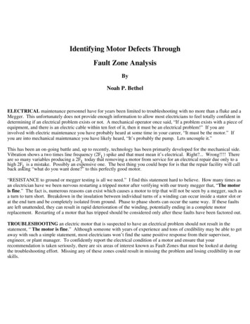
J Periodontol November 2012McGuire, Scheyer, Nunn[CEJ]) used at baseline were nolonger present, preventing thepossibility of quantitative 10year follow-up data. For eachof the four known patients inthis group, there was no evidence that dental restorativetherapy was required to compensate for residual or worsening gingival recession. Overall,loss to follow-up was a randomevent unrelated to treatment oroutcomes so that the only problem presented was a reductionin sample size.Summary of OriginalSurgerySurgical protocol for testtreatment with CAF EMD. After root preparation, a sulcularincision was made at the recession site and extended horizontally into the adjacent interdentalregions. Bilateral vertical releaFigure 1.sing incisions, connected to thePatient 20. A) At baseline, a maxillary cuspid randomized to receive test (EMD) treatment. B)horizontal incision, were extenIntraoperative measurements after full-thickness flap elevation beyond the mucogingival junction.ded out into the lining mucosaC) EMD applied to the root surface. D) Mucogingival flap coronally advanced to the level of the CEJ andsecured with sutures. E) A 12-month follow-up, test site with no evidence of GR. F) A 10-year follow-up,and a full-thickness mucopertest site with no GR and an increase of 4 mm of KT from both baseline and 12 months. (This patientiosteal flap elevated until thereceived virtually no maintenance care after the completion of the initial study.)mucogingival junction was passed (Figs. 1A and 1B). The periosteum was then cut, followedMATERIALS AND METHODSby blunt dissection into the vestibular lining mucosaStudy Populationto eliminate muscle tension. The facial aspects ofOf 17 patients completing the original study, nine werethe interdental papillae were de-epithelialized, creatingavailable for follow-up 10 years after the originala connective tissue bed for suturing the CAF.surgery. The 18 evaluated teeth were distributed amongThe exposed root surface was then conditionedincisors, cuspids, and bicuspids. The follow-up patientwith 24% EDTA‡ for 2 minutes, rinsed with saline,population, ranging from 44 to 74 years of age (meanand then dried. EMD§ was then applied onto the rootage: 55.4 years), included four males and five femalessurface and the mucoperiosteal flap coronally adand was 100% white. Seven never smoked, and twovanced to the level of the CEJ and sutured to thehad not smoked for 39 years. Of the nine patients, fivede-epithelialized papillae (Figs. 1C and 1D).had no reported medical problems, whereas four reSurgical protocol for control treatment withported generalized anxiety-related symptoms.CAF CTG. The control procedure was identical tothe test procedure, with the following exceptions:Patient Population Lost to Follow-Up1) a partial-thickness mucosal flap was developedEight of the original 17 patients were lost to follow-up.in lieu of a full-thickness mucoperiosteal flap; andTwo patients chose not to participate, and two could2) a subepithelial CTG from the premolar region ofnot be contacted. Of the remaining four patients, onethe palate was obtained and secured over the denudedfractured the test tooth and had it removed, two paroot surface in lieu of placement of EMD (Figs. 2A andtients had the test and control teeth prosthetically2B). As in the test group, the mucosal flap was pascrowned for reasons other than residual recession,sively coronally advanced to the level of the CEJ.and one patient had multiple teeth with deep abfracPostoperative oral hygiene instructions designed totive lesions restored, including the test and controlteeth. In each of the latter three patients, the mea‡ PreGel, Straumann, Andover, MA.surement reference points (cemento-enamel junction§ Emdogain, Straumann.1355
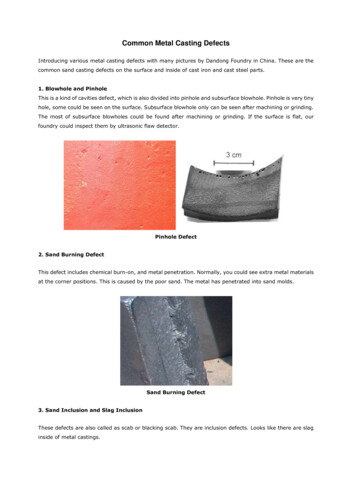
Results 10 Years After Root Coverage with CAF CTG or EMDVolume 83 Number 11Figure 2.Patient 20. A) At baseline, the contralateral cuspid randomized to receive control (CTG) treatment. B) Subepithelial CTG (control) is sutured over the denudedroot surface. C) A 12-month follow-up, control site with no evidence of recession and a 4-mm increase in KT. D) A 10-year follow-up, control site withno change in GR or KT. (This patient received virtually no maintenance care after the completion of the initial study.)minimize trauma at the gingival margins were given,and the patients were examined 1 week after surgeryand at months 1, 2, 3, 6, 9, and 12.Clinical Evaluation 10 Years After OriginalSurgeryAs performed for the original RCT 10 years earlier, thetreated sites were clinically examined, the measurementswere recorded, and clinical photographs were taken.The same examiner (Carol Waring, RDH, PerioHealthProfessionals, Houston, Texas) who recorded theoriginal study measurementswas still maskedandperformed the follow-up 10-year examinations afterbeing recalibrated for measurement accuracy andconsistency. The primary efficacy parameter wasthe change in the depth of the recession defect. Secondary efficacy parameters included the following: 1)PD; 2) CAL; 3) keratinized tissue width (wKT); 4) percentage of root coverage; 5) root dentin hypersensitivity; 6) clinician rating of color (compared toadjacent tissue), texture (compared to adjacent tissue), and contour (compared to adjacent tissue) oftreatment sites; and 7) patient satisfaction at 10 years.At baseline, there were no significant differencesobserved between test and control sites. Recessiondepth, using a periodontal probe,i was measured fromthe CEJ to the free gingival margin. A periodontalprobe¶ measured mid-buccal PD from the gingivalmargin to the base of the defect. CAL was measuredmid-buccal from the CEJ to the base of the defect.wKT measurements, measured with a periodontalprobe,# extended from the free gingival margin to themucogingival junction.Qualitative efficacy parameters were assessed asfollows. Root dentin hypersensitivity was assessedusing a conventional blast of air for 3 seconds at the1356exposed root surface. The hypersensitivity was recorded as present (yes) or not present (no). Gingivalcolor, texture, and contour were assessed by comparing test and control grafts to surrounding tissues andscoring through questionnaires, i.e. more red, less red,equally red; more firm, less firm, or equally firm; ormore contour, less contour, or equal contour.Patient satisfaction at 10 years was assessed by responses to the following two questions: 1) Which procedure do you prefer to correct your gum recession,the procedure with the EMD or the CTG and why?and 2) Are you equally satisfied with the esthetic results of the two sites treated or are you more satisfiedwith one treated site versus the other?Statistical MethodsQualitative measures, including root hypersensitivity,soft-tissue color (compared to adjacent tissue), softtissue texture (compared to adjacent tissue), and softtissue contour (compared to adjacent tissue), weredichotomized for statistical analysis. Specifically, roothypersensitivity was dichotomized into ‘‘yes’’ or‘‘no,’’ indicating that either there was root hypersensitivity present or there was no root hypersensitivitypresent. Soft-tissue measures were dichotomized intoeither ‘‘equal’’ (i.e., equivalent to adjacent tissue) or‘‘not equal’’ (i.e., not equivalent to adjacent tissue).McNemar test for paired dichotomous outcomes wasused to test for differences in qualitative outcomesbetween control and test sites.Within-treatment comparisons across time and between-treatment comparisons at each point in timewere made using non-parametric tests. Likewise, alli UNC-15 periodontal probe, Hu-Friedy, Chicago, IL.¶ Florida periodontal probe, Florida Probe Corp., Gainesville, FL.# UNC-15 periodontal probe, Hu-Friedy.
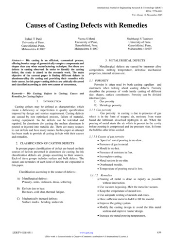
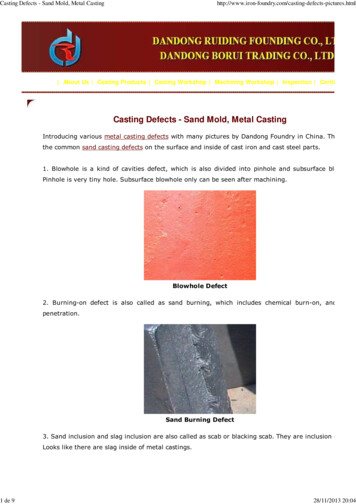
J Periodontol November 2012McGuire, Scheyer, Nunnstudy by McGuire and Nunnhad incorrect tooth designation.Instead of maxillary cuspids,bicuspid sites were originallygrafted. This error has beencorrected here.) Moderate rootdentin hypersensitivity continued to exist in a minority ofthe test (n 3) and control(n 1) sites, although themajority of sites exhibited nosensitivity to a conventional3-second blast of air to the rootFigure 3.surface. Although control sitesPatient 13. A) At baseline, a bicuspid site randomized to receive control (CTG) treatment. B) A 12-monthtended to be less likely to exfollow-up after CTG. There has been a 1-mm increase in KT from baseline. Mild marginal inflammationhibit root hypersensitivity compresent. C) The results at 10 years after CTG remain the same as at 12 months.pared to test sites (one of ninecontrol sites with root hypersensitivity versus three of ninetest sites with root hypersensitivity), the difference betweentest and control sites failed toachieve statistical significance(P 0.157). Test sites were morelikely to exhibit equivalent texture to surrounding tissues compared to control sites (eight ofnine test sites with equivalenttexture to surrounding tissuesversus five of nine control siteswith equivalent texture to surrounding tissues), although theFigure 4.difference falls short of achievPatient 13. A) At baseline, the contralateral bicuspid randomized to receive test (EMD) treatment. B)ing statistical significance (P A 12-month follow-up with no recession and 2 mm of wKT. C) At 10 years after EMD treatment, there isno GR evident and no increase in KT from baseline and 12 months.0.083). When examining colorequivalence of the surgical sitesto adjacent tissues, eight of ninechange comparisons (baseline versus 1 year andtest sites and six of nine control sites were judgedbaseline versus 10 years) both within and betweento have equivalent color to surrounding tissues, altreatments were made using non-parametric tests.though this difference between test and control sitesIn particular, for continuous outcomes (recessionfailed to achieve statistical significance (P 0.157).depth, PD, CAL, wKT, and percentage root coverage),For equivalence of tissue contour, test sites werea (paired) Wilcoxon signed-rank test was used. Forfound to be significantly superior, with eight of eightthe binary outcome of patients with 100% root covertest sites having equivalent tissue contour to adjaage versus those without 100% root coverage, Fisher’scent tissue, whereas only one of eight control sitesexact test comparing two binomial proportions washad equivalent tissue contour to adjacent tissueused.(P 0.008).RESULTSTen-Year Assessment of Qualitative ParametersAt 10 years after the original surgeries, clinical photoswere taken and a number of qualitative parameterswere examined (Figs. 1F, 2D, 3C, and 4C). (To avoidselection bias, only those grafted site images includedin the McGuire and Nunn24 publication are includedhere. Note that patient 13, Figures 9 and 10, of theTen-Year Assessment of Quantitative ParametersMean differences in quantitative clinical measuresbetween control and test sites were not significantlydifferent from zero at baseline. Similarly, mean differences in quantitative clinical measures between control and test sites were not significantly different fromze
dence that dental restorative therapy was required to com-pensate for residual or worsen-ing gingival recession. Overall, loss to follow-up was a random event unrelated to treatment or outcomessothattheonlyprob-lem presented was a reduction in sample size. Summary of Original Surgery Surgical protocol for test treatmentwithCAF EMD.Af-