Transcription
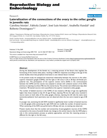
Reproductive Biology andEndocrinologyBioMed CentralOpen AccessResearchLateralization of the connections of the ovary to the celiac gangliain juvenile ratsCarolina Morán1, Fabiola Zarate1, José Luis Morán1, Anabella Handal1 andRoberto Domínguez*2Address: 1Department of Biology and Toxicology of Reproduction; Science Institute BUAP, Mexico and 2Biology of Reproduction Research Unit;FES Zaragoza UNAM, Av. 14 sur 6301, San Manuel, Puebla, Pue. CP 72570, MexicoEmail: Carolina Morán - moranraya@yahoo.com.mx; Fabiola Zarate - biosfabiola@hotmail.com;José Luis Morán - luis.moran@icbuap.buap.mx; Anabella Handal - ahandals@yahoo.com.mx; Roberto Domínguez* - rdcasala@hotmail.com* Corresponding authorPublished: 21 May 2009Reproductive Biology and Endocrinology 2009, 7:50doi:10.1186/1477-7827-7-50Received: 15 August 2008Accepted: 21 May 2009This article is available from: http://www.rbej.com/content/7/1/50 2009 Morán et al; licensee BioMed Central Ltd.This is an Open Access article distributed under the terms of the Creative Commons Attribution License (http://creativecommons.org/licenses/by/2.0),which permits unrestricted use, distribution, and reproduction in any medium, provided the original work is properly cited.AbstractDuring the development of the female rat, a maturing process of the factors that regulate thefunctioning of the ovaries takes place, resulting in different responses according to the age of theanimal. Studies show that peripheral innervation is one relevant factor involved.In the present study we analyzed the anatomical relationship between the neurons in the celiacsuperior mesenteric ganglia (CSMG), and the right or left ovary in 24 or 28 days old female prepubertal rats. The participation of the superior ovarian nerve (SON) in the communicationbetween the CSMG and the ovaries was analyzed in animals with unilateral section of the SON,previous to injecting true blue (TB) into the ovarian bursa. The animals were killed seven days aftertreatment. TB stained neurons were quantified at the superior mesenteric-celiac ganglia.The number of labeled neurons in the CSMG of rats treated at 28 days of age was significantlyhigher than those treated on day 24. At age 24 days, injecting TB into the right ovary resulted inneuron stains on both sides of the celiac ganglia; whereas, injecting the left side the stains wereexclusively ipsilateral. Such asymmetry was not observed when the rats were treated at age of 28days.In younger rats, sectioning the left SON resulted in significantly lower number of stained neuronsin the left ganglia while sectioning the right SON did not modify the number of stained neurons.When sectioning of the SON was performed to 28 days old rats, no staining was observed.Present results show that the number and connectivity of post-ganglionic neurons of the CSMGconnected to the ovary of juvenile female rats change as the animal mature; that the SON plays arole in this communication process as puberty approaches; and that this maturing process isdifferent for the right or the left ovary.Page 1 of 7(page number not for citation purposes)
Reproductive Biology and Endocrinology 2009, 7:50BackgroundThe innervation of the ovary involves sympathetic, parasympathetic and sensorial components of the autonomicnervous system and reaches the ovary through the ovarianplexus, the superior ovarian nerve (SON), and the vagusnerve [1-3]. The terminals of such nerves release a series ofneurotransmitters to the interior of the gland; some ofwhich have been considered regulators of steroidogenesis,early follicular development and ovulation [4-7].The postganglionic perikarya of the ovarian fibers travelalong the SON and the ovarian plexus nerve and comefrom the pre-vertebral ganglia of the celiac-superiormesenteric ganglia (CSMG) and other small ganglialocated close to the origin of the ovarian and renal artery.The right and left parts of the CSMG are asymmetrical [1].The vagus nerve has an important amount of synapses tothe main CSMG [2].Large neurons, small granular cells, described as smallintensively fluorescent cells, (SIF); glial cells (Schwannand satellite cells); capilaries; mast cells; and fibroblastscan be observed in the CSMG. Fluorescence histochemicalmethods have localized catecholamines in sympatheticganglia in ganglion cells bodies and SIF cells [8]. Severalneuropeptides, including substance P (SP), vasointestinalpeptide (VIP), gastrin and enkephalin [9] are localized innerve fibers. This group of cells forms a barrier that surrounds the main neurons axonal cone [10,11].In mammals, the CSMG is the main nervous informationrelay site between the gonads and the central nervous system; conversely, the link between the gonads and the central nervous system present modifications during thedevelopment of the ovaries [12,13]. The presence of neuropeptides, the topographic organization and maturingpatterns of the neurons at the celiac ganglia of guinea pigsare set up at the end of the fetal period. After this period,neurons acquire their electro-physiological and morphological features [14].http://www.rbej.com/content/7/1/50similar to that of the adult rat. Immunoreactive fiberswere conspicuous in the intermediolateral and both thoracic and sacral levels. After day 14th of age, a further overall increase in the density of innervations occurs [21]. Inrats the ovarian innervation appears before birth and precedes the postnatal initiation of folliculogenesis, suggesting that the initial formation of follicles is, at least in part,facilitated by signals of neural origin [22]. The ovary of therhesus monkey undergoes changes in nerve fibers densityreaching the ovaries, increasing gradually since the postnatal stage, while during the pre-pubertal stage the densityof nerve fibers increases at twice the rate [23]. D'Albora etal. [24] found changes in number, morphology and distribution of ovarian neurons in the Wistar rat, depending onthe age of the animal.Flores et al. [25] showed that in denervated pre-pubertalrats treated with guanethidine, the number of ova releasedduring the first estrus was higher than in control rats.Guanethidine treatment to adult rats resulted in a significant reduction in the number of ova shed, suggesting thatthe ovarian noradrenergic innervation regulating ovulation plays different roles in pre-pubertal and adult rats.Chavez et al. [26,27] showed that ovulation processes areimpacted when the SON is sectioned suggesting that theparticipation of the innervation on ovarian regulation varies during the estrous cycle and is asymmetric.Previously we have shown that sensorial denervationthrough capsaicin treatment at birth, previous to folliculogenesis, results in a reduction of steroidogenesis and anincrease in the number of 100–350 μm follicles. Most ofthese follicles were normal when the animals were studiedon day 24 of age, and atretic when animals reached 28days of age [28].The changes of the autonomic ganglia require neuralinteractions with its target organ. The nerve growth factor(NGF) and steroid hormones such as 17-β estradiol[15,16] control this regulation. Estradiol plays a criticalrole in regulating neuron survival and the way in whichdifferent neuron populations respond during the prenatal and perinatal development [17,18]. According toPatrone et al. [19], during the early post-natal development estrogens increase the survival of dorsal root ganglianeurons.In the spinal cord of the rat the mediolateral dendrites ofpreganglionic sympathetic neurons are detectable as earlyas the 15th embryonic day [20]; and in the post- natal inthe14th day, the pattern of noradrenergic innervation wasFigureTheleft 1and right CSMGThe left and right CSMG. 4 .Page 2 of 7(page number not for citation purposes)
Reproductive Biology and Endocrinology 2009, 7:50http://www.rbej.com/content/7/1/50Similar differences on the effects of denervation between24 and 28 days old rats, were observed in rats with unilateral or bilateral sectioning of the SON or vagus nerve [2931], suggesting that innervation plays different roles onthe regulation of ovarian development. It has been suggested that at age 28 days the system would be starting toprepare to reach the sexual mature stage [28].To our knowledge, no quantitative morphological studyof ovarian innervation has been conducted throughoutthe juvenile period of the rat, and thus, it is not clearwhether the observed changes reflect metabolic or structural alterations. The aim of the present study was todetermine whether structural remodeling of ovarianinnervation in CSMG occurs during the pre-pubertalperiod and if the neural information amount between theovaries and the CSMG is different between the left andright sides.MethodsThe injection of TB was performed following previouslydescribed procedures [32]. In brief, the animals were etheranesthetized between 10:00 am and 12:00 PM, and a unilateral incision was performed 1 cm below the last rib,affecting skin, muscle, and peritoneum. The left or rightovary was exposed and 3–5 μL of TB (Sigma, St. Louis,Missouri, USA) solution at 4%, diluted in distilled water,was injected into the ovarian bursa. To prevent the leakageof the tracer, the needle was kept in the bursa for 5 minafter injection treatment. Subsequently, the ovary wascarefully cleaned, dried, and returned to the abdominalcavity. The possibility that TB leaked into the abdominalcavity was assessed by exposing the cavity to a fluorescentlight, and the animals with TB leakage were excluded fromthe experiment.In other groups, the animals were treated as previously,but before the TB treatment into the bursa, the ipsilateralSON to the ovary to be treated was sectioned, followingpreviously described methodologies [27].Seven days after surgery the rats were anesthetized withsodium pentobarbital (40 mg/Kg) IP and intracardiac perfused with 250 mL of cold saline solution (0.9%), followed by the injection of 150 mL solution of 4%70706060true blue positive neuronstrue blue positive neuronsThe study was conducted with virgin adult female rats ofthe CIIZ-V strain, reproduced and maintained in the"Claude Bernard" Animal House in the UniversidadAutónoma de Puebla. Animals were kept under controlledlighting conditions (lights on from 07:00 to 19:00 h),with free access to the mother until weaning (day 21) andto food (Purina S.A., Mexico) and tap water thereafter. Theexperiments were carried out in strict accordance with theMexican Guide for Care and Use of Laboratory Animals atthe National Academy of Science Animals and Treatment.Protocols were approved by the FES Zaragoza, UNAM. Atbirth (day 1) the animals were randomly allotted ingroups of 5 females and 1 male to one of the experimentalgroups described below.5040302010504030201000 NumbertheFigureleft or2ofrightpositiveovarianneuronsbursatoattrue24 orblue28 d seven daysganglialater of the animals injected with the true blue inNumber of positive neurons to true blue in the celiac superior-mesenteric ganglia of the animals injected withthe true blue in the left or right ovarian bursa at 24 or 28 days old, and sacrificed seven days later. * p 0.05 vs.contralateral CSMG.Page 3 of 7(page number not for citation purposes)
Reproductive Biology and Endocrinology 2009, uedaysrightoldovaryBlue3ratslabeled(c,injectedd) neuronswith TB(whitein thearrows)left (a,inb)theor CSMGinto the24True Blue labeled neurons (white arrows) in theCSMG 24 days old rats injected with TB in the left (a,b) or into the right ovary (c, d). Left CSMG a and c; RightCSMG b and d. Scale bar 100 injectedc) neuronswith TB(whitein thearrows)left (a,inb)theor CSMGinto the28True Blue labeled neurons (white arrows) in theCSMG 28 days old rats injected with TB in the left (a,b) or into the right ovary (b, c). Left CSMG a and c; RightCSMG b and d.paraformaldehyde. After perfusion of the fixative solution, the pre-vertebral CSMG, ovaries, and uterus were dissected and kept in the fixative solution overnight (approx18 h). The ganglia were cryoprotected successively in 10,20, and 30% sucrose in phosphate buffer and were seriallysectioned at twenty micrometer with the aid of a cryostatkept at -20 C. The sections were analyzed following thesame procedure previously described [32]. In brief, eightto ten images of the right or left CSMG, from the animalsinjected with TB, were used to count the number of positively labeled cells. Positive TB cells are defined as cells inwhich fluorescence is present when the sections wereexposed to UV light. Only principal neurons were labeledwith TB and SIF neurons are not labeled with the tracer.ResultsIn figure 1 the left CSMG and right CSMG are showing.Number of positive neurons to TB in the CSMG of the animals injected with the tracer in the left or right ovarianbursa (Figure 2).24 days old. The injection of TB into the right ovarianbursa resulted in stained neurons in both, left and rightThe pictures were obtained with a Digital Camera(Optronics 60300, USA) and analyzed with a KS-300Imaging System 3.0 (Carl Zeiss Vision GmbH, Germany).The imaging system was programmed to generate binaryregions, automatically count and integrate them.Statistical AnalysisThe mean number of True Blue positive cells was analyzedby a multivariate analysis of variance (MANOVA), followed by Tukey's test. A p 0.05 was assumed as significant.FigureDiferencesof24 and5 28indaysthe intensitityold rats of TB positive cells in the CSMGDiferences in the intensitity of TB positive cells in theCSMG of 24 and 28 days old rats.Page 4 of 7(page number not for citation purposes)
Reproductive Biology and Endocrinology 2009, 7:50http://www.rbej.com/content/7/1/5024 days old. When the left SON was sectioned, thenumber of TB stained neurons in the left CSMG diminishes significantly. This effect was not observed in theright CSMG when the right nerve was sectioned (Table 1,and Figure 6).28 days old. When the SON was sectioned at this age, nostained neurons were observed in the ipsilateral CSMG,independently of the side in which the section-TB injection was applied (Table ry6intoratslabeledwiththe(a) ) leftofarrows)theSONrightinjectedin theSONCSMGwithinjectedTBof 24inTrue Bleu labeled neurons (white arrows) in theCSMG of 24 days old rats with section of the leftSON injected with TB in the left ovary (a) or withsection of the right SON injected with TB into theright ovary (b).CSMG. When the tracer was injected into the left ovarianbursa, stained neurons were observed only in the leftCSMG (Figure 3), and the number of stained neurons inthe left CSMG was significantly higher than when thetracer was injected into the right ovarian bursa.28 days old. The number of TB stained neurons was significantly higher than in rats injected at 24 days of age. Significant differences in the number of TB positive cells inthe left and right CSMG were not observed. Unlike whatwas observed in animals treated at 24 days, injecting TBinto the right or left ovary to 28 days old rats resultedexclusively in stains neurons of the ipsilateral CSMG thanthe treated ovary (Figure 4).In figure 5 we show the differences in the fluorescenceintensity in the TB positive cells in the CSMG of 24 and 28days old rats.Number of TB positive neurons in the CSMG of animalswith unilateral sectioning of the SON, injected with TB inthe left or right ovarian bursa.The results obtained in the present study show that in rats,during the juvenile and pre-pubertal period arrangementsin the post-ganglionic neurons reaching the ovaries thatarrive from the pre-vertebral ganglia, take place. Theresults also suggest that the SON plays an important rolein the communication process between the CSMG and theovaries as puberty approaches.Nerve terminals are present in the rat's ovaries since veryearly stages of embryonic development, increasing innumber and density until puberty onset; and many ofthem are catecholaminergic [13,22,33-35].Present results support the idea of the presence of two celialic ganglions (left and right) and that the left celialic ganglion is larger than the right one [1]; and differ fromBurden and coworkers [3,33] who described the presenceof a single celialic ganglion. The difference if the numberof celialic ganglions reported by Burden et al and thoseobserved in this investigation may be explained by the different rat strains used.In this study, differences in the number of TB stainedCSMG neurons, between rats treated at the age 24 and 28days, suggest that at these two development stages there aredifferences in the level of information transmitted betweenthe pre-vertebral ganglia and the ovaries. This differencemay be related to the maturation processes because thenumber of innervations observed in the CSMG in 28 daysold animals is similar to those observed in adult cyclic rats.In a previous study rats injected with TB directly into theovaries, where animals treated throughout the estrous cycleshowed an increase in the number of stained neurons fromTable 1: Number of positive neurons in CSMG of the rats injected with TB in the 6 left or right ovarian bursa posterior to the sectionof the ipsilateral SON.GroupOvarian bursa injectedControlAfter SON section24 days old24 days old28 days old28 days oldLeftRightLeftRight19.8 3.65.2 1.3*55.8 1.564.6 15.77.7 2.4 5.6 1.100* p 0.05 vs. rats injected with TB in the left ovarian bursa p 0.05 vs. control rats injected with TB in the left ovarian bursaPage 5 of 7(page number not for citation purposes)
Reproductive Biology and Endocrinology 2009, 7:50diestrus 1 to proestrus, with a small drop in animals treatedon estrus. These results were interpreted as suggesting thatthe number of active neurons in the CSMG varies duringthe estrous cycle. Such changes could be related to oscillating estradiol plasma levels and to changes in the neurons'ability to be stimulated by estrogens throughout the estrouscycle [32].There is evidence of functional asymmetry between pairedorgans [36-38]. In cyclic rats the left ovary releases moreova than the right one [39,40]. Morphological asymmetryin the intensity of supraespinal innervation of the left andright ovary has been showed by Tóth et al. [41]. The neuralconnections between the left ovary and brain structures,such as the nucleus of the solitary tract, the dorsal nucleusof the vagus, the A5 noradrenergic cell group, the caudalraphe nuclei, the hypothalamic paraventricular nucleus,and the lateral hypothalamus, are more abundant thanthe number of neural connections between these cellgroups and the right ovary [41].Previously we have shown that the unilaterally sectioningthe SON of pre-pubertal rats results in puberty onset delayand lower number of oocytes released by the denervatedgonad [42,28]. The sequential injection of gonadotropinsto these animals did not reestablish ovulation in the denervated ovary [30]. In the ovaries, asymmetric functionsdid not seem to depend on the gonadotropic stimuli, buton the extrinsic innervations received by each ovary [39].In the CSMG of rats injected with TB into the right ovarianbursa at 24 days of age, the number of stained neurons inthe left and right ganglia was lower than in animalsinjected with the tracer in the left ovary; a difference notobserved in 28 days old animals. In the adult rat the highest number of stained cells was observed when the tracerwas injected into the left ovary on proestrus. The numberof stained cells was significantly different between theright and left CSMG of animals injected on proestrus, andbilateral staining in the CSMG was more pronouncedwhen the tracer was injected into the left ovary. Such communication could partially explain the response differences to peripheral denervation observed in the right andleft ovaries [30].In 24 days old animal TB treatment in the right ovarian bursaresulted in a bilateral staining of neurons in the CSMG, whiletreatment in the left ovary resulted in staining of neurons onthe ipsilateral side only. Such behavior supports the idea of aneural communication between the ovaries, partiallyexplaining the difference in the response of the left and rightovaries to unilateral denervations [42,28,11].The CSMG is the origin of the two catecholaminergicpathways innervating the ovary, the SON and the ovarianhttp://www.rbej.com/content/7/1/50plexus nerve. The fact that in the 28 days old animal sectioning the SON eliminated the presence of TB stainedneurons in the CSMG, we presume that the neuron subpopulation in the ganglia projecting its axons to thegonad through the SON has been developed.Given that in 24 days old animals sectioning of the leftSON results in a decrease of TB stained neurons in theCSMG, though not completely eliminating the presenceof stained cells, it is proposed that during the first part ofthe juvenile period the neural connection between the leftCSMG and the left ovary is carried through the nerve ofthe ovarian plexus and the SON. The neural connectionbetween the right CSMG and the right ovary is carriedonly by the nerve of the ovarian plexus, because the section of the right SON did not modify the number oflabeled cells.Because the amount of tracer (TB and horseradish peroxidase) detectable in the cell body is correlated with functional activity of the neuron [43], and there is evidencethat the capacity of the ovary to release noradrenalineincreases when the rat reaches puberty [13], we presumethat the activity of the CSMG cells related with the ovariesincreases its activity when the animal reaches the puberty.Present results support the hypothesis that the participation of the peripheral nerves on ovarian performance varies according with the age, and is asymmetric[28,42,44,45].Taken together, present and previous results, suggest thatin the rat: 1) The variations in response of both ovaries along their development- are due to the rearrangement ofthe neurons and the fibers that convey information fromthe prevertebral ganglia to the gonads. 2) That the participation of the SON becomes more important for suchcommunication as puberty approaches.Competing interestsThe authors declare that they have no competing interests.Authors' contributionsCM and RD devised the study, participated in the discussion of results and in the planning of experiments; CM,and FZ, conducted the descriptive and quantitative histological studies; JLM and AH participated in the discussionof results. All authors read and approved the final manuscript.AcknowledgementsWe thank MSc Alvaro Domínguez-González for assistance in English revision.This research was supported by PROMEP/103.5/07/2208 folio BUAP-EXB634 and DGAPA-UNAM Grants IN 200405 and IN209508.Page 6 of 7(page number not for citation purposes)
Reproductive Biology and Endocrinology 2009, 16.17.18.19.20.21.22.23.Baljet B, Drukker J: The extrinsic innervation of the abdominalorgans in the female rat. Acta Anat 1979, 104:243-267.Berthoud HR, Powley TL: Interaction between parasympathetic and sympathetic nerves in prevertebral ganglia: morphological evidence for vagal efferent innervation of ganglioncells in the rat. Micros Res Tech 1996, 35:80-86.Klein CM, Burden HW: Anatomical localization of afferent andpostganglionic sympathetic neurons innervating the ratovary. Neurosc lett 1988, 85:217-222.Domínguez R, Cruz ME, Chávez R: Differences in the ovulatoryability between the right and left ovary are related to ovarian innervation. In Growth Factors and the Ovary Edited by: HirshfieldAM. New York: Plenum Press; 1989:321-325.Domínguez R, Riboni L: Failure of ovulation in autograftedovary of hemispayed rat. Neuroendocrinology 1971, 7:164-170.Dissen GA, Lara HE, Fahrenbach WH, Costa ME, Ojeda SR: Immature rat ovaries become revascularized rapidly afterautotransplantation and show a gonadotropin-dependentincrease in angiogenic factor gene expression. Endocrinology1994, 134:1146-54.Mayerhofer A, Dissen GA, Costa ME, Ojeda SR: A role for neurotransmitters in early follicular development: induction offunctional follicle-stimulating hormone receptors in newlyformed follicles of the rat ovary.Endocrinology 1997,138:3320-3329.Dail WG, Barton S: Structure and organization of mammaliansympathetic ganglia. In Autonomic Ganglia Edited by: Elfvin LG.New York: John Wiley & Sons Ltd; 1983:13-25.Gabella G: Autonomic Nervous system. In The rat nervous systemEdited by: Paxinos G. New York: Academic Press; 1995:81-102.Cardinali DP, Gejman PV, Ritta MN: Further evidence of adrenergic control of translocation and intracellular levels ofestrogen receptors in rat pineal gland. Endocrinology 1983,112:492-498.Aguado LI: Role of the central and peripheral nervous systemin the ovarian function. Micros Res Tech 2002, 59:462-473.Dees WL, Hiney JK, McArthur NH, Johnson GA, Dissen GA, OjedaSR: Origin and ontogeny of mammalian ovarian neurons.Endocrinology 2006, 147:3789-96.Ricu M, Paredes A, Greiner M, Ojeda SR, Lara HE: Functionaldevelopment of the ovarian noradrenergic innervation.Endocrinology 2008, 149:50-56.Anderson RL, Morris JL, Gibbins IL: Neurochemical differentiation of functionally distinct populations of autonomic neurons. J Comp Neurol 2001, 429:419-435.Korsching S, Thoenen H: Developmental changes of nervegrowth factor levels in sympathetic ganglia and their targetorgans. Dev Biol 1988, 126:40-6.Thoenen H, Barde YA: Physiology of nerve growth factor. Physiol Rev 1980, 60:1284-335.Levi-Montalcini R: The nerve growth factor 35 years later. Science 1987, 237:1154-62.Chávez-Genaro R, Crutcher K, Viettro L, Richeri A, Coirolo N, Burnstock G, Cowen T, Brauer MM: Differential effects of oestrogenon developing and mature uterine sympathetic nerves. CellTissue Res 2002, 308:61-73.Patrone C, Andersson S, Korhonen L, Lindholm D: Estrogen receptor-dependent regulation of sensory neuron survival indeveloping dorsal root ganglion. Proc Natl Acad Sci USA 1999,96:10905-10.Markham JA, Vaughn JE: Migration patterns of sympathetic preganglionic neurons in embryonic rat spinal cord. J Neurobiol1991, 22:811-22.Rajaofetra N, Poulat P, Marlier L, Geffard M, Privat A: Pre- and postnatal development of noradrenergic projections to the ratspinal cord: an immunocytochemical study. Brain Res Dev BrainRes 1992, 67:237-46.Malamed S, Gibney JA, Ojeda SR: Ovarian innervation developsbefore initiation of folliculogenesis in the rat. Cell Tissue Res1992, 270:87-93.Schultea TD, Dees WL, Ojeda SR: Postnatal development ofsympathetic and sensory innervation of the rhesus monkeyovary. Biol Reprod 1992, 43.44.45.D'Albora H, Anesetti G, Lombide P, Dees WL, Ojeda SR: Intrinsicneurons in the mammalian ovary. Microsc Res Tech 2002,59:484-9.Flores A, Ayala ME, Domínguez R: Does noradrenergic peripheral innervation have a different role in the regulation ovulation in the pubertal and the adult rat? Med Sci Res 1990,18:817-818.Chávez R, Sánchez S, Ulloa-Aguirre A, Domínguez R: Effects onoestrous cyclicity and ovulation of unilateral section of thevagus nerve performed on different days of the oestrouscycle in the rat. J Endocrinol 1989, 123:441-4.Chavez R, Domínguez R: Participation of the superior ovariannerve in the regulation of compensatory ovarian hypertrophy: the effects of its section performed on each day of theoestrous cycle. J Endocrinol 1994, 140:197-201.Morán C, Morales L, Chavira R, Domínguez R: Efectos de la denervación sensorial inducida por la administración de capsaicinaal nacimiento sobre la esteroidogénesis y el desarrollo folicular durante la etapa juvenil de la rata. XXVI Reunión Anualde la Academia de Investigación en Biología de la Reproducción, A.C. Acapulco Gro México 2001.Morán C, Morales L, Quiróz U, Domínguez R: Effects of unilateralor bilateral superior ovarian nerve section in infantile rats onfollicular growth. J Endocrinol 2000, 166:205-11.Morales L, Chávez R, Ayala ME, Domínguez R: Effects of unilateralor bilateral superior ovarian nerve section in prepubertalrats on the ovulatory response to gonadotrophin administration. J Endocrinol 1998, 158:213-9.Morales-Ledesma L, Betanzos-García R, Domínguez-Casalá R: Unilateral or bilateral vagotomy performed on prepubertal ratsat puberty onset of female rat deregulates ovarian function.Arch Med Res 2004, 35:279-83.Morán C, Franco A, Morán JL, Handal A, Morales L, Domínguez R:Neural activity between ovaries and the prevertebral celiacsuperior mesenteric ganglia varies during the estrous cycleof the rat. Endocrine 2005, 26:147-52.Lawrence IE, Burden HW: The origin of the extrinsic adrenergicinnervation to the rat ovary. Anat Rec 1980, 196:51-59.Bahr JM, Ben-Jonathan N: Preovulatory depletion of ovarian catecholamines in the rat. Endocrinology 1981, 108:1815-20.Dees WL, Hiney JK, Schultea TD, Mayerhofer A, Danilchik M, DissenGA, Ojeda SR: The primate ovary contains a population of catecholaminergic neuron-like cells expressing nerve growthfactor receptors. Endocrinology 1995, 136:5760-8.Sherman GF, Galaburda AM: Neocortical asymmetry and openfield behavior in the rat. Exp Neurol 1984, 86:473-82.Gerendai I, Kocsis K, Halász B: Supraspinal connections of theovary: structural and functional aspects. Microsc Res Tech 2002,59:474-83.Gerendai I, Banczerowski P, Halász B: Functional significance ofthe innervation of the gonads. Endocrine 2005, 28:309-18.Dominguez R, Morales L, Cruz ME: Ovarian Asymmetry. Ann RevBiol Sciences 2003, 5:95-104.Dominguez R, Cruz ME, Morán C: Differential effects of ovarianlocal anaesthesia during pro-oestrus on ovulation by theright or left ovary in normal and hemi-ovar
FES Zaragoza UNAM, Av. 14 sur 6301, San Manuel, Puebla, Pue. CP 72570, Mexico Email: Carolina Morán - moranraya@yahoo.com.mx; Fabiola Zarate - biosfabiola@hotmail.com; José Luis Morán - luis.moran@icbuap.buap.mx; Anabella Handal - ahandals@yahoo.com.mx; Roberto Domínguez* - rdcasala@hotmail.com * Corresponding author Abstract