Transcription
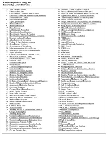
Campbell Reproductive Biology SiteEndocrinology Lecture .50.51.52.53.54.55.56.57.58.59.What is EndocrinologyEndocrine FunctionsComponents of Communication SystemsEndocrine Analogs of Communication ComponentsKnown Hormonal ClassesDefinition of a HormoneSignal System TypesHormonal SourcesHormonal Sources IIBrain AnatomyLimbic System AssociationHypothalamic Nuclei FunctionHypothalamic Anatomy & FunctionHypothalamus & Posterior Pituitary AssociationsNeurophypophysis CirculationAdenohypophysis CirculationPituitary & Hypothalamic AnatomyGross Pituitary HistologyGross Anatomy of the AdrenalMicroanatomy of the Adrenal CortexInformation Content and Signal FluctuationSignal PulsatilityControls on Bioavailable Hormone LevelsHierarchical Systems of ControlPostive and Negative Control LoopsReceptor TypesProperties of ReceptorsReceptor NotesTransduction System PropertiesTransduction NotesNotes on Transduction SystemsHormone and Receptor EvolutionHormone-Receptor PromiscuityInsulin Family StructuresInsulin Molecular StructureEvolution of Insulin Family Hormones and ReceptorsRelative Affinities in the Insulin H-R GroupAssessment of Endocrine FunctionSerpentine ReceptorsCytokine/Growth Factor ReceptorsAcetyl Choline ReceptorIntracellular ReceptorsNuclear Receptor Response ElementsBinding of Nuclear Receptors to DNABioassay Dose-ResponseBiphasic Dose Response of GHBioassay NotesChemical Assay NotesAssay ParametersAntibody Binding to Epitopes from DavidsonCollege, MA CampbellAntibody Assay NotesCompetititve Immunoassay CharacteristicsCompetitive Immunoassay Error DistributionsCompetititve Immunoassay Estimation ErrorsImmunoassay Precision ProfileCompetitive Immunoassay Precision ProfileCompetitive Immunoassay, ParallelismNoncompetitive Immunoassay CharacteristicsCell and Receptor Sizes60. Adjusting Cellular Response Sensitivity61. Receptor Binding and Numbers of Receptors62. Impact of Losing Receptors on Biological Response63. Hypothalamic Sources of Releasing Hormones64. Adenohypophysial Hormones and Regulators65. Protein Hormone Production66. Images for Review of Cell Physiology and Biochemistry67. Endoplasmic Reticulum Role in Protein Synthesis68. Golgi Actions in Protein Synthesis69. LH, FSH, TSH, hCG Introduction70. More on Glycoprotein Hormone Comparisons71. Yet More on Glycoproteins72. LH Bioassay Setup73. Proopiomelanocortin Metabolism74. TSH Control75. ACTH Control76. Adrenal Function77. Adrenal Function & Regulation78. MSH Control79. FSH Control80. LH Control81. GH Control82. PRL Control83. GH and PRL Gene Properties84. Pituitary Testituclar Axis85. Spelling Is Important!86. Large G Proteins and Protein Kinase A Cascade87. A cAMP Cartoon88. Guanylyl Cyclase Activation89. Large G Proteins and Protein Kinase C Cascade90. Glyceride Chemistry91. Phosphoinositide Metabolism92. Small G Proteins and Tyrosine Kinase Cascades93. Growth Factor/ Tyrosine Kinase Pathway (Examples)94. Transduction Mechanism Networks95. Insulin and Related Receptor Mechanisms96. Oncogenesis Notes97. Cell Cycle Control Points98. Restriction Point Switch99. Cancer Genes100. DNA Replication101. DNA Replication Fork102. Lipoprotein Metabolism103. Receptor Mediated Endocytosis104. Steroid Structure105. Steroid Synthesis106. Steroid Hormones of the Reproductive System107. C21 Metabolic Pathways108. C19 & C18 Metabolic Pathways109. Cellular Steroidogenesis110. STAR Protein111. Enterohepatic Circulation112. Introduction for Reproduction113. Images from Veterinary Reproductive Endocrinology114. Cell Division Notes115. Meiosis116. Prophase Meiosis I117. Meiosis I and II beyond Prophase I118. Gametogenesis Outline119. Male Reproductive Anatomy
2120. Testis Anatomy121. Seminiferous Tubule Gross Histology122. Seminiferous Tubule Microanatomy123. Seminiferous Tubule Closeup124. Seminiferous Tubule SEM125. Seminiferous Tubule Architecture126. ABP Notes127. Stages of Spermatogenesis128. Spermatogenesis129. Sperm Cytology130. Epididymal Sperm Notes131. Capacitation and Acrosome Reaction Notes132. Female Reproductive Anatomy133. Menstrual Cycle134. Fertile Phase135. Oogenesis136. Ovarian Germ Cell Numbers137. Female Gamete Development138. Folliculogenesis139. Primordial Follicle Histology140. Primary Follicle Histology141. Secondary Follicle Histology142. Graafian Follicle Histology143. Follicle Dynamics144. Follicular Estrogen Synthesis: 2 Cell Model145. Corpus Luteum Histology146. Proliferative Phase Uterine Histology147. Secretory Phase Uterine Histology148. Vaginal Epithelial Histology149. Gamete and Zygote Transport in the Oviduct150. Fertilization Site151. Fertilization152. Fertilization: An Illustrated Outline153. Sperm-Egg Fusion154. Initial Stages of Zygote Division & Development155. Luteal Lifespan & Luteolysis: Nonfertile Cycle156. Counter-Current Delivery of Prostaglandins to the Ovaryfrom the Endometrium157. Maternal Recognition of Pregnancy; Luteal Lifespan: FertileCycle158. Nidation, Early Stages159. Nidation, Late Stages160. Normal Profiles of Hormones of Pregnancy161. Steroidogenesis by the Maternal-Feto-Placental Unit162. Embryology & Organogenesis in the Primate163. Sex Determination in Mammals is a Process164. SRY Is the Sex Determining Gene in Mammals165. Molecular Biological Cascade Involved in GonadalFormation166. Gonadal Differentiation167. Differentiation of the Internal Reproductive Phenotype168. Development of the External Reproductive Phenotype169. Term Placenta Villi Histology170. Prostaglandin Metabolism & Childbirth Initiation171. Pregnancy & Childbirth172. Parturition173. Descriptive Anatomy of the Breast174. Hormonal Control of Breast Development175. Cellular Organization of the Breast Alveolus176. Progesterone Inhibition of Milk Production in Pregnancy177. Nonlactating Breast Histology178. Lactating Breast Histology179. Initiation of Puberty & LH Changes during Adolescence180. GONADOSTAT Theory of Pubertal Onset181. Normal Thyroid & Goiter Anatomy182. Schematic of Thyroid Cellular Anatomy183. Biosynthesis of Thyroid Hormones by the Thyroid FollicularEpithelial Cells184. Chemistry of Thyroid Hormone Biosynthesis185. Thyroid Hormone Mechanism of Action186. Schematic of Gross Pancreatic Anatomy187. Pancreatic Histology188. Schematic of Pancreatic Islet189. Islets of Langerhans Histology190. Hormones from the Pancreatic Islets191. Notes on Pancreatic Hormones192. Simplified Schematic of Glucose Homeostasis193. Hormonal Impacts on Glucose Homeostasis194. Some Introductory Notes on Diabetes195. Satiety196. Satiety 2003197. Homeostasis of Blood Pressure Control, Water & SodiumBalance198. The Juxtaglomerular Apparatus & Renin Production199. Metabolism of Angiotensinogen & Angiotensin200. The Physiological Problem of Glucocorticoid Binding toMineralocorticoid Receptors201. Kallikrein Metabolism202. Integration of Kinin and Renin Metabolism203. Effectors of Aldosterone Action204. Calcium Homeostasis205. Metabolism of Cholecalciferol206. Bone Cellular Anatomy, Sagittal View207. Bone Cellular Anatomy, Cross Section208. Calcium Movements Associated with Osteoid CellsPowerpoint Presentations1.2.3.4.5.6.7.8.9.10.11.Introduction to Basic, Hypothalamic, and Hypophysial EndocrinologyEvolution of Basic Digestive Physiology and EndocrinologyDiabetes: Basics and DrugsIntroduction to EndocrinologyOverview of Male EndocrinologyOverview of Female EndocrinologyEndocrine DisruptorsOpportunities and Active Research Areas in EndocrinologyTraining in Endocrinology & Other Biomedical SciencesThyroid PhysiologyRBI Sigma Descriptions of Apoptotic Pathways
3Endocrinology Intercellular Chemical CommunicationThis is a course about communication systems and information transfer.It covers the chemistry, biochemistry, molecular biology, genetics, cell biology,cell physiology, organismal physiology, whole animal biology, and behaviorassociated with chemical communication.Endocrine Functions Maintain Internal HomeostasisSupport Cell GrowthCoordinate DevelopmentCoordinate ReproductionFacilitate Responses to External StimuliElements of a Communication System SenderSignalNondestructive MediumSelective ReceiverTransducerAmplifierEffectorResponse (2ndary signal)Elements of an Endocrine System Sender Sending CellSignal HormoneNondestructive Medium Serum & Hormone BindersSelective Receiver Receptor ProteinTransducer Transducer Proteins & 2ndary MessengersAmplifier Transducer/Effector EnzymesEffector Effector ProteinsResponse (2ndary signal) Cellular Response (2ndary hormones)Known Hormonal Classes1.2.3.4.Proteins and peptidesLipids (steroids, eicosanoids)Amino acid derivatives(thyronines, neurotransmitters)Gases (NO, CO)
4HormoneA molecule that functions as a message within an organism; its only function isto convey information.Because of this function, physical descriptions of a chemical suspected of beinga hormone are inadequate to indicate the molecule's role in a biological system.A molecule is a hormone only when described in the context of its role in abiological communication system. Definition of a hormone requires testing ofthat molecule in a biological response system, running a bioassay.The existence of endocrinology is totally dependent on the existence and use ofbioassays. (This is also true for pharmacology and toxicology.)Forms of Intercellular Communication1. Endocrine: secretion of a hormone by one cell with transmission via theblood, lymph, or intercellular fluid to a second, target, cell.2. Paracrine: secretion of a hormone by one cell with transmission viaintercellular fluid to a second, nearby cell.3. Autocrine: secretion of a hormone by one cell with reception and responseby the same cell.4. Pheromonal: secretion by one organism and sensation and response by asecond.
5 2000 Kenneth L. Campbell
612) Hypothalamic Nuclei Function http://cal.man.ac.uk/student projects/2000/mnby6kas/function.htm13) Hypothalamic Anatomy & Function oendo3b.htm
7
8
9
10
11Information Content is Greatest When Levels of aSignal Fluctuate Fluctuations with distance provide cues to the position of thesource, e.g., developmental gradients.Fluctuations with time prevent saturation, desensitization,down regulation, habituation.
12Hormones Usually Released as Pulses Pulses reflect the packaging of proteins, peptides, andneurotransmitters.Pulses reflect the coordinated actions of groups of releasingcells.Each pulse has an amplitude and period.Groups of pulses have a frequency.Basal levels of hormone often reflect the spacing betweenpulses and its relation to hormonal clearance.Biologically Available Hormone Levels Can VaryDue To: Changes in synthesisChanges in secretionChanges in clearanceChanges in degradationChanges in binding proteinsAgeGenderDevelopmental stageReproductive statusStage of temporal rhythm
13Control Loop Types1. Negative These maintain hormonal balance and are often linked to homeostatic processes. If the multiplicative effect of the several links in a control loop is negative, the entirecontrol loop is negative.2. Positive These cause physiologic changes in the system involved. If the multiplicative effect of the several links in a control loop is positive, the entirecontrol loop is positive.
14Receptor Characteristics ProteinsHighly Specific for HormoneHigh Affinity for Binding (high Ka, low Kd)Saturable (low numbers per cell)Localized in responding tissues and necessary for specificcellular responses
15Hormone ReceptorsBinding to hormones is noncovalent and reversible.Membrane Receptors Imbedded in the cell membrane, integral proteins, glycoproteinsAt the surface of target cells for protein molecules and charged hormones (peptides orneurotransmitters)Three major groups Serpentine (7 transmembrane domains)Growth factor, cytokine receptors (1 transmembrane domain)Ion channelsNuclear Receptors In the nucleus for small, neutral, hydrophobic molecules (steroids, thyroid hormones)Often involve cycling of R from cytoplasm where it resides as an inactive complex with heatshock proteins (HSPs) until binding of H and/or phosphorylation or dephosphorylation of theR or HSPs to the nucleus where the HR complex binds to specific Hormone RecognitionElements (HREs) or sequences on the DNA in the promoter regions of target genes. The HRoften binds as a homodimer to palindromically oriented HREs and bends the DNA at the siteof binding to allow transcriptional enzymes access to gene sequences. The HR maydisaggregate as H levels fall or the complex may be phosphorylated or dephosphorylated soas to cause H loss and movement of the R back to the cytoplasm where HSPs can again bind.Transduction Systems: Function to translate information contained in hormonal messages, when these aresensed by a receptor, into a language that can be interpreted and acted upon bytarget cells. For proteins, peptides, and hormones with a significant ionic charge at neutral pH,receptors are usually integral membrane proteins located at the cell surface. Whenhormones bind to the receptors, the receptors interact with membrane-bound orintracellular transducer proteins to begin the cascade of events leading to cellularresponse. Some membrane receptors, e.g., the acetyl-choline receptor, act as ion channelsthat open or close in response to hormone binding and induce changes viachanges of the intracellular ion/charge balance.
16 For many lipophilic hormones, e.g., steroids or thyronines, receptors areintracellular, usually intranuclear, proteins. When their specific ligands bind, thehormone-receptor complexes undergo conformational changes that allow them tointeract with specific hormone recognition sites (HREs) in the DNA of the regulatoryregions of certain genes. These transduction processes involve allosteric changes in receptor and/ortransducer protein shape. The signaling cascades of membrane-bound receptorsalmost universally involve protein phosphorylation by kinases. These kinases maybe activated initially via generation of secondary messengers produced byallosteric activation of enzymes like adenylyl or guanylyl cyclase or via unmaskingof kinase activities that are part of the cytoplasmic portions of the receptor proteinsthemselves. Both allosteric changes and/or phosphorylation, which often changes proteincharge, alter protein shape and/or intercellular location and protein function. Theseare exactly the kind of changes that would be needed to trigger a biochemical andcellular response. This may occur without the intervention of protein synthesis andtherefore may be very rapid, milliseconds to minutes. Opening or closing of ion channels will also precipitate rapid responses eitherdirectly or via the intervention of phosphorylation cascades with their associatedchanges in protein functions. Intranuclear receptor transduction also involves allosteric and phosphorylationchanges. It normally triggers changes in gene transcription and subsequentprotein production and frequently modulates changes during a longer time courseof minutes to days.Transduction Systems2 Features of transduction that both alter protein shape and function:1. Allosteric changes2. PhosphorylationMembrane Receptors usually for proteins and charged moleculesrapid response systems, sec-minIntranuclear Receptors lipids and hydrophobic hormoneslonger term responses, min-days
17Hormones are often evolutionarily and geneticallyclosely related to other hormones.Receptors are often related to one another insimilar ways.
18Genetic relatedness among hormones orreceptors leads to promiscuity amonghormone/receptor systems. GH, PRLInsulin, IGF-I, IGF-IIhCG, TSH35) Insulin Molecular Structure http://c4.cabrillo.cc.ca.us/projects/insulin tutorial/index.html
19Relative Affinities for Receptors of the InsulinFamily to Family MembersReceptor Relative AffinitiesInsulinIGF IIGF IIRelaxinNGFInsulin Proinsulin (10%) IGF II IGF I Relaxin( 0)IGF I IGF II Insulin ProinsulinIGF II IGF I Insulin ProinsulinRelaxin NGF Proinsulin IGF Insulin ( 0)NGF (only)
20
21
22
23
24
25Chemical Assays Include Those Using Biochemical Reagents Like Receptors or AntibodiesCompetitive Binding Assays1. Isolate/prepare a receptor suspension, or a serum binding protein, or an antibody2. Isolate/prepare a small amount of purified hormone1. Need to label part of it so it can be visualized2. Need pure hormone for standards or references, analytical references.3. In each of a series of tubes or wells place the same small amount of label and binder - thebinder will be the limiting reagent in this assay4. In part of this series of tubes add increasing amounts of unlabeled hormone, one amountin each tube or well starting at zero additional.5. In the rest of this series of tubes add a measured volume of the unknowns, one unknownper tube.6. Incubate for a period of hours7. Separate the hormone bound to the binder from the remaining free hormone by somesimple method.8. Quantify the amount of label that is bound to the binder; it will be inversely proportionalto the amount of unlabeled hormone present since the binder is the limiting reagent - thisis a molecular game of musical chairs.Analytical Assay Performance Parameters Specificity: uniqueness of the detected species, lack of cross-reactivities, parallelism of diluted samples Precision: reproducibility of measurements Accuracy: ability to estimate the "correct," known, amount ofanalyte contained in standards or reference samples; lack of Bias:comparability of results with other assays for the same analyte Sensitivity: (analytically) slope of the assay response curve, heightof the response/amount of analyte yielding the response; also,(diagnostically) limit of detection or the lowest measurable, nonzero,amount of analyte
26
27
28
29
30
31Adjusting Cellular Sensitivity to Hormone1. Number of ReceptorsSpare Receptors - Multiple Hypotheses1. Cell has 2 pools, 1 "true" or active, 1 "spare" or inactive2. "Spare" R only reflect those not coupled to the response being monitored3. Spare R [R] [H] [R] [HR], favors [HR] and responses to HR, allows regulation without altering Ka2. Location of ReceptorsExpression on part of the cell surface regulates exposure to hormone,may allow directional or polar responses to hormone3. Biochemical Status of Receptors1. Receptor aggregation alters surface location and may trigger cellular internalization of HR with proteolysisof H and/or R (receptor - mediated endocytosis; associated with at least temporary R inactivation andinaccessibility to H)2. Receptor aggregation may trigger tyrosine kinase activities that alter receptor association with hormone(Ka), other receptors or with transducers3. Transducer/Effector molecules may phosphorylate receptors and alter receptor associations as in 3b.4. Number of TransducersInteractions with multiple transducer systems may deplete available molecules.5. Location of TransducersIntracellular localization of transducers in membranes, near receptors make them more available than if they mustdiffuse along cytoskeletal elements in the cytoplasm; recruitment to one end of the cell by a highly localizedreceptor may make them unavailable for coupling to receptors at the other end of the cell.6. Biochemical Status of Transducers1. Allosteric interactions mediated by binding to GTP/GDP, ATP, IP3,DAG, or other small molecules mayactivate or inactivate transducer elements.2. Phosphorylation, or other covalent biochemical modifications may activate or inactivate transducerelements.7. Number of EffectorsAs for transducers.8. Location of EffectorsProximity to receptor/transducer elements helps speed modulations of activity.9. Biochemical Status of EffectorsAs for transducers, except that availability of suitable substrates may also alter activity levels and these may beaffected by activities of transducers.
The existence of endocrinology is totally dependent on the existence and use of bioassays. (This is also true for pharmacology and toxicology.) Forms of Intercellular Communication 1. Endocrine: secretion of a hormone by one cell with transmission via the blood, ly
