Transcription
B The Author(s), 2010Published Online: 8 September 2010Mol Imaging Biol (2011) 13:898Y910DOI: 10.1007/s11307-010-0386-xRESEARCH ARTICLEArticulated Whole-Body Atlases for Small AnimalImage Analysis: Construction and ApplicationsArtem Khmelinskii,1 Martin Baiker,1 Eric L. Kaijzel,2 Josette Chen,3 Johan H. C. Reiber,1Boudewijn P. F. Lelieveldt1,41Division of Image Processing (LKEB), Department of Radiology, Leiden University Medical Center (LUMC), Leiden, The NetherlandsDepartment of Endocrinology, Leiden University Medical Center (LUMC), Leiden, The Netherlands3Mouse Imaging Center (MICe), Hospital for Sick Children, Toronto Center for Phenogenomics, Toronto, Canada4Department of Mediamatics, Delft University of Technology, Delft, The Netherlands2AbstractPurpose: Using three publicly available small-animal atlases (Sprague–Dawley rat, MOBY, andDigimouse), we built three articulated atlases and present several applications in the scope ofmolecular imaging.Procedures: Major bones/bone groups were manually segmented for each atlas skeleton. Then,a kinematic model for each atlas was built: each joint position was identified and thecorresponding degrees of freedom were specified.Results: The articulated atlases enable automated registration into a common coordinate frameof multimodal small-animal imaging data. This eliminates the postural variability (e.g., of thehead, back, and front limbs) that occurs in different time steps and due to modality differencesand nonstandardized acquisition protocols.Conclusions: The articulated atlas proves to be a useful tool for multimodality imagecombination, follow-up studies, and image processing in the scope of molecular imaging. Theproposed models were made publicly available.Keywords: Small animal imaging, C57BL/6, C3H mouse, SD rat, Articulated atlas, Imageprocessing, Registration, microCT, BLI, microMRIIntroductionIn preclinical research, different imaging modalities areused for the in vivo visualization of functional andanatomical information. Structural imaging modalities suchas magnetic resonance imaging (MRI), computed tomography (CT), and ultrasound provide detailed depictions ofanatomy; positron emission tomography (PET), singlephoton emission computed tomography (SPECT), andspecialized MRI protocols add functional information. Inaddition, optical imaging modalities, such as biolumines-Artem Khmelinskii and Martin Baiker contributed equally to this work.Correspondence to: Boudewijn Lelieveldt; e-mail: b.lelieveldt@lumc.nlcence imaging (BLI) and near-infrared fluorescence imaging, offer a high sensitivity in visualizing molecularprocesses in vivo. In combination, these modalities enablethe visualization of the cellular function and the follow-up ofmolecular processes in living organisms without perturbingthem.Due to the high number of existing imaging modalities, anew, different challenge emerged: how to best combine andanalyze all these data? The problem is shifting from dataacquisition to data organization, processing and analysis,and the main difficulty of this task is the enormous dataheterogeneity and volume/throughput. The above-mentionedimaging techniques provide 2D, 3D, or 4D images depending on modality and are used in follow-up and crosssectional studies using different animals (according to strain,size, age, body fat percentage, population). One other very
Khmelinskii et al.: Articulated whole-body atlases for small animal image analysisFig. 1. Illustration of the postural variability (limbs, head) thatoccurs in follow-up and cross-sectional molecular imagingstudies: top and middle mouse—same subject S1, two timesteps T0 and T1; bottom mouse—different subject S2.important factor is the postural variability: there is nostandardized protocol for imaging. If a subject is imagedusing different imaging modalities and protocols, duringfollow-up studies or if different animals are used, the subjectis positioned in different ways and postural variations occur(e.g., of the head, back, and front limbs, etc.; Figs. 1, 3, 4).Although there are some multimodality animal holders, todate, they are not widely used, and even with the use of theholders, there are still significant differences in animalposture between different time points. All these factorscontribute to the large data heterogeneity.One way of handling this problem is to use atlases. Inbiomedical imaging research, anatomical atlases have proven to be useful for defining a standard geometric referencefor further subject analysis and meaningful comparisons.Atlases may consist of a 3D, sometimes 4D, whole-body ororgan-based geometric representations. This enables mapping functional activity and anatomical variability amongindividuals and populations. Considering the issues men-899tioned above, having such a model allows for a moreeffective way to combine, structure, and execute all sorts ofcomparisons and correlations within the data. For example,it is possible to make population brain studies in a specifictime frame. For that, brain images from each individual,obtained through MRI, PET, and other imaging techniques,are spatially warped to a brain template. After combining thedata, inferences are made about tissue identity at a specificlocation by referring to the atlas or looking for variability ofthose locations within that population.There are a large number of clinical atlases that areavailable and widely used in population imaging, imagesegmentation, image registration, and in shape differencesand follow-up studies. Three of the most well-known andused atlases within the clinical research scope are theTalairach brain atlas [1], the Visible Human Project wholebody atlas [2], and the 4D NCAT torso phantom [3]. TheTalairach atlas consists of a standard 3D coordinate spacewith labeled regions and structural probability maps and isavailable for clinical use. This atlas is not only used forstereotactic and functional neurosurgery but also in humanbrain mapping, neuroradiology, medical image analysis, andneuroscience education. The Visible Human Project consistsof manually annotated MRI, CT, and cryosection images forboth male and female human bodies. The available datasetswere designed to serve as a reference for the study of humananatomy and have been applied to a wide range of educational, diagnostic, treatment planning, virtual reality, andartistic, mathematical, and industrial uses [2]. The 4D NCATphantom on the other hand provides a more realistic modelof the human anatomy and motions because it does notsacrifice any flexibility to model the anatomical variationsand patient motion and has been used in SPECT simulations[3]. For a more detailed survey on computational anatomicaland physiological models, see [4].Within the scope of preclinical molecular imagingresearch, there are various mouse and rat atlases withdifferent characteristics and purposes, acquired using different techniques (CT, MRI, cryosectioning, etc.). Many ofthose are thoroughly described and published in literatureand are publicly available: the LONI Rat atlas published bythe UCLA Laboratory of Neuro Imaging [5] and other brainfocused atlases [6–10], the Edinburgh Mouse Atlas Project[11] that describes and presents a 3D model of the mouseembryo, the MRI Atlas of Mouse Development from theCalifornia Institute of Technology [12], the Mouse CochleaDatabase made by the University of Minnesota [13], andwhole-body small animal atlases like MOBY mouse [14]and Digimouse [15] and the high resolution Sprague–Dawley (SD) rat [16,17].However, these mouse and rat atlases are either specific,organ-dedicated atlases (brain, hypothalamus, heart, etc.),low-resolution, or cannot deal with the large posturalvariations that occur within the scans acquired using different imaging modalities during follow-up studies (differenttime steps) or if different animals are used because mice are
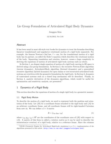
900Khmelinskii et al.: Articulated whole-body atlases for small animal image analysispositioned in different ways when scanned and there is nostandardized acquisition protocol.The work described here addresses the abovementionedproblems by introducing articulations in three existingwhole-body atlases: (1) Digimouse [15], (2) MOBY mouse[14], and (3) high-resolution SD rat [16,17]. A kinematicmodel is built for each atlas where bones in each skeletonare manually segmented and labeled, and the correspondingdegrees of freedom (DoFs) for each joint are defined.Mapping to this articulated atlas has the advantage that allthe different imaging modalities can be (semi) automaticallyregistered to a common anatomical reference; postural variations can be corrected, and the different animals (according tostrain, size, age, body fat percentage) can be scaled properly.The goals of this work are to:1. Introduce the concept of the articulated whole-body smallanimal atlas;2. Present and discuss several implemented applicationexamples: atlas to microCT data registration, follow-upmicroCT studies, cross-sectional microCT studies, multimodality atlas to BLI and microCT image registration andanalysis, and atlas to microMRI data approximation; and3. Make these three articulated whole-body small animalatlases publicly available.The final atlas consists of the 3D volume (in which the voxelsare labeled to define the anatomical structures listed above) withco-registered PET, X-ray CT, and cryosection images and can beused in 3D BLI simulations and PET image reconstruction [15].High-Resolution SD Rat Atlas Xueling et al. built a highresolution 3D anatomical atlas of a healthy adult SD rat from9,475 horizontal cryosection images (at 20 μm thickness).Coronal and sagittal section images were digitized from thehorizontal sections and anatomical structures under the guidanceof an experienced anatomist. The 3D computerized model of therat anatomy was generated using a parallel reconstructionalgorithm and interactive atlas-viewing software was developedthat offers orthoslice visualization, featuring zoom, anatomicallabeling, and organ measurement. Also, an interactive 3D organbrowser based on a virtual reality modeling language wasdeployed on a website. The models of each organ and tissueconstructed from the images were used for calculations ofabsorbed dose from external photon sources [16,17].Figure 9 in the Appendix provides a visual comparison betweenthe original atlases described above. While the MOBY and Digimouseatlases are quite similar in content, they differ in terms of the species ofthe mouse, the types of organs defined, resolution, and in the modalitiesfrom which they were constructed. Also, the MOBY atlas includes amodel of cardiac and respiratory motion. In Table 4 in the Appendix, anoverview of the main differences between these three atlases is presented.Articulated Atlas ConstructionMethodsAtlas DescriptionsPresently, in the work described here, three small animal atlases areused. In this section, a brief description of each one is presented.MOBY (Mouse Whole-Body) Atlas Segars et al. generated arealistic 4D digital mouse phantom based on high-resolution 3DMRI data from Duke University. The organs of this atlas were builtusing non-uniform rational b-spline (NURBS) surfaces, which arewidely used in 3D computer graphics.The final package includes a realistic 3D model of the mouseanatomy and accurate 4D models for the cardiac and respiratorymotions. Both the cardiac and respiratory motion models weredeveloped based on cardiac gated black-blood MRI and respiratorygated MRI data from the University of Virginia. It has been used insimulation studies in SPECT and X-ray CT [14].Digimouse Atlas Dogdas et al. constructed a 3D whole-bodymultimodal mouse atlas from co-registered X-ray microCT and colorcryosection data (anatomical information) of a normal nude malemouse. It also includes PET data (functional information) representing the distribution of a mixture of the tracers [18F] fluoride and 2deoxy-2-[18F]fluoro-D-glucose within the mouse. The image datawere co-registered to a common coordinate system using the fiducialsand resampled to an isotropic 0.1 mm voxel size. Using interactiveediting tools, several organs were segmented and labeled.In all the abovementioned atlases, the included skeletons do notdistinguish between single bones and joints. To render the registrationperformance independent of the data acquisition protocol and largepostural variations due to postural heterogeneity between scans, wepresent a segmentation of the skeleton into individual bones and addanatomically realistic kinematic constraints to each joint.Segmenting the Skeleton The first step was to manually segmentthe following bones/bone groups in each atlas from the skeletonusing the Amira V3.1 software [18], guided by anatomical textbooks [19,20], and a high resolution CT scan of a real mouse:scapula, humerus (upper front limb), clavicula (collarbone, ratonly), ulna-radius (lower front limb), manus (front paw), femur(upper hind limb), tibia-fibula (lower hind limb), pes (hind paw),caput (skull), columna vertebralis (spine), costae (ribs), sternum (chest bone), and pelvis. The resulting labeled skeletons foreach atlas can be seen in Fig. 2.Introducing Joint Kinematics In the second step, a kinematicmodel for each atlas was built, i.e., each joint position wasidentified and the corresponding DoFs were specified. Two typesof joints were distinguished: ball joints and hinge joints.In Table 1, the DoFs for the ball and hinge joints can be seen.These DoFs are anatomically correct and were defined according toexpert specifications described in literature [19,20].
Khmelinskii et al.: Articulated whole-body atlases for small animal image analysis901Fig. 2. Illustration of the three segmented skeletons for each atlas: top row—before partitioning, bottom row—afterpartitioning. a MOBY, b Digimouse, and c SD Rat.Atlas-Based Whole-Body Registration/Segmentation of Small Animals DatasetsThe skeleton is the rigid frame of the animal, in the sense of tissuestiffness. Besides the articulations of individual bones with respect toeach other, little deformation takes places in the bones themselveswithin the same animal. This is in contrast to, e.g., organs, whichhighly vary in shape, depending on the posture of the animal.Therefore, a robust registration strategy should be based on theskeleton. Although there are approaches in literature that perform smallanimal whole-body image registration based on the entire skeleton[23,24], these methods may fail if large postural variations amongdifferent animals or among the same animal in a follow-up study occur.Therefore, we propose an approach that employs the articulatedskeleton model as described above for registration of the skeleton
902Khmelinskii et al.: Articulated whole-body atlases for small animal image analysisTable 1. Joint types in the atlas skeleton and the correspondent DoFs [21],pictograms from [22]Joint typesModeled jointDoFs of thearticulated boneShoulderWristHipAnkle3 translations3 rotations3 scalingsElbowKnee3 translations1 rotation3 scalingsBall jointHinge jointin a first step. Organs are non-rigidly matched in a second step,initialized by the result of the skeleton matching.Skeleton Registration The more distal a given bone is in theskeleton, the more variable its position between acquisitions is.Therefore, if datasets of several mice are globally aligned to eachother, the location of the skulls is more similar than for instancethat of the paws. Given that the entire atlas skeleton is coarselyaligned to a target dataset in a first step, all bones can subsequentlybe matched individually by executing the registration fromproximal to distal bone segments. The registration of a distalsegment is thereby constrained by the joint type of the proximalbone it connects to. For example, for the tibia, the registration isconstrained by the DoFs of the knee joint. The deformation modelthat is required for the individual bones depends on the type ofstudy and may vary between rigid (intra-subject) and non-rigid(inter-subject) deformation models. The selected registration criterion depends on the modality of interest. It can be a point-based(e.g., Euclidean distance), surface-based (e.g., Euclidean distanceand surface curvature), or volume-based registration criterion (e.g.,Normalized Mutual Information). In this paper, we limitedourselves to a surface-based registration measure, i.e., the Euclidean distance between two surfaces. Since the registration has todeal with large articulations, potentially pathological data (as aresult of bone resorption) and inter-subject data, a rigid transformation model including non-isotropic scaling was chosen. Thisrenders the registration robust to pathological cases while stilltaking different bone sizes into account. The registration wasembedded in the Iterative Closest Point [25] framework andoptimized using an interior-reflective Newton method.Organ Registration The registered skeleton allows us to initializethe registration of several other major organs, because their location isstrongly dependent on the animal posture. To realize this, thetransformation model should be chosen such that it can handle thelarge deformations that can occur for soft tissues. Many methods havebeen proposed for registration of individual organs (see e.g., [26,27]for reviews), which are not discussed further here. In the applicationsdescribed next, we selected thin-plate-spline (TPS) interpolation [28].The required anatomical landmarks that define the TPS mapping areprimarily derived from the registered skeleton. To this end, wecompute a sparse set of initial correspondences on the animal skin byselecting the skin points closest to a set of anatomical landmarks on theskeleton (e.g., the joints). From this sparse set of skin points, a denserset of point correspondences is calculated by means of an iterativematching of local distributions of geodesic distances [21]. This resultsin a set of correspondences on the skin and on the skeleton, which incombination define the TPS interpolants.Evaluation Metrics for Registration AccuracyTo evaluate the accuracy of the registration algorithm for the skeleton,skin, and organs, three different error metrics were defined [21]: Joint localization error is calculated as the Euclideandistance between corresponding anatomical landmarks(point-to-point distance). To this end, the locations of theupper lower limb and the lower limb-paw joints of alldatasets were indicated manually using the extractedskeleton surfaces. For validation, the manually determinedjoint locations were compared to those automaticallydetermined by registration of the skeleton. Euclidean point-to-surface distance was determined toquantify border positioning errors. It was used to evaluatethe registration error over the surface of the entire skeletonand skin. Dice coefficients of volume overlap [29] were computedto assess the organ interpolation performance. The Dicecoefficient is widely used in literature to assess segmentation accuracy by evaluation of the spatial overlap of amanual and an automated segmentation. It is a voxelbased measure and therefore includes differences inobject sizes as well as spatial misalignment [30]. Giventhe absolute volumes of a manual segmentation result Vmand an automated segmentation result Va, the Dicecoefficient is defined as the intersection of the volumes,divided by the average volume:Dc ¼2jVm \ Va jjVm j þ jVa jð1ÞApplicationsIn this section, two application examples are presented thatemploy the articulated skeleton model for analysis of followup, cross-sectional, and multimodality small animal imagingstudies. Each application was quantitatively validated.Atlas to microCT Registration for Follow-Upand Cross-Sectional microCT StudiesWhole-Body Segmentation Based on Articulated SkeletonRegistration Anatomical referencing of molecular eventsinside the animal using non-contrast-enhanced microCT is
Khmelinskii et al.: Articulated whole-body atlases for small animal image analysisTable 2. Skeleton, lungs, and skin registration results. The results areseparated in two columns: before registration, i.e.: after the initializationalone and after registration. For the bone, the used error metric is the jointlocalization error, while for the lungs, skin, and the whole skeleton, it is thepoint to surface distanceBefore registrationRight kneeRight ankleLeft kneeLeft ankleRight elbowRight wristLeft elbowLeft wristEntire skeletonLungsSkinAfter registrationJoint localization error (mm)14.29 5.510.75 0.2918.70 5.871.82 1.0116.61 4.800.77 0.2619.93 5.151.69 1.145.66 2.111.31 0.4415.56 4.491.27 0.535.23 2.961.23 0.3918.04 6.471.21 0.56Euclidean point to surface distance (mm)3.68 0.770.58 0.031.27 0.260.47 0.0311.06 8.490.75 0.53difficult, because although the skeleton can be extracted easilyfrom the data as a whole it is often required to know exactly inwhich bone the molecular event takes places and because thepoor soft-tissue contrast in the abdomen complicates organlocalization and renders registration very difficult. Above that,microCT is often used in oncological studies to assess metastaticactivity in bone, and since the locations where possiblemetastases can develop greatly varies, a very flexible dataacquisition protocol with respect to animal positioning in thescanner is required. For such applications, animal posture, shape,and limb position may vary substantially.To deal with the challenges specific to microCT, weemploy the fully automated articulated atlas-based skeleton903and organ segmentation method for non-contrast-enhancedwhole-body data of mice [21] described in the section above.The skeleton is represented with a surface, derived from themodified MOBY atlas.To test the proposed method, data acquired during a study ofthe metastatic behavior of breast cancer cells were used. Breastcancer has a preference to metastasize to bone, and at thelocation of a metastatic lesion, osteolysis occurs, causingstructural damage in the skeleton (fractures or completelyresorbed bones). The subject was injected with luciferasepositive human MDA-MB-231 breast cancer cells into thecardiac left ventricle. The animal was scanned 40 days after cellinjection to screen for possible small amounts of photo-emittingtumor cells in bone marrow/bone mimicking microCT-metastatic spread. Nine anesthetized mice (Balb/c, Charles RiverWIGA, Sulzfeld, Germany), 6–9 week old, eight female, onemale, with a mean weight of 22.23 2.18 g, were acquired witha Skyscan (Kontich, Belgium) 1178 microCT scanner.Fourteen 3D data volumes of the nine mice were acquired withstep size 1 , 50 keV X-ray voltage, an anode current of 200 μA,an aluminum filter of 0.5 mm thickness, an exposure time of640 ms, and without using a contrast agent. The reconstructeddatasets covered the range between 1,000 (air) and 1,000(bone) Hounsfield units. Neither cardiac nor respiratory gatingwas used. The mice were scanned in arbitrary prone and supinepostures and arbitrary limb positions.Table 2 shows the joint localization and point to surfaceerrors for before and after registering the articulated atlasskeleton, lungs, and skin to the data. Subsequently, the brain,heart, liver, kidneys, spleen, and stomach were mapped fromthe atlas to the subject using TPS interpolation [21]. The resultFig. 3. Registration results between the atlas (red) and two different subjects (gray) after coarsely aligning the skeleton (top),after the articulated registration (middle), and after organ approximation (bottom).
904Khmelinskii et al.: Articulated whole-body atlases for small animal image analysisFig. 4. Skeleton registration and organ approximation using the same subject, at five different time points (4 weeks). Theanimal was put into the acquisition device arbitrarily, in supine (a–c) and prone (d–e) position, respectively, and the posturalvariations of the head, back, and front limbs are clearly visible.is a segmentation of the animal body into individual bones andmajor organs. This can be used for qualitative assessment ofmorphology at a single point in time in one or more animals—across-sectional study (Fig. 3), or to follow morphologicalchanges over time—a follow-up study (Fig. 4). To facilitate thecomparison of cross-sectional and follow-up data, also visualization concepts were developed that are based on mapping thedata to a common reference frame and present the resultssimultaneously (Fig. 5).Multimodality Registration, Visualization,and Analysis Combination of BLI and SegmentedmicroCT DataBLI is an imaging technique that has found widespreadapplication in preclinical research over the past years. It isused to track cells and monitor the function of specific genesand processes in the cellular biochemistry with a highsensitivity in living animals. A typical application domain isFig. 5. Demonstration of mapping the registered bones of four different animals from the corresponding target domain to acommon reference domain (the MOBY atlas domain). The large postural differences of the animals (left) are not present anymore (right), enabling a more intuitive comparison of different time points.
Khmelinskii et al.: Articulated whole-body atlases for small animal image analysisoncology, where researchers aim at monitoring the development of metastases using a highly sensitive optical modality,BLI, and relate it to morphological changes using ananatomical modality like microCT [31,32].Since BLI does not show anatomical information, it is oftenoverlaid on multiple 2D photographs from different anglesaround the animal. This, however, has the disadvantage thatanatomical referencing is limited to the animal skin and,therefore, allows only coarse source localization. Thus, acombination with a real 3D anatomical modality like microCTis preferable. This requires a BLI to CT registration approach.The BLI data in this work were acquired using theXenogen IVIS Imaging System, 3D series scanner byCaliper LifeSciences (Alameda, USA). The data were905collected from a study with two experiments in mice on themetastatic behavior of breast cancer cells as to visuallyverify the reconstructed BLI sources with the injection siteas seen in the microCT data. One hundred thousand RC21luc cells-luciferase expressing human renal carcinoma cellline and 100 μl 100,000 KS483-HisLuc cells-luciferaseexpressing murine mesenchymal stem cell line were injectedunder the renal capsule and into the left heart ventricle,respectively, and scanned after 3 to 4 weeks (time for thecarcinoma to establish).Two alternative ways have been worked out to performthe BLI to CT registration. A semi-automated method,which requires manual selection of at least three anatomicallandmarks both on the photographs, and the CT data wereFig. 6. Overview of the steps towards a combined visualization of fully segmented whole-body microCT and BLI data. TheMOBY atlas is registered to the microCT data and subsequently, the microCT data are registered to the BLI data usingthe photographs either by using manually selected landmarks or fully automatically using a 3D distance map (see text). In theresulting visualization, the BLI source (red) is shown and can be related to the skeleton and organs.
906Khmelinskii et al.: Articulated whole-body atlases for small animal image analysisimplemented. Subsequently, these corresponding landmarksare used to map one data domain to the other. As a secondapproach, a fully automated way to perform this registrationwas implemented. Based on the skin contours on thephotographs, a 3D distance map is derived and used forregistration of the animal skin, derived from CT [33]. Inaddition, the atlas to CT mapping as described above can beapplied as well. The result is a fully segmented animal thatserves for anatomical referencing if combined with aqualitative BLI source localization algorithm (e.g., [34]) asshown in Fig. 6. The quantitative results for the articulatedskeleton atlas to microCT registration are the following:entire skeleton—before registration 4.25 12.25 mm, afterregistration 0.63 1.04 mm; lungs—before registration 1.27 2.44 mm, after registration 0.50 1.35 mm.Atlas to μMRI ApproximationOrgan and Bone Approximation for Ex Vivo Mouse Data SincemicroMRI data provide greater contrast between the differentsoft tissues of the body but poorer bone contrast than CT data, itcan be used to closely follow the changes in phenotype in studiesthat require genetic modifications.Fig. 7. Organ and bone approximation results for microMRI mouse data: a, c, e manual organ segmentation. b, d, f organ andbone approximation results. Coronal and sagittal planes, respectively. yellow lungs, red heart, green spleen, cyan stomach,cream bone, gray skin, white liver. Reproduced from [35] with permission.
Khmelinskii et al.: Articulated whole-body atlases for small animal image analysis907Natick, USA) and took always 6 min of runtime in a 2.40 GHzIntel Quad Core , with 4 GB of RAM, Windows PC.Discussion and ConclusionsFig. 8. Top Atlas-based bone and organ approximation.Bottom Manual organ segmentation.A novel semi-automated organ approximation method forμMRI mouse data that considerably reduces the required usereffort compared to manual segmentation was implemented. Itincludes the limbs and provides a shape approximation of thebones in MR data. To derive the set of skin correspondences, theuser interactively points out the joints/bone landmarks guided byanatomically realistic kinematic constraints imposed by thearticulated atlas. Given this set of dense skin correspondences,the organ approximation is performed using the TPS approximation as described in the “Methods” section. The boneapproximation is performed by (1) automatically identifyingall the joints out of the manually indicated landmarks and (2)applying a scaling and rotation to the atlas bone surfaces [35].This MRI segmentation method was tested on female C3Hmice perfusion fixed with
Talairach brain atlas [1], the Visible Human Project whole-body atlas [2], and the 4D NCAT torso phantom [3]. The Talairach atlas consists of a standard 3D coordinate space with labeled regions and structural probability maps and is available for clinical use. This atlas is not only used for stereotactic and functional neurosurgery but also in .