Transcription
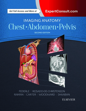
Get Full Access and More atExpertConsult.comFEDERLE I ROSADO-DE-CHRISTENSONRAMAN I CARTER I WOODWARD I SHAABANELSEVIER
SECOND EDITIONFEDERLE ROSADO-DE-CHRISTENSONRAMAN CARTER WOODWARD SHAABAN
ii
SECOND EDITIONMichael P. Federle, MD, FACRProfessor and Associate Chair for EducationDepartment of RadiologyStanford University School of MedicineStanford, CaliforniaMelissa L. Rosado-de-Christenson, MD, FACRSection Chief, Thoracic RadiologyDepartment of RadiologySaint Luke’s Hospital of Kansas CityProfessor of RadiologyUniversity of Missouri–Kansas City School of MedicineKansas City, MissouriSiva P. Raman, MDAssistant Professor of RadiologyDepartment of RadiologyJohns Hopkins University School of MedicineBaltimore, MarylandBrett W. Carter, MDAssistant Professor of RadiologyDepartment of Diagnostic RadiologyThe University of Texas MD Anderson Cancer CenterAssistant Professor of RadiologyDepartment of Diagnostic and Interventional ImagingThe University of Texas Medical School at HoustonHouston, TexasPaula J. Woodward, MDDavid G. Bragg, MD and Marcia R. Bragg Presidential EndowedChair in Oncologic ImagingProfessor of RadiologyDepartment of Radiology and Imaging SciencesUniversity of Utah School of MedicineSalt Lake City, UtahAkram M. Shaaban, MBBChProfessor of RadiologyDepartment of Radiology and Imaging SciencesUniversity of Utah School of MedicineSalt Lake City, Utahiii
1600 John F. Kennedy Blvd.Ste 1800Philadelphia, PA 19103-2899IMAGING ANATOMY: CHEST, ABDOMEN, PELVIS, SECOND EDITIONISBN: 978-0-323-47781-9Copyright 2017 by Elsevier. All rights reserved.No part of this publication may be reproduced or transmitted in any form or by any means, electronic or mechanical, includingphotocopying, recording, or any information storage and retrieval system, without permission in writing from the publisher. Details onhow to seek permission, further information about the Publisher’s permissions policies and our arrangements with organizations such asthe Copyright Clearance Center and the Copyright Licensing Agency, can be found at our website: www.elsevier.com/permissions.This book and the individual contributions contained in it are protected under copyright by the Publisher (other than as may benoted herein).NoticesKnowledge and best practice in this field are constantly changing. As new research andexperience broaden our understanding, changes in research methods, professional practices,or medical treatment may become necessary.Practitioners and researchers must always rely on their own experience and knowledge inevaluating and using any information, methods, compounds, or experiments describedherein. In using such information or methods they should be mindful of their own safetyand the safety of others, including parties for whom they have a professional responsibility.With respect to any drug or pharmaceutical products identified, readers are advised to checkthe most current information provided (i) on procedures featured or (ii) by the manufacturerof each product to be administered, to verify the recommended dose or formula, themethod and duration of administration, and contraindications. It is the responsibility ofpractitioners, relying on their own experience and knowledge of their patients, to makediagnoses, to determine dosages and the best treatment for each individual patient, and totake all appropriate safety precautions.To the fullest extent of the law, neither the Publisher nor the authors, contributors, oreditors, assume any liability for any injury and/or damage to persons or property as a matterof products liability, negligence or otherwise, or from any use or operation of any methods,products, instructions, or ideas contained in the material herein.Publisher Cataloging-in-Publication DataNames: Federle, Michael P. Rosado-de-Christenson, Melissa L. Raman, Siva P. Carter, Brett W. Woodward, Paula J. Shaaban, Akram M.Title: Imaging anatomy. Chest, abdomen, pelvis / [edited by] Michael P. Federle, Melissa L.Rosado-de-Christenson, Siva P. Raman, Brett W. Carter, Paula J. Woodward, andAkram M. Shaaban.Other titles: Chest, abdomen, pelvis.Description: Second edition. Salt Lake City, UT : Elsevier, Inc., [2016] Includesbibliographical references and index.Identifiers: ISBN 978-0-323-47781-9Subjects: LCSH: Diagnostic imaging--Handbooks, manuals, etc. Chest--Anatomy--Handbooks,manuals, etc. Abdomen--Anatomy--Handbooks, manuals, etc. Pelvis--Anatomy--Handbooks,manuals, etc. Imaging systems in medicine--Handbooks, manuals, etc. MESH: Thorax-anatomy & histology--Atlases. Abdomen--anatomy & histology--Atlases. Pelvis--anatomy& histology--Atlases. Magnetic Resonance Spectroscopy--Atlases. Tomography, X-RayComputed--Atlases.Classification: LCC RC78.7.D53 D534 2016 NLM WE 17 DDC 616.07’54--dc23International Standard Book Number: 978-0-323-47781-9Cover Designer: Tom M. Olson, BACover Art: Lane R. Bennion, MSPrinted in Canada by Friesens, Altona, Manitoba, CanadaLast digit is the print number: 9 8 7 6 5 4 3 2 1iv
DedicationsTo Ric Harnsberger, whose vision, enthusiasm, charisma, and hard work createdand sustained this entire enterprise. Your legacy will endure long after all of ournames are forgotten.MPFTo my family, especially my husband, Paul, and my daughters, Jennifer andHeather, whose love, encouragement, and support have seen me through thisproject. To my dear friend and coauthor, Dr. Brett Carter, from whom I continueto learn every day.MRDCTo my wife, Janani Venkateswaran, whose support throughout my career hashelped sustain me and drive me forward.SPRI dedicate this project to my parents, Ralph and Joy Carter, without whom thiseffort would not have been possible.BWCTo Miguel Flores.The stalwart patriarch of the Flores/z clan, whose staunch work ethic remainsan inspiration to me. You started in this world with so little and have done somuch. Thank you not only for your example but also for your hijo.PJWTo my parents, I truly owe you everything.To my wife, Inji, son, Karim, and daughters, May and Jena, the jewels of my life,thanks for your understanding and tremendous support.AMSv
vi
Additional Contributing AuthorsGerald F. Abbott, MD, FACRAssociate Radiologist, ThoracicMassachusetts General HospitalAssociate ProfessorHarvard Medical SchoolBoston, MassachusettsRania Farouk El Sayed, MD, PhDAssistant ProfessorGenitourinary and MR Pelvic Floor Imaging UnitDepartment of Radiology, Faculty of MedicineCairo University HospitalsCairo, Egyptvii
viii
PrefaceIn his elegant foreword to the first edition of this book, Professor Morton Meyersrelated the evolution of our understanding of human anatomy, from Vesalius’ Fabricathrough the many contributions of surgeons, such as Harvey Cushing, who wrote that“.from the publication of the Fabrica almost to the present day the intimate pursuitof.anatomy has constituted the high road for entry into the practice of surgery.”Meyers went on to write, “Today it is the radiologist who is most facile with highlydetailed anatomy, and who.demonstrates this in vivo. Dissectional anatomy has beensuperseded by cross-sectional anatomy.”In the decade since the publication of the first edition of this book, our ability to definenormal and abnormal anatomy of the chest, abdomen, and pelvis has continuedto advance. An example is the major improvement in MR evaluation of the pelvis,depicting in multiple planes with unprecedented detail the pertinent anatomicalalterations that may result in pelvic floor laxity, urinary and fecal incontinence, andperianal fistulas. Similar advances have been made in the multimodality depiction ofanatomic structures throughout the body, and these have been incorporated into thissecond edition of Imaging Anatomy.As with the first edition, we feature what Meyers deemed “exquisite, museum-qualityillustrations,” paired with the imaging modalities most relevant to the understandingof human anatomy in health and disease.We hope that the efforts of our radiologist authors and talented medical illustratorswill make the anatomy of the chest, abdomen, and pelvis “come alive” for our readers.Michael P. Federle, MD, FACRProfessor and Associate Chair for EducationDepartment of RadiologyStanford University School of MedicineStanford, Californiaix
x
AcknowledgmentsText EditorsArthur G. Gelsinger, MANina I. Bennett, BATerry W. Ferrell, MSKaren E. Concannon, MA, PhDMatt W. Hoecherl, BSMegg Morin, BAImage EditorsJeffrey J. Marmorstone, BSLisa A. M. Steadman, BSIllustrationsLane R. Bennion, MSRichard Coombs, MSLaura C. Sesto, MAArt Direction and DesignTom M. Olson, BALaura C. Sesto, MALead EditorLisa A. Gervais, BSProduction CoordinatorsAngela M. G. Terry, BARebecca L. Hutchinson, BAEmily Fassett, BAxi
xii
SectionsSECTION 1: CHESTSECTION 2: ABDOMENSECTION 3: PELVISxiii
TABLE OF 2SECTION 1: CHEST636Chest OverviewMelissa L. Rosado-de-Christenson, MD, FACRLung DevelopmentMelissa L. Rosado-de-Christenson, MD, FACRAirway StructureBrett W. Carter, MD and Gerald F. Abbott, MD, FACRVascular StructureMelissa L. Rosado-de-Christenson, MD, FACRInterstitial NetworkBrett W. Carter, MD and Gerald F. Abbott, MD, FACRLungsMelissa L. Rosado-de-Christenson, MD, FACRHilaMelissa L. Rosado-de-Christenson, MD, FACRAirwaysBrett W. Carter, MD and Gerald F. Abbott, MD, FACRPulmonary VesselsMelissa L. Rosado-de-Christenson, MD, FACRPleuraBrett W. Carter, MD and Gerald F. Abbott, MD, FACRMediastinumMelissa L. Rosado-de-Christenson, MD, FACRSystemic VesselsMelissa L. Rosado-de-Christenson, MD, FACRHeartMelissa L. Rosado-de-Christenson, MD, FACRCoronary Arteries and Cardiac VeinsAkram M. Shaaban, MBBChPericardiumMelissa L. Rosado-de-Christenson, MD, FACRChest WallBrett W. Carter, MD and Gerald F. Abbott, MD, FACR666SECTION 2: ABDOMEN448484508528550592608xivEmbryology of AbdomenMichael P. Federle, MD, FACR and Siva P. Raman, MDAbdominal WallSiva P. Raman, MD and Michael P. Federle, MD, FACRDiaphragmSiva P. Raman, MD and Michael P. Federle, MD, FACRPeritoneal CavitySiva P. Raman, MD and Michael P. Federle, MD, FACRVessels, Lymphatic System and Nerves, AbdominalSiva P. Raman, MD and Michael P. Federle, MD, FACREsophagusMichael P. Federle, MD, FACR and Siva P. Raman, MDGastroduodenalSiva P. Raman, MD and Michael P. Federle, MD, FACR708732778804834860882920Small IntestineSiva P. Raman, MD and Michael P. Federle, MD, FACRColonSiva P. Raman, MD and Michael P. Federle, MD, FACRSpleenSiva P. Raman, MD and Michael P. Federle, MD, FACRLiverSiva P. Raman, MD and Michael P. Federle, MD, FACRBiliary SystemSiva P. Raman, MD and Michael P. Federle, MD, FACRPancreasSiva P. Raman, MD and Michael P. Federle, MD, FACRRetroperitoneumSiva P. Raman, MD and Michael P. Federle, MD, FACRAdrenalSiva P. Raman, MD and Michael P. Federle, MD, FACRKidneySiva P. Raman, MD and Michael P. Federle, MD, FACRUreter and BladderSiva P. Raman, MD and Michael P. Federle, MD, FACRSECTION 3: PELVIS946Vessels, Lymphatic System and Nerves, PelvicPaula J. Woodward, MD and Akram M. Shaaban, MBBChMALE974Male Pelvic Wall and FloorPaula J. Woodward, MD and Akram M. Shaaban, MBBCh1000 Testes and ScrotumPaula J. Woodward, MD and Akram M. Shaaban, MBBCh1018 Prostate and Seminal VesiclesPaula J. Woodward, MD and Akram M. Shaaban, MBBCh1036 Penis and UrethraPaula J. Woodward, MD and Akram M. Shaaban, MBBChFEMALE1050 Female Pelvic FloorPaula J. Woodward, MD, Rania Farouk El Sayed, MD, PhD,and Akram M. Shaaban, MBBCh1078 UterusPaula J. Woodward, MD and Akram M. Shaaban, MBBCh1104 OvariesPaula J. Woodward, MD and Akram M. Shaaban, MBBCh
This page intentionally left blank
This page intentionally left blank
SECOND EDITIONFEDERLE ROSADO-DE-CHRISTENSONRAMAN CARTER WOODWARD SHAABAN
This page intentionally left blank
SECTION 1ChestChest OverviewLung DevelopmentAirway StructureVascular StructureInterstitial NetworkLungsHilaAirwaysPulmonary VesselsPleuraMediastinumSystemic VesselsHeartCoronary Arteries and Cardiac VeinsPericardiumChest Wall4446486106118148178200232258294336380402422
ChestChest OverviewGENERAL ANATOMY AND FUNCTIONChest WallStandard Chest Radiography Anatomy Spine Sternum Ribs Clavicles Scapulae Skeletal muscles Chest wall nerves and vessels Skin and subcutaneous fat Function Protects lungs, cardiovascular structures, andintrathoracic organs Participates in bellows-like process of respiration Imaging study of choice for initial assessment ofcardiopulmonary disease PA and lateral chest radiographs Orthogonal views (at right angles to each other) Analysis of orthogonal views for precise anatomiclocalization of imaging abnormalitiesPleura Anatomy Thin, continuous membrane Parietal pleura: Lines nonpulmonary surfaces Visceral pleura: Lines pulmonary surfaces Pleural space: Potential space between pleural surfaces Function Production and absorption of normal pleural fluid– Lubrication of pleural surfaces– Facilitation of lung motion during respiration Clearance of abnormal pleural fluidAirways Anatomy Trachea Mainstem bronchi Lobar bronchi Segmental/subsegmental bronchi Bronchioles Distal airways and alveoli Function Gas exchange during respiration Protective mechanisms against foreign particles– Mucociliary escalator– Cough reflex Gas exchange to and from alveolar-capillary interfaceHeart and Great Vessels Anatomy Venae cavae Right atrium Right ventricle Pulmonary arteries Capillary network Pulmonary veins Left atrium Left ventricle Thoracic aorta and branches Function Pump action for systemic and pulmonary circulations Transport of deoxygenated blood to capillary-alveolarinterface Transport of oxygenated blood to tissues4CHEST RADIOGRAPHYStandard Radiographic Positioning Upright patientFull inspiration and breath hold near total lung capacityNo rotation or motionAttempt to minimize overlying osseous structuresArea of interest closest to image receptor (IR)Radiographic technique Source-to-image receptor distance (SID): 72 inches tominimize magnification Central x-ray beam centered on thorax Beam collimation to include outer chest wallRadiographic Projections Posteroanterior (PA) chest radiography Term PA: Describes PA direction of x-ray beam traversingchest toward IR Anterior chest against IR Head vertically positioned and chin on top of grid device Dorsal wrists on hips, and elbows rotated anteriorly tomove scapulae laterally Shoulders moved caudally and squarely against IR tobring clavicles below apices Left lateral chest radiography Term left lateral: Denotes that left lateral chest wall isagainst IR X-ray beam traverses chest from right to left toward IR Arms above head to move upper extremities away fromlungs and mediastinum Anteroposterior (AP) chest radiography Term AP: Describes anteroposterior direction of x-raybeam traversing chest toward IR Supine and bedside (portable) radiography and imagingof sitting and semiupright patients– Neonates, infants, and young children– Debilitated and unstable patients– Critically ill and bed-ridden patients Distinctive features– Magnification of anterior structures (heart andmediastinum) farthest from IR; shorter SID– Clavicles course horizontally and partially obscureapices– Ribs assume horizontal course Lateral decubitus chest radiography Recumbent position with right or left side down Elevation of chest on radiolucent support Frontal radiograph (AP or PA) with horizontal x-ray beam Indications– Evaluation of pleural fluid in dependent pleural space(x-ray beam tangential to fluid-lung interface)– Evaluation of air in nondependent pleural space (x-raybeam tangential to visceral pleura-air interface)
Chest OverviewRadiographic Interpretation Assessment of patient's identity and location of right/leftmarkers Assessment of entire thorax Frontal radiography– Inclusion of all thoracic structures from larynx to lungbases– Full inspiration Diaphragm below posterior 9th-10th ribs or at 5th7th anterior ribs at mid clavicular line Lateral radiography– Inclusion of anteroposterior extent of chest wall– Inclusion of apical lung and posteriorcostodiaphragmatic sulci Assessment of appropriate radiographic positioning No rotation– Spinous process of T3 (posterior structure) centeredbetween medial clavicles (anterior structures) onfrontal radiography– Superimposition of right and left ribs posterior tothoracic vertebrae on lateral radiography Medial aspects of scapulae lateral to lungs on frontalradiography Arms above thorax without superimposition on lung andmediastinum on lateral radiography Appropriately exposed radiograph Assessment of peripheral pulmonary vasculature Visualization of pulmonary vessels through heart anddiaphragm on frontal radiography Visualization of vertebrae through heart on frontalradiography Systematic evaluation Assessment of multiple superimposed structures andtissues Assessment of all visible structures including portions ofneck, shoulders, and upper abdomen Anatomic localization of abnormalities Assessment of associated findings Comparison to prior studies Challenges Evaluation of retrocardiac lung Evaluation of retrodiaphragmatic lung Evaluation of apical lungRadiographic Densities 5 radiographic densities Air Water (fluid, blood, and soft tissue) Fat Bone Metal (contrast, metallic portions of medical devices,metallic foreign bodies) Silhouette sign Intrathoracic process (mass, consolidation, pleural fluid)that touches mediastinum or diaphragm obscuresvisualization of their borders on radiography Critical for radiographic diagnosis of– Atelectasis– Consolidation– Pleural effusionChest Apical lordotic (AP or PA axial) chest radiography Superior angulation of x-ray beam from horizontal planeof 15-20 Distinctive features– Clavicles and first anterior ribs project above apices– Ribs course horizontally– Magnification (foreshortening) of mediastinum Indications– Radiographic visualization of apex, superiormediastinum and thoracic inlet– Enhanced visualization of minor fissure in suspectedmiddle lobe atelectasis Expiratory radiography Evaluation of air-trapping Evaluation of pneumothorax (limited value)– No clear difference in sensitivity or specificity fordiagnosisCOMPUTED TOMOGRAPHYGeneral Concepts Imaging based on x-ray absorption by tissues with differingatomic numbers Display of differences in x-ray absorption in cross-sectionalformat Excellent spatial resolution Enhanced visualization of structures of different tissuedensity based on display of wide range of Hounsfield unit(HU) measurements Window width refers to number of HU displayed Window level refers to median (center) HU Window width and level for assessment of thoracic CT– Lung window (width of 1,500 HU; level of -650 HU) Evaluation of lungs, airways, and air-containingportions of gastrointestinal tract Identification of pneumomediastinum,pneumoperitoneum, and soft tissue gas– Soft tissue (mediastinal) window (width of 400 HU;level of 40 HU) Evaluation of vascular structures and soft tissues ofmediastinum and chest wall– Bone window (width of 2,000 HU; level of 400 HU) Evaluation of skeletal and calcified structures Evaluation of metallic objects and medical devices– Liver window (width of 150 HU; level of 50 HUwithout contrast and 100 HU with contrast) Evaluation of upper abdomen: Liver, kidney Evaluation of pleural effusions for solid pleuralnodulesConventional CT Evaluation, localization, and characterization ofabnormalities detected on radiography Localization of lesions in preparation for CT-guidedbiopsy/drainageContrast-Enhanced CT Administration of intravenous contrast Evaluation of vascular structures Evaluation of vascular abnormalities Distinction of vascular structures from adjacent softtissues (e.g., lymph nodes)5
ChestChest Overview Determination of lesion/tissue enhancement Administration of enteric contrast Evaluation of gastrointestinal perforations/leaksCT Angiography Vascular imaging Timing of contrast bolus Imaging of specific vascular structures– CT pulmonary angiography for evaluation ofpulmonary thromboembolic disease– CT aortography for evaluation of traumatic aorticinjury or aneurysm– CT aortography for evaluation of acute aorticsyndrome Baseline unenhanced images required prior tocontrast administration to document intramuralhematomaHigh-Resolution CT Technique Thin sections to minimize partial volume effects High-resolution reconstruction algorithm Indications Evaluation of diffuse infiltrative lung disease Evaluation of patients with unexplained dyspnea andnormal radiographs Special techniques Prone imaging for evaluation of peripheral basilar lungdisease Expiratory imaging for evaluation of small airwaysdiseasePostprocessing Techniques Multiplanar reformations Multiplanar evaluation of axially oriented structures andabnormalities Evaluation of anatomic location of lung lesions in relationto interlobar fissures Evaluation of chest wall and mediastinal involvement byadjacent pulmonary lesions Coronal and sagittal lesion measurements Maximum-intensity projection (MIP) Evaluation of vascular structures Increased conspicuity of pulmonary nodules Minimum-intensity projection (minIP) Evaluation of central airways and air-trapping Surface-rendered displays Volume-rendered techniques for problem solving andeducation Virtual bronchoscopyMAGNETIC RESONANCE IMAGINGGeneral Concepts Application of radiofrequency to excite protons withinmagnetic field Detection of signal emitted by nuclei as they relax to theiroriginal alignment with generation of image depicting theirspatial distribution Advantages of MR Excellent contrast resolution Multiplanar imaging6 Intrinsic vascular "contrast" Increased soft tissue contrastTechnique Spin-echo sequences typically used in chest imaging T1-weighted images T2-weighted images Bright blood sequencesIndications Imaging of heart and great vessels Distinction of vascular structures from adjacent soft tissueswithout use of contrast Evaluation of mediastinum and hila Diagnostic imaging of thymus Assessment of chest wall and diaphragmANGIOGRAPHYPulmonary Angiography Venous catheterization Cannulation of pulmonary arterial system Indications Evaluation of congenital and acquired pulmonaryvascular abnormalities Management of selected cases of thromboembolicdiseaseAortography Arterial catheterization Cannulation of proximal aorta Indications Evaluation of traumatic aortic and great vessel injury Assessment of congenital arterial vascular anomalies Evaluation of caliber and integrity of aortic and greatvessel luminaBronchial Artery and Intercostal Arteriography Arterial catheterization Selective cannulation of bronchial/intercostal arteries Indications Diagnosis and treatment of hemoptysisOTHER CHEST IMAGING MODALITIESScintigraphy Positron-emission tomography Determination of metabolic activity of lesions Staging of malignant neoplasms Use of integrated PET/CT imaging Ventilation-perfusion imaging Evaluation of thromboembolic disease– Study of choice in pregnant subjects Evaluation of pre- and postoperative lung functionUltrasound Evaluation of pleural effusion Free vs. loculated Thoracentesis/biopsy planning and thoracostomy tubeplacement Evaluation of diaphragmatic motion in cases of suspectedparalysis
Chest OverviewChestCHEST OVERVIEWChest wall skeletalstructuresAirwaysThoracic great vesselsLungHeartChest wall muscleChest wall subcutaneoustissuePleuraGraphic shows the complex and diverse structures and organs of the thorax. The chest wall skeletal and soft tissue structures surroundand protect the primary organs of respiration, the thoracic cardiovascular system, and the proximal gastrointestinal tract. The apposedpleural surfaces create a potential space that normally contains a small amount of fluid, which lubricates the pleural surfaces andreduces friction during respiratory motion. The airways deliver oxygen to the alveolar-capillary interface and carry carbon dioxide out tothe environment. The heart and vessels deliver deoxygenated blood to the capillary-alveolar interface and oxygenated blood to theperipheral organs and tissues.7
ChestChest OverviewPA CHEST RADIOGRAPHYChest wall skeletalstructuresAortaChest wall soft tissuesAirwaysInterlobar pulmonaryarteryHeartLungNormal posteroanterior (PA) chest radiograph helps illustrate inherent challenges regarding interpretation of radiographs of thethorax. Chest radiography displays a wide range of structures and tissue types with significant superimposition of structures of differentradiographic densities. Portions of the lung may be obscured by overlying mediastinal soft tissues and skeletal structures. Attention toradiographic image quality is of paramount importance for accurate diagnosis of subtle abnormalities.8
Chest OverviewChestLATERAL CHEST RADIOGRAPHYThoracic aortaTracheaLungSternumThoracic vertebraHeartHemidiaphragmsLateral chest radiography is orthogonal (at 90 ) to PA chest radiography. The lateral chest radiograph is a complementary radiographicprojection that allows visualization of the retrocardiac left lower lobe and the retrodiaphragmatic lung bases, and it allows evaluationof the thoracic vertebrae. As on the PA chest radiograph, multiple structures of various densities are superimposed and must beevaluated in a systematic manner. Evaluation of both PA and lateral chest radiographs allows anatomic localization andcharacterization of thoracic abnormalities and the formulation of an appropriate differential diagnosis.9
ChestChest OverviewPA CHEST RADIOGRAPHY, POSITIONING AND COLLIMATIONChin over grid deviceVertical image receptorX-ray beam travels inposteroanterior directionScapula rotated off lungHand pronated with dorsalwrist on hipCollimation of x-ray beamLung apex includedX-ray beam centered on chestLeast magnification of heartand mediastinumCostophrenic angle included(Top) Graphic shows proper positioning for PA chest radiography. The patient is upright with the anterior chest against the verticalimage receptor, the chin over the top of the device, the arms flexed with the backs of the hands on the hips, and the shoulders internallyrotated to move the scapulae off the lungs. The x-ray beam travels through the patient in a posteroanterior direction. (Bottom) Graphicshows proper PA chest radiographic collimation for imaging the lungs and mediastinum. The white target sign shows the centering ofthe x-ray beam. The blue overlay represents the collimated x-ray beam that extends from the cervical airway superiorly to below thecostophrenic angles inferiorly and includes the left and right skin surfaces. The anterior structures of the chest (shown in color) areclosest to the image receptor and experience the least magnification.10
Chest OverviewChestPA CHEST RADIOGRAPHYSpinous process of T3Medial claviclesMedial left scapulaRetrocardiac thoracic vertebraRetrocardiac pulmonaryvesselsRetrodiaphragmaticpulmonary vesselsSpinous process of T3Medial right clavicleMedial left clavicleMedial right scapulaIncreased opacity of lefthemithorax(Top) Well-positioned normal PA chest radiograph shows that the scapulae are rotated off the lungs and the spinous process of T3 islocated equidistant from the medial clavicles. Proper collimation spans from the cervical trachea superiorly to below the costophrenicangles inferiorly and includes the lateral aspects of the chest wall. Optimal exposure allows visualization of the peripheral pulmonaryvessels, the vertebral bodies (visible through the mediastinum), and the retrocardiac and retrodiaphragmatic pulmonary vessels.(Bottom) Poorly positioned PA chest radiograph shows marked rotation to the right. The left medial clavicle overlies the spinousprocess of T3, and the right medial clavicle is displaced to the right of midline. Increased density of the left hemithorax results from xray penetration of a greater thickness of left-sided chest wall soft tissues, when compared to the right, due to rotation.11
ChestChest OverviewLEFT LATERAL CHEST RADIOGRAPHY, POSITIONING AND COLLIMATIONArms extended upwardVertical image receptorX-ray beam travels from rightto leftLeft lateral chest against griddeviceUpper lung includedUpper extremities rotated offupper lungx-ray beam centered on chestLeast magnification of leftsided structuresCostophrenic angle includedCollimation of x-ray beam(Top) Graphic shows proper positioning for left lateral chest radiography. The patient is upright with the left lateral chest against thevertical image receptor and the arms extended upward for unobstructed visualization of the upper lungs. The x-ray beam travelsthrough the patient from right to left for a left lateral chest radiograph. (Bottom) Graphic shows proper left lateral chest radiographiccollimation for imaging the lungs and mediastinum. The white target sign shows the centering of the x-ray beam. The blue overlayrepresents the collimated x-ray beam that extends from the cervical airway superiorly to below the costophrenic angles inferiorly andincludes the anterior and posterior skin surfaces. The structures of the left chest (shown in color) are closest to the image receptor andexperience the least magnification.12
Chest OverviewChestLEFT LATERAL CHEST RADIOGRAPHYAnterior scapular bordersHilaIntervertebral diskLeft posterior ribLeft costophrenic angleRight posterior ribRight costophrenic angleHumeriUpper extremity soft tissuesLeft posterior ribRight posterior ribLeft costophrenic angleRight costophrenic angle(Top) Well-positioned normal left lateral chest radiograph shows that the upper extremities are not visible. The hila are centrallylocated. The thoracic intervertebral disks are visible. The posterior ribs are superimposed and project behind the vertebrae. There isminimal magnification of the left posterior
Get Full Access and More at ExpertConsult.com FEDERLE I ROSADO-DE-CHRISTENSON RAMAN I CARTER I WOODWARD I SHAABAN ELSEVIER . SECOND EDITION FEDERLE ROSADO-DE-CHRISTENSON RAMAN CARTER WOODWARD SHAABAN IA2_CAP_Frontmatter_Sept.08.2016.indd 1 9/14/16 12:25 PM. ii


