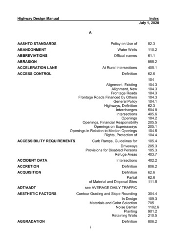
Transcription
21104 (2003)y. Phys. France0 EDP Sciences, Les UlisDOI: 10.1051/jp4:200300021CohérentX-raywithimaginga nergyRecoveryopportunitiesLinac(ERL)Q. Shen1 6, D. H. Bilderback'5,K. D. Finke) ste ! n\I. V. Bazarovand S. M. Gruner1Corne/y/-//ghEnergy Synchrotron Source (CHESS), Corne/y University, Ithaca, NY 14853,U. S. A.2 Laboratory for Elementary Particle Physics, Comell University, Ithaca, NY 14853, U.S.A.3 Physics Department, Corne/y University, Ithaca, NY 14853, U.S. A." Laorafor/ofAfom/c anof So//d Sfafe Pnys/'cs, Corne/y L/n/t/ers/fy,/aca, MY M853, L/.S. A.5 School of Engineering and Applied Physics, Comell University, Ithaca, NY 14853, U.S. A.6 Department of Materials Scienceand Engineering, Corne/y University, Ithaca, NY 14853,U. S. A.Abstract. A proposedEnergyRecoveryLinac (ERL) x-ray sourceat Cornettwouldbe a 5-7 GeV synchrotronlinac. Becauseof its ultra-smallroundfacility basedon the energy-recoveryprinciplewith a superconductingélectron source and ultra-short but flexible bunch structure, the ERL has the potential to produce subpicosecondultra-bright diffraction-limited hard x-ray beamsthat are superiorto thosetrom existing storagerings,and would enablenew scientificexperimentsthat aredifficult or impossibleto performtoday. In this paper, we discussthe transversecoherencepropertiesof the ERL sourceand show how thèsepropertiescouldbenefitvariousx-ray microscopyapplicationsthatrequirea highly cohérentand intensex-raybeam. Thèseapplications includefull cohérentillumination of wide-aperturezone-plateopticsfor scanningx-ray microscopy,diffraction-limited cohérentphase-contrastmicroscopyand tomography,fully coherent hard x-ray diffractionmicroscopy,and large-coherence-areax-rayholographytechniques.1. INTRODUCTIONX-ray microscopy has made substantial advances in récent years [1], especially in the hard x-ray régime. Thèse advances can be mainly attributed to two factors : improvements in focusing x-ray opticalcomponents and the availability of bright third génération synchrotron sources. Focusing optics haveproduced sub-tOOnm focal spot sizes for hard x-rays [2], and condensing optics have reached 50nm spatial resolution [3]. Present-day storage-ring synchrotron sources are essentially diffraction-limitedfor 8keV photons in the vertical direction, with source sizes in the 10-20 micrometer range [4]. The combination of thèse two factors appears to be the key for x-ray microscopic applications using hard x-rays, ascan be seen from thèse proceedings. Compared to other forms of microscopy, x-ray microscopes havethe advantage of less radiation damage and greater pénétration depth than électron probes, and of muchbetter spatial resolution than optical microscopes [5].Further advances in spatial resolution in x-ray microscopy require substantial improvements in boththird-generation synchrotron sources mayx-ray optics and x-ray sources. Although some present-day 102-10be diffraction-limitedvertically, the horizontal emittance istimes larger, resulting in an essentially incohérent hard x-ray source. The extremely anisotropic, flat-beam characteristic also limits efficient throughput for x-ray optics and is thus undesirable for two-dimensional microscopic and imagingapplications. Although not every form of microscopy requires high degree of coherence, almost all focusing optics benefit from a diffraction-limitedsource [6], which is the case for all scanning x-ray microscopes (SXM).In principle, x-ray microscopy should be able to reach sub-nanometer spatial resolution comparableto the x-ray wavelength. In practice, however, this ultimate resolution may be difficult to achieve byfocusing optics only, as current fabrication techniques may reach a limit of 10 nm. To go beyond the 10
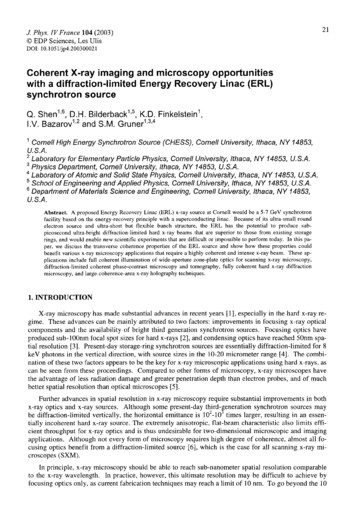
22JOURNAL DE PHYSIQUE IVnm fabrication barrier, coherence-based x-ray imaging and microscopic methods [7-11] become extremety attractive since the spatial resolution in thèse methods dépend only on diffraction resolution in acohérent diffraction pattern. Such horizon methods require not only a fully diffraction-limitedx-raysource for high diffraction resolution applications, but also orders-of-magnitude increase in cohérentintensity compared to what is possible at present third-generation hard x-ray sources.2. PROPOSEDERL X-RAYSOURCEThe recently proposed energy recovery linac (ERL) synchrotron source [12-17] would be an idéalsource for ultimate resolution and cohérent x-ray microscopic applications. Such a facility proposed atCornell would be a 5-7 GeV synchrotron source (Table 1) based on closed-loop energy recovery withsuperconducting linear accelerators and small-gap short-period undulators. It offers significant advantages over storage ring sources, both in terms of the possible x-ray beam quality and, once the technology is developed, cost-effectiveness. The basic idea behind an ERL was suggested long ago for beamcolliding machines [18] and the feasibility of operating an ERL has recently been demonstrated with ahighly successful free electron laser at the Thomas Jefferson National Accelerator Facility.Table 1 : Preliminary design parameters for the Cornell ERL source.ERL High-ERL High-Compared tofluxcoherenceESRF [4]BeamenergyEc (GeV)Beam current 1 (mA)Hori. emittance (nm-rad)Vert. emittance y (nm-rad)gxsy(nm-rad) 2Bunch length fwhm 7 (ps)Undulator length L (m)Undulator période (cm)Energy @ 1 S ianceB(phs/s/0.1%/mm/mr)5. 3100 tO5. 362000. 150. 150. 0150. 0) 540. 0)0. 020. 3-50. 00020. 3-50. 043525251.7 1.8.0 8. 071.5. 10' " 1. 5. 10'''53. 58. 031. 101513. 10 52. 10 31. 10Hori. divergence fwhm (rad)Vert. divergence fwhm () irad)9. 19. 16. 26. 226. 810.4Hori. source size fwhm (jm)Vert. source size, fwhm (m)Cohérent f [uxF. (phs/s/0. 1%)1031031.8. 10'24. 524. 53. 1 10' "87913.91. 8. 10'Cohérent intensity (iT) I. O-IO' " 6. 6. 10' 1. 9-IO' "(phs/s/mm/0.1 %/1:t focus)The basic machine parameters for the proposed Corne) ! ERL are listed in Table 1, which is takenfrom Ref. [14]. The product of the vertical and horizontal emittances is important, since the brilliance,and, hence the coherence, scales with the inverse of this product. As can be seen from the Table, theERL would produce round synchrotron x-ray beams with average brilliance more than one order ofmagnitude higher than the existing storage rings, making the ERL comparable in this regard to proposedprototype 4 "' génération x-ray free-electron laser (XFEL) sources. For x-ray microscopy applications,the high coherence ERL would improve the source properties in two ways : by reducing the source size
X-RAY MICROSCOPY 2002Diffractionlimifed 5 8keVERL emittance(0.015nm)ESRF emittance(dnm x o :ü1nm)Figure 1. Schematicillustration of transverseemittancesof the proposedERLsynchrotronsource,comparedto the emittancesat a third-generationstoragering,ESRF. A) soshownis the diffraction limitedemittanceareaat 8 keV for theERL.by one-to-two orders of magnitude in the horizontal direction to make essentially a round beam as illustrated in Fig. 1, and by providing more than two orders-of-magnitude higher cohérent flux as comparedto third génération storage ring sources. The combination of the two factors is reflected in the cohérentintensity or flux density at the focal spot assuming a 1 : t focusing ratio, leading to a potentially 103 increase in cohérent flux delivered onto a specimen.3. X-RAY MICROSCOPYAPPLICATIONSThe intense, ultra-small, round x-ray beam produced by the proposed ERL source would offer substantial advantages for x-ray microscopy applications. Hère we briefly discuss several of thèse potentialadvantages.(1) Full coherent illumination of wide-aperture optics:A typical scanning x-ray microscope uses a Prèsne ! zone plate to demagnify an x-ray source. The ef-x-rayfect of coherence illumination of a wide-aperture zone plate has been studied extensively by Winn et al. [6].It bas been found in order to achieve thé best possiblespatial resolution, thé usablewavelength normalized<1, 'source phase space area 2jre/Â. bas to be limited towhere s is the source émittance. The correspondingsource, with e /2jt, is usually referred as an almostdiffractionfllimited source, as comparedto E /4jr forpa fully diffraction limited source. Because all existingthé difhard x-ray synchrotron sources are far fromfraction limit, it is often necessaryto insert an aperturein front of the zone plate optic to sélect a small hori--,. otozontal portion of the undulator radiation in orderachieve the desired spatial resolution for a SXM. Thistrade-off of course limits the available x-ray intensityto a sample.Based on the preliminary design parameters aslisted in Table 1, the proposed high-coherence ERLup to 6. 6source would be fully diffraction-limitedkeV, and almost diffraction-limited up to 13 keV. AsX-rayPhotonEnergyphotonEnergy (kev(keV);a.o1:013.3 a3S. 5 3ss3:2 : 26.zs.z.iFaSs" i §tp4where,e is thé sourc9.9qr wewa LW9& F'In 49a ,ôd.0Horiz. e pp(,)p, 2 ! r)Figure 2. Focal spotsize of a tbcusingoptic verX/2X.sus the acceptedphasespaceareain units of Mn.Given the fixed ERL emittanceof 0.015 nm-rad, itshowsthat full undulatorradiationwithin the centralcône canbe acceptedup to 13 keV without significantdégradationof resolutionfor SXM applications.
24JOURNAL DE PHYSIQUE IVshown in Fig. 2, this implies that at thèse énergies, full undulator radiation within the central cône canbe utilized for SXM applications without significant dégradation of spatial resolution determined by theoptic, thus minimizing alignment problems from différent components and increasing the x-ray flux to aspecimen.(2) Phase-contrast imaging and microscopy :Phase-contrastmicroscopyhas severalforms [5]. The Zernike phase contrast method[19], based on phase shifting the zeroth orderwave by jr/2 in transmission x-ray microscopy,does not require full coherence in the incidentbeam. On the other hand, high coherence is required for several other types of phase-contrasttechniques, which include phase-contrast imagoing in the near-field Fresnel régime, coherentscattering microscopy, and x-ray holographieapplications.RcR<: Ro :neon-fieldR»e:'fGr-fjsd) (u, v)! ----.,P (x. y)""Phase-contrast imaging in the Fresnel réFigure3. Schematicillustrationillustrationof ofphase-contrastphase-contrastimaggime (Fig. 3), normally referred to simply asthéin thenear-fieldor Fresneldiffraction,andthe far-fieldphase-COl2tYastimaging is a form of Gabor in-' " ë' "ing[20-22]. It makes use of theor theFraunhoferdiffraction régimes.,.,,, holographyinterférence between the direct beam and therefracted x-rays from a close-by region in thespecimen. The degree of cohérence required dépends on the spatial frequency of interest, which is theinverse of average spacing between the inhomogeneities in the sample that one wants to study. For agiven detector to sample distance R and x-ray wavelength },, the incident x-ray beam should be transversety cohérent over the size of the first Fresnel zone,and not necessarily over the whole spécimen [23]. A higher degree of coherence would allow quantitative holographie phase reconstruction of a phase object. Inthis regard, the larger coherence width and much higher coherent flux from an ERL source would make phase-contrastimaging an attractive technique for time-resolved dynamics ! ! ! ! SMstudies of structural features in alloys and other materials ::science specimens. It may also be possible to visualize1w!*Saphase imaging with x-ray microscopy, which would improves'spatial resolution by projection [24].'isa'S 5ux:'ï(3) Cohereht diffraction x-ray microscopy :z,eIn phase-contrast imaging described above, the spatialresolution is in practice limited by the pixel size of a twodimensional detector. To overcome this limit, one can moveT'the detector further into the far-field or Fraunhofer diffraction régime to record a complète diffraction pattern from thé,'specimen as illustrated in Fig. 4. When the specimen is il. : 6e. R3luminated by fully cohérent radiation, the diffraction patternis equivalent to a continuous Fourier transform intensity mapfrom the specimen and can be inverted by advanced phasingFigure 4.4. CohérentCoherentx-rayscatteringfrommethods to reconstruct the original object tyieldsyieldsa continuoustransformFourier transformintensityintensitypattern,pattern,There has been considerable scientific interest and re-tinuous Fourierfrom ch activity m récent years devoted to the topic of coher-by by itérativephasingtechnique.aent scattering and phase reconstruction. The original idea
X-RAY MICROSCOPY200225was proposed by Sayre [25] in the early 1980's, but not until recently the principle of the so-called oversampling or itérative phasing algorithm has been demonstrated by Miao et al [7] in 1999 and by He eta'/. [10] in 2002. The same itérative phasing method has been used to reconstruct images of goldnanocrystallites by Robinson et al. [8]. Very recently an Escherichia coli bacteria real space image hasbeen reconstructed to 30 nm resolution using the technique [H], showing the potential of single particlediffraction to study biological structures in high resolution without the need for crystallization.In principle, the technique of cohérent diffraction does not have any intrinsic resolution limit. Inpractice, it is only limited by the weak scattering signal at high angles and by the radiation damage ofthe specimen especially for biological samples. The proposed ERL source offers a factor of 10 increase in cohérent flux density compared to present-day facilities, yet the overall flux density is similarto the existing sources. Therefore it would be an idea) x-ray source for further advances in the area ofcohérent diffraction microscopy from noncrystalline and nanocrystalline specimens.(4) X-ray holographic techniques:X-ray holographic imaging refers to imaging techniques that make deliberate use of a separate beamas the référence wave for interférence pattern recording [26-29]. The référence wave can be a simplepoint source generated by a focusing optic [26, 29], or a complex wave produced by a nearby objectwith known structure [27, 28]. In either case, the specimen is placed in the vicinity of the référencesource, within the transverse coherence width of the incident beam. Thèse techniques therefore requirea highly cohérent x-ray source such as the proposed ERL. Although x-ray holography is in its early devefopment stage, further research and advances can be expected in this exciting area of x-ray imaging ifa highly cohérent source becomes available.4. CONCLUSIONSIn summary, we have briefly described the basic parameters and properties of the proposed energyrecovery linac (ERL) synchrotron source at Cornell. This ultra-high brilliance source would offer substantial improvements in transverse coherence area, in transverse beam shape, in degree of coherence,and in cohérent x-ray intensities for a specimen. We believe that thèse source properties would open upnew research and application opportunities in x-ray imaging and microscopy. Potential advantages include full cohérent illumination of wide-aperture focusing optics, dynamic studies using phase-contrastimaging and microscopy, cohérent diffraction microscopy on noncrystalline specimens, and development of x-ray holographic techniques.5. ACKNOWLEDGMENTSWe would like acknowledge our Wilson Laboratory colleagues, Charlie Sinclair, Maury Tigner, andother members of the collaborative ERL project. This work is supported by Cornell University. CHESSis supported by the US National Science Foundation and National Institute of Gênera) Médical Scienceunder NSF grant DMR 97-13424.Références[1][2][3][4][5]X-ray Microscopy 2002, the VIIth International Conférence on X-ray Microscopy,France, July 29-August 2, 2002 ; Thèse proceedings.Yun, W. et al., Rev. Sci. Instrum. 70 (1999) 2238-2241.Bilderback, D. H., et al., Science 263 (1994) 201-203.European Synchrotron Radiation Facility, http ://www. esrf. fr/.Kirz, J., Jacobsen, C. and Howells, M., Quarterly Rev. Biophys. 28 (1995) 33-130.Grenoble,
26[6][7][8][9][10][t 1][12][13][14][15][ ! [29]JOURNAL DE PHYSIQUE IVWinn, B. et al, J Syn. Rad. 7 (2000) 395-404.Miao, J. et al., Nature 400 (1999) 342-344.Robinson, I. K. et a/., Phys. Rev. Lett. 87 (2001) 195505.Miao, J. et al., Phys. Rev. Lett. 89 (2002) 088303.He, H. et al., Acta Cryst 24 (2002), in press.Miao, J. et al., Proc. Nat. Acad. Sci. (2002), in press.Gruner, S., Bilderback, D. H. and Tigner, M., Internai Report (2000) ; http ://erl. chess.comell. edu/papers/White Paperv41. pdf.Gruner, S. and Tigner, M., eds., ERL Study (2001) ; http ://erl. chess.cornell. edu/papers/ERLStudy. pdf.Shen, Q., Chess Technical Memo Report No. 01-002 (2001) ; http ://erl. chess.cornell. edu/Papers/ERL CHESSmemo01002.pdf.Bilderback, D. H. et a/., Synchrotron Radiation News 14 (2001) 12.Shen, Q. et a/., SPIE Proceedings 4501 (2001) 14-23.Gruner, S. et a/., Rev. Sci. Instrum. 73 (2002) 1402-1406.Tigner, M., Nuovo Cimento 37 (1965) 1228.Born, M. and Wolf, E., Principles of Optics, 6th ed., Pergamon Press (New York, 1980).Pagany, A., Gao, D. and Wilkins, S.W., Rev. Sci. Instrum. 68 (1997) 2774-2782.Paganin, D. and Nugent, K. A., Phys. Rev. Lett. 80 (1998) 2586-2589.Cloetens, P. et al., Appl. Phys. Lett. 75 (1999) 2912-2914.Cloetens, P. etal., J Phys. D : 24ppl. Phys. 29 (1996) 133-146.Allman, B. E. et a/., J Opt. Soc. w. A 17 (2000) 1732-1743.Sayre, D., in Imaging Processes and Coherence in Physics, Springer Lecture Notes in Physics,vol. 112 (1980) 229-235, Schlenker, M. et al., eds. (Springer, Berlin).McNulty, I., Nucl. Instum. Meth. A 347 (1994) 170-176.Howells, M. R. et al., Nucl. Instum. Meth. X 467-8 (1994) 864-867.Szoke, A., J : Imaging Sci. Tech. 41 (1997) 332-341.Leitenberger, W. and Snigerev, A., J Appl. Phys. 90 (2001) 538-544.
The basic idea behind an ERL was suggested long ago for beam colliding machines [18] and the feasibility of operating an ERL has recently been demonstrated with a highly successful free electron laser at the Thomas Jefferson National Accelerator Facility. Table 1 : Preliminary design parameters for the Cornell ERL source.