Transcription
Neurotoxicology and Teratology 26 (2004) 253 – 260www.elsevier.com/locate/neuteraLead (Pb2 ) modulation of potassium currentsof guinea pig outer hair cellsG.-H. Lianga, L. Järlebarka, M. Ulfendahla, J.-T. Bianb, E.J. Mooreb,c,*aCenter for Hearing and Communication Research, Department of Clinical Neuroscience, Karolinska Institutet, S-171 76 Stockholm, SwedenbDepartment of Molecular Pharmacology and Biological Chemistry (S215), Northwestern University Feinberg School of Medicine,303 E Chicago Avenue, Chicago, IL 60611-3008, USAcNorthwestern Drug Discovery Program, Northwestern University Feinberg School of Medicine, 303 E Chicago Avenue, Chicago, IL 60611-3008, USAReceived 9 July 2003; received in revised form 26 November 2003; accepted 1 December 2003AbstractOuter hair cells (OHC) are mechanosensitive sensory cells of the inner ear cochlea and are involved in modulating the activity of innerhair cells in the transduction of an acoustic stimulus. Potassium (K ) currents play an important role in the sensory transduction process. K currents were recorded from acutely dissociated OHC obtained from the guinea pig organ of Corti. The whole-cell patch clamp technique wasemployed. We identified a channel that exhibited outward current of the delayed rectifier type (Kv). Kv channels mediating inward currentscarried by potassium ions were also identified and took on the appearance of a previously described inwardly rectifying current. Lead (Pb2 )acetate at concentrations of 0.1, 1.0, 10, and 100 AM was bath applied. Time to activation for outward-going current was not affected byPb2 . The time course of Pb2 effects was seen as a dose-dependent reduction of K current over time, with very little or no recovery afterwashout. Pb2 inhibited the outward Kv relative current with values of 0.10, 0.14, 0.18, and 0.30 at 0.1, 1.0, 10, and 100 AM, respectively.Pb2 did not modulate time to activation, peak current, or inactivation of inward IK. The effects of Pb2 on the potassium currents of OHCare not remarkable and therefore OHC are probably not a major cause of purported peripheral hearing loss observed in Pb2 -exposed animalsand humans.D 2004 Published by Elsevier Inc.Keywords: Outer hair cells, OHC; Potassium channels; Outward K current; Inward K current; Lead (Pb2 ) acetate; Patch clamp technique1. IntroductionLead (Pb2 ) is an insidious environmental toxicant thatcauses irreversible medical health effects, including centralauditory processing problems [11,29], cognitive dysfunctioning [5], psychologic and classroom deficits [8,31],changes in auditory evoked potentials [15,22,36], and purported decreased hearing sensitivity [35,41,42,44,49]. Ablood lead level (BLL) as low as 0.5 AM (10.34 Ag/dl) hasbeen reported to cause decreased hearing sensitivity [44]. Ithas been shown that the threshold of the maximum voltageof the N1 peak of the compound action potential (AP)* Corresponding author. Department of Molecular Pharmacology andBiological Chemistry (S215), Northwestern University Feinberg School ofMedicine, 303 E Chicago Avenue, Chicago, IL 60611-3008, USA. Tel.: 1312-503-1736; fax: 1-312-503-1700.E-mail address: mooreer@northwestern.edu (E.J. Moore).0892-0362/ – see front matter D 2004 Published by Elsevier Inc.doi:10.1016/j.ntt.2003.12.002recorded from the auditory nerve in the guinea pig injectedwith 100 mg/kg of Pb2 was elevated by about 15 dB SPL,and the latency of the N1 peak of the AP was increased [50].Contrary to these findings are reports of essentially normalhearing sensitivity in a group of children from the Andesexposed to BLLs of 5.32 AM (110 Ag/dl) [7]. Thus, it is not atall clear that Pb2 causes significant peripheral hearing lossfrom acute or chronic exposure, and if so, the underlyingmechanisms at the membrane level responsible for thesupposed sensory deficit remain undefined.In an attempt to better understand basic underlying mechanisms for Pb2 effects at the membrane level, a number ofinvestigations have been conducted on ion channels usingseveral biological preparations [3,4,21,28,30,33,34,37,39,43,48]. K channels have been the focus of several investigations in rat dorsal root ganglia [9], neurons of Lymnaeastagnalis L [45], of cloned neurons [24], as well as hippocampal neurons [25,47,52]. K channel kinetics underlie the
254G.-H. Liang et al. / Neurotoxicology and Teratology 26 (2004) 253–260shape, frequency, and duration of ionic conductances in theseand a variety of other cells. The effects of Pb2 on K currents of hair cells, however, have not been the focus of anyprevious investigations. Thus, the basis for acute differentialmodulation of outer hair cells (OHC) by various concentration of Pb2 is unknown and warrants investigation.Different types of potassium conductances [delayedrectifier K channels, IK(dr); calcium-activated K channels,IK(Ca); and inward rectifier leak current, IK(n)] of OHC ofguinea pig have been reported and are purported to play arole in frequency tuning and maintaining the cell restingmembrane potential [1,2,16,17,18,26,27]. K channels havebeen proposed as a drug target in the treatment of myasthenia gravis, multiple sclerosis, Huntington chorea, and Alzheimer disease [40]. Furthermore, the effects of Pb2 onOHC K channels were of primary interest since the stereocilia of OHCS are bathed in endolymph, which has a highconcentration of K ions, and pores conducting K conductances are present near the stereocilia bundle [10]. Thus,this study sought to identify more precisely the effects oflead acetate on guinea pig solitary OHC voltage-sensitive(Kv) potassium channels [i.e., IK(dr) and IK(Ca)]. To identifythe effects of Pb2 on guinea pig OHC K channels, thewhole cell patch clamp technique was employed and theresults formed the bases for the present findings.2. Materials and methods2.1. Dissociated OHCPigmented adult guinea pigs were rapidly decapitated(Animal permit N7c/98, Karolinska Institutet, Stockholm,Sweden). A midline incision was made through the head;the temporal bones were rapidly removed and placed on ice.The bony labyrinth was chipped away to expose the sensoryepithelium. The spiral organ of Corti was visualized afterremoval of the stria vascularis. The sensory epithelium wasisolated and incubated in a 150-Al solution using trypsin(0.5 mg/ml, type VI; Sigma, St. Louis, MO) and/or collagenase (0.125 – 0.5 mg/ml, type I; Sigma) in minimumessential medium (MEM) for 5 – 15 min at 23 – 25 jC.Enzyme use was followed by rinsing three times usingMEM. The cells were dissociated mechanically from thesensory epithelium by gently pipetting the media using aconstricted glass micropipette. A small amount of the cellsuspension was added to approximately 0.8 ml of MEM ina recording chamber. The cells were allowed approximately15 min to attach to the bottom of the chamber as well asrecover from the mechanical dissociation. Most cellsobtained were from the middle and apical turns of thecochlea, and therefore their cell length ranged from approximately 40 to 90 Am. The mean cross-sectional diameter ofthe cells was approximately 10 Am, but varied from 9.0 to13.5 Am in accordance to the decreasing length of the cell.The cells were maintained in good physiological conditionby employing a number of standard but significant cellculture practices, such as rapid isolation, dissections on ice,filtration of media and solutions, and maintenance ofisotonic tension.2.2. Electrophysiologic recordingsIonic currents were recorded using the whole-cell patchclamp technique. Patch pipettes were made from 1.5 mmstandard wall borosilicate glass capillary tubes (GC150F-10,Clark Electromedical Instruments, Pangbourne, England).The pipettes were pulled on a two-stage puller (Mecanex,Switzerland) to have a resistance of 8 –12 mV and a narrowbore of 1.0 – 3.0 Am. Two recording protocols wereemployed, both from a holding potential (VH) of70mV. The first protocol consisted of a 300-ms duration pulsethat was stepped from VH to 130 mV a total of five times,separated by an interstimulus interval (ISI) of 60 s. Thesecond protocol was similar to the first protocol except thatthe pulse was stepped from the VH to 30 mV a total of fivetimes, with each pulse separated also by an ISI of 60 s.Voltage clamp command pulses were applied through thepipette and a pellet/3.0 M KCl—agar bridge was used as thereference electrode. The pipette potentials were corrected fora liquid junction potential of approximately7 mV.Membrane current passing through the pipette was recordedby a patch clamp amplifier (EPC 7, List-Medical, Darmstadt, Germany). Data were acquired using software(pClamp 7, Axon Instruments, Burlingame, CA) with a16-bit A/D converter (Digidata 1200, Axon Instruments).Some fast capacitive transients were subtracted manually iftime permitted. Series resistance was compensated forbetween 70% and 80%. Currents were sampled at 20 kHz(total data throughput) and filtered with a three-pole Besselfilter from DC—5 kHz.2.3. External and internal solutionsThe external solution consisted of (in mM) the following:NaCl 140, KCl 5, MgCl2 1, CaCl2 2, HEPES 10, Glucose10, TTX 0.2 AM, with pH adjusted to 7.4 with 1 N NaOHand osmolality of 300 mOsm/kg. In certain experiments,tetraethylammonium chloride (TEA-Cl) (25 mM), cadmiumchloride (CdCl2) (100 AM), or 4-aminopiridine (4-AP) (100AM) was used to suppress potassium currents sensitive tothese compounds in order to verify K currents. Theinternal solution consisted of (in mM) the following: KCl140, HEPES 5, EGTA 0.5, MgCl2 6H2O 2.0, ATP-Na 1.0,GTP-Na2 0.1, pH adjusted to 7.3 with 1 N KOH, andosmolality of 290 mOsm/kg. All experiments were conducted at a room temperature of 23 – 25 jC. Individualdonor animals contributed no more than one cell pertreatment group. The data are therefore based on observations of three to four OHC at 0.1, 1.0, 10, and 100 AM Pb2 that were well clamped and in good physiological condition.Lead acetate [Pb(C2H3O2)2 3H2O] was made in double
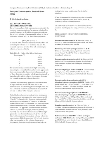
G.-H. Liang et al. / Neurotoxicology and Teratology 26 (2004) 253–260distilled H2O as a stock solution of 10 mM and stored at20 jC until further use.3. ResultsAll cells from which K currents were obtained displayed a basally located nucleus without appreciable blebs,a prominent cuticular plate, were phase bright, exhibited noBrownian motion and possessed a readily identifiable stereocilia bundle. Ohmic leakage current (IL) of from approximately 50 to 400 pA that shifted the baseline from zero wascorrected offline. IL was present for the much longer haircells ( 70 Am) than the shorter cells ( 40 Am) and as therecording time of the experiments progressed. The maximum time that cells remained completely healthy for recordings was approximately 20 – 40 min. A compromised cellcondition was readily detectable in the form of a more255positive resting potential (ranged from 25 to 55 mV).All recordings were terminated when Vrest declined to V 25mV, when prominent Brownian motion occurred, a visibleswollen membrane occurred, a grossly deformed morphology was present, or the giga-ohm seal was lost.3.1. Effects of 0.1, 1.0, 10, and 100 lm Pb2 The effects of 0.1 AM Pb2 (Fig. 1A), 1.0 AM Pb2 (Fig. 1B), 10 AM Pb2 (Fig. 1C), and 100 AM Pb2 (Fig.1D) on Kv outward currents and a lack of effects on K inward currents are presented. The rapid current spikes atthe onset and offset of the voltage pulses at each concentration (except 100 AM Pb2 ) are indicative of a mixtureof the electromotor activity of the OHC and membranecapacitive transients [1]. In each instance, the outwardgoing current displayed kinetics of the delayed rectifiertype (Kv) by its slow onset and its failure to inactivateFig. 1. Potassium currents (Kv) and block by Pb2 elicited by hyperpolarization and depolarizing voltage steps in guinea pig OHC. Voltage steps of 300 msduration were used at a holding potential of 70 mV, with voltage stepped to 130 mV (inward traces) and 30 (outward traces) mV. (A) Pb2 acetate bathapplied at 0.1 AM for 5.0 min. (B) Pb2 acetate bath applied at 1.0 AM for 5.0 min. (C) Pb2 acetate bath applied at 10 AM for 5.0 min. (D) Pb2 acetate bathapplied at 100 AM for 5.0 min.
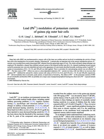
256G.-H. Liang et al. / Neurotoxicology and Teratology 26 (2004) 253–260over the period of the 300-ms pulse. The control currentscould be partially suppressed with the organic cations of25 mM TEA or 100 AM 4-AP applied to the bath (fourcells, data not shown). The application of 100 AM CdCl2further suppressed the outward currents suggesting a calcium component (e.g., IK(Ca)) as part of these currents (datanot shown).The application of Pb2 applied to the bath was investigated on three or four OHC for each concentration. Thecell motility/capacitive spikes, present at the three lowestconcentrations, were largely unaffected by the application ofPb2 (Fig. 1). The ratio of the difference between controlrecordings and the application of Pb2 for the outwardcurrent for the individual cells of Fig. 1 was 0.10 at 0.1AM, 0.14 at 1.0 AM, 0.18 at 10 AM, and 0.30 at 100 AM.Thus, the outward current was inhibited while the inwardcurrent due to Pb2 treatment was unaffected. That is, Pb2 only slightly decreased the peak amplitude of the inwardcurrent, and the steady state end current amplitude of theinward K current was not inhibited at any of the concentrations. The inward IL was unaffected by the externalapplication of Pb2 . Pb2 did not shift the reversal potentialfor any of the Pb2 concentrations tested (data not shown).Given that Pb2 did not affect the inward currents, the timecourse of these currents will not be displayed.3.2. Time-dependent effects of Pb2 The time-dependent effect by which bath-applied 1.0 AMPb2 inhibited OHC Kv was investigated in three OHC. Fig.2 shows the time inhibition of the mean outward peakcurrents including F 1.0 S.E.M. Control recordings wereobtained in which outward currents were recorded to fivepresentations of the positive pulses at 1-min intervals from aFig. 2. Time course of 0.1 AM Pb2 to outward Kv peak current of threeOHC. The first five data points are control recordings in normal externalsolution; the second five data points were obtained in the presence of 0.1AM Pb2 . The final 10 data points were obtained during wash with normalexternal solution.Fig. 3. Time course of 1.0 AM Pb2 to outward Kv peak current of threeOHC. The first five data points are control recordings with normal externalsolution; the second five data points were obtained in the presence of 1.0AM Pb2 . The final 10 data points were obtained during wash with normalexternal solution.VH of 70 mV stepped to 30 mV. This was followed byusing the same stimulus protocol in the presence of 0.1 AMPb2 . Two 5-min washes were employed in an attempt toreverse the effects of Pb2 application. The second washfollowed the first one by 6.0 min, in which only two cellswere tested, as one cell did not survive the period of thesecond wash. The inhibition was not reversible with the twoconsecutive periods of washout with normal external solution. The average fraction of current inhibition for the peakonset current was 0.10 for 0.1 AM Pb2 . The increasedvariability of the recordings as time increased is reflected bythe increased size of the standard error bars.Fig. 3 shows the mean effects with F 1.0 S.E.M. of theapplication of 1.0 AM Pb2 on the outward K currents.The effect of 1.0 AM Pb2 on the peak outward currentsover time was 0.14. From the first to the fifth pulse duringtreatment with Pb2 , the current continued to decrease. Twoconsecutive washes with normal external solution did notcause appreciable recovery of the amplitude of the currentwhen compared to the control current. Since the outwardcurrent contains a mixture of IK(Ca), the Pb2 may beblocking Ca2 entry through the voltage-sensitive calciumchannels. Upon the application of Pb2 , the variability ofthe current recordings increased as reflected in the increasedsize of the error bars. This was particularly noteworthy forthe two 5.0-min washes.The inhibition effects of adding 10 AM Pb2 to the bathwere investigated in four OHC (see Fig. 4). Outward goingKv exhibited an average ratio reduction of 0.18. An initial5.0-min wash failed to return the currents to the preexposurelevels. There was partial recovery of the currents to thesecond 5.0-min wash. Inactivation kinetics could not becalculated since the duration of the pulses of 300 ms failedto yield any appreciable inactivation.
G.-H. Liang et al. / Neurotoxicology and Teratology 26 (2004) 253–260257Table 1Distribution of statistical values for Pb2 at 0.1, 1.0, 10, and 100 AMPb2 AXc0.11.010.01000.100.140.180.30XPb2 10.0010 *0.0002 * 0.0001 * 0.0001 *Pb2 A is the concentration of lead, Xc XPb2 is control minus the leadtreatment, t is the value of the t test, df is degrees of freedom, S.E. is thestandard deviation, and P is the two-tailed probability. * Statisticalsignificance beyond the 0.01 level.3.3. Maximum effects of Pb2 on outward KvFig. 4. Time course of 10 AM Pb2 to outward Kv peak current of fourOHC. The first five data points are control recordings with normal externalsolution; the second five data points were obtained in the presence of 10AM Pb2 . The final 10 data points were obtained during wash with normalexternal solution.The maximum inhibition for the amount of Pb2 used inthe present investigation was calculated. The current amplitude was expressed as the percentage of the control current(mean F S.E., n 3– 4 cells). The average maximum inhibition for the outward Kv was 30% at 100 AM Pb2 . Giventhat the maximum current inhibition was less than 50%, anIC50 was not computed.3.4. Statistical analysesFig. 5 displays the effects of bath application of 100 AMPb2 to three cells. The control recordings reflect very littlevariability of the five different recordings that establishedthe baseline. When 100 AM Pb2 was applied to the bath,there was an average ratio reduction of 0.30. The variabilityof the recordings, however, increased with the application ofPb2 . A 5.0-min wash did not return the current amplitudeto the preexposure levels, and the variability of the currentsincreased as reflected in the increased size of the standarderror bars.A t test for correlated samples was used to probe for thesignificance of the difference between the amplitude of thecurrent of the control data and the amplitude of the current ofthe Pb-treated data at each of the Pb2 concentrations. Table1 summarizes these data. The Pb-treated inhibitions of theK current were significantly different from the controlcurrent at the 0.01 levels for the four concentrations ofPb2 used.4. DiscussionFig. 5. Time course of 100 AM Pb2 to outward Kv peak current of threeOHC. The first five data points are control recordings with normal externalsolution; the second five data points were obtained in the presence of 100AM Pb2 . The final 10 data points were obtained during wash with normalexternal solution.We have confirmed [1,2] the existence of a distinct Kvchannel in guinea pig OHC that generate current of theoutward type—the delayed rectifier current with a mixtureof calcium-activated components. Channels mediating inward currents carried by potassium channels were alsoidentified and took on the appearance of a fast partialinactivating type and a slow steady state non-inactivatingtype of the IK(n) type [2,13,16,17,18,23,26,27,32]. Outwardgoing Kv were partially blocked by 25 mM TEA, 100 AM4-AP, and by 100 AM CdCl2 (data not shown), indicatingthat the currents obtained were K currents and that parts ofthe current were Ca activated. These several compounds hadno effect on the inward IK(n). Thus, it would appear that theinward IK(n) does not contain a mixture of the Ca-activatedcomponent as seen in the outward current.Cell electromotility has been observed previously tohyperpolarizing and depolarizing pulses [1] and was observed in the present investigation. The electromotility ofthe cells occurs to the changing electric field [1,6,9] whenthe cells are stimulated. The motile responses of the cells
258G.-H. Liang et al. / Neurotoxicology and Teratology 26 (2004) 253–260were unaffected by Pb2 at any of the concentrations.However, a few cells did not exhibit the motility-dependent variation in cell shortening and lengthening as attributed to the kinetics of K channels [16,26,27] prior totreatment. Quinine, another ototoxic agent with propertiessimilar to heavy metals, applied in vitro on OHC at aconcentration of 5.0 mM, substantially decreased activeforces of cell elongation and shortening [19]. These resultsare indicative that Pb2 perhaps has different actions oncell membrane properties and motility mechanisms thanthat of quinine.The results suggest that OHC outward components canbe slightly modulated by various concentrations of Pb2 .The decrease of outward current ranged from 10% to 30%at 0.1 – 100 AM. Previous reports have reported thatchronic exposure to Pb2 , however, substantially increasedcalcium currents in both invertebrate and mammalian cells[4]. The inward current was not reduced by Pb2 . Thismay indicate that separate perhaps independent channelsare generating the inward currents when compared to theoutward currents. The blocked currents were not totallytime-dependent reversible after washing. These results arein agreement with several mammalian studies in whichPb2 action proved irreversible to varying degrees inseveral biological preparations [33,34,48], but not inagreement with other results of various K currents [9,24,25,45,47,52].Kv was recorded in all OHC tested, and it is not at alluncommon that slowly inactivating or noninactivating conductances can be found in the same cell [40]. Kv is moreslowly activated, and unless a very long pulse is used in therange of several seconds, it will not inactivate. It is interesting to speculate that Pb2 may be acting on OHCsimilarly to the effects of the anesthetic agent ether[(CH3CH2)2O] of the Shaker mutant in which Kv is reduced, and thus membrane currents become electricallyunstable [9,25,52].Another important consideration of the present data isthat our isolation technique is biased toward OHC locatedfrom the apical and middle part of the cochlea or the mid- tolow-frequency regions of the cochlea. Typically, this is aregion that has been for the most part insensitive to mostototoxic agents. To what extent our selection of cells thatwere biased more toward the apical end of the cochlea canexplain the small effects observed here is unknown and willhave to await a more extensive in vitro electrophysiologicalsurvey of OHC from different regions of the sensoryepithelium. Another factor important for consideration isthat the observations of the present study were obtained atroom temperature. Thus, the small effects of the Pb2 maybe explained based on the fact that room temperature is not,of course, the normal physiological condition of the cochlea. These data are consistent, however, with the effects ofPb2 on IK(Ca) since the outward currents contain a mixtureof K channel conductances that include the calciumcomponent [37].While OHC are not directly coupled to inner hair cells,but since they are purported to modulate inner hair cellactivity via mechanical properties, it is suggested that thepresent data may account for the underlying results foundfor the slight voltage threshold modifications of the compound AP in the guinea pig by Pb2 [50]. That is, amodification of K conductances by Pb2 may interferewith the resting membrane potential, the repolarizationphase, and after hyperpolarization of the IHC receptorpotentials, perhaps causing an abnormal release of neurotransmitter substance(s) and a subsequent modification ofAP transduction in afferent auditory nerve fibers [51]. Theseresults may suggest also that at higher doses, Pb2 molecules may get stuck in the channel responsible for theoutward currents and cannot be expelled at least within areasonable time or expulsion may be voltage dependent,which was not tested under these conditions. The fact thatthe effects of Pb2 are not completely reversible by washingperhaps is indicative that palliative chelation therapy thatprevents or reverses the binding of metallic cations tocellular ligands may not hold excellent promise to preventor reverse peripheral or central effects of the auditory systemdue to the effects of Pb2 [20]. The association of BLL to anumber of neurodevelopmental conditions [12,14,38,46]should not be minimized, however, in spite of the relativelybenign effects of Pb2 observed in vitro on hair cells in thepresent investigation, albeit hair cells have an epithelialorigin and may therefore react differently than nerve cells.However, based on the present observations, we cannotreconcile differences between acute effects of lead on ionchannel function as measured here and potential effects ofchronic developmental exposure, which occur in the humanpopulation as a cause of hearing loss [5,7,29,31].AcknowledgementsThis research was supported in part by grants from theSwedish Research Council, the Swedish Council for WorkingLife and Social Research, the Foundation Tysta Skolan, andthe Foundation Wenner-Grenska Samfundet. EJM wassupported by funds from the Northwestern Drug DiscoveryProgram. The authors wish to thank Dr. S. Allen Counter ofHarvard University and Dr. Bo Rydqvist of the KarolinskaInstitutet for insightful comments about the manuscript.References[1] J.F. Ashmore, A fast motile response in guinea-pig outer hair cells: thecellular basis for the cochlear amplifier, J. Physiol. (Lond.) 388 (1987)323 – 347.[2] J.F. Ashmore, R.W. Meech, Ionic basis of membrane potential in outerhair cells of guinea-pig cochlea, Nature 322 (1986) 368 – 371.[3] W.D. Atchison, T. Narahashi, Mechanism of action of lead on neuromuscular junctions, Neurotoxicology 5 (1984) 267 – 282.[4] G. Audesirk, T. Audesirk, The effects of inorganic lead on voltage-
G.-H. Liang et al. / Neurotoxicology and Teratology 26 (2004) e calcium channels differ among cell types and among channelsubtypes, Neurotoxicology 14 (1983) 259 – 266.D. Bellinger, A. Leviton, C. Waternaux, H. Neddleman, M. Rabinowitz,Longitudinal analyses of prenatal and postnatal lead exposure and earlycognitive development, N. Engl. J. Med. 316 (1987) 1037 – 1043.W.E. Brownell, Observation on a motile response in isolated hair cells,in: W.R. Webster, L.M. Aiken (Eds.), Mechanisms of Hearing, Monash Univ. Press, Melbourne, 1983, pp. 5 – 10.S.A. Counter, M. Vahter, G. Laurell, L.H. Buchanan, F. Ortega, S.Skerfving, High lead exposure and auditory sensory-neural function inAndean children, Environ. Health Perspect. 105 (1997) 522 – 526.M.R. Cullen, J.M. Robins, B. Eskenazi, Adult inorganic lead intoxication: presentation of 31 new cases and a review of recent advancesin the literature, Medicine 62 (1983) 221 – 247.X.Q. Dai, D. Ruan, J. Chen, M. Wang, L. Cai, The effects of lead ontransient outward currents of acutely dissociated rat dorsal root ganglia, Brain Res. 904 (2001) 327 – 340.P. Dallos, Overview: cochlear neurobiology, in: P. Dallos, A.N.Fay, R.R. Fay (Eds.), Springer Handbook of Auditory Research: thecochlea vol. 8, Springer, New York, 1996, pp. 1 – 43.K. Dietrich, P. Succop, O. Berger, R. Keith, Lead exposure and thecentral auditory processing abilities and cognitive development ofurban children: the cincinnati lead study cohort at age 5 years, Neurotoxicol. Teratol. 14 (1992) 51 – 56.E. Ernst, Heavy metals in traditional Indian remedies, Eur. J. Clin.Pharmacol. 57 (2002) 891 – 896.P.A. Fuchs, Synaptic transmission at vertebrate hair cells, Curr. Opin.Neurobiol. 6 (1996) 514 – 519.A. Gemmel, M. Tavares, S. Alperin, J. Soncini, D. Daniel, J. Dunn, S.Crawford, N. Braveman, T.W. Clarkson, S. McKinlay, D.C. Bellinger,Blood lead level and dental caries in school-age children, Environ.Health Perspect. 110 (2002) A625 – A630.Y. Holdstein, H. Pratt, M. Goldsher, G. Rosen, R. Shenhav, S.Linn, A. Mor, A. Barkai, Auditory brainstem evoked potentials inasymptomatic lead-exposed subjects, J. Laryngol. Otol. 100 (1986)1031 – 1036.G.D. Housley, J.F. Ashmore, Ionic currents of outer hair cells isolatedfrom the guinea-pig cochlea, J. Physiol. (Lond.) 448 (1992) 73 – 98.D.J. Jagger, J.F. Ashmore, Effect of intracellular calcium release onionic currents in outer hair cells isolated from the guinea-pig cochlea,J. Physiol. (Lond.) 489P (1995) 48P.D.J. Jagger, J.F. Ashmore, Regulation of ionic currents by proteinkinase A and intracellular calcium in outer hair cells isolated fromthe guinea-pig cochlea, Pflügers Arch. 437 (1999) 409 – 416.J. Jarboe, R. Hallworth, The effect of quinine on outer hair cell shape,compliance and force, Hear. Res. 132 (1999) 43 – 50.C. Klaassen, Heavy metals and heavy-metal antagonists, in: J.G.Hardman, L.E. Limbird, P.B. Molinoff, R.W. Ruddon, A.G. Gilman (Eds.), Goodman and Gilman’s, The Pharmacological Basisof Therapeutics, McGraw-Hill, New York, 1996, pp. 1649 – 1671.K. Kostial, V.B. Vouk, Lead ions and synaptic transmission in thesuperior cervical ganglion of the cat, Br. J. Pharmacol. 12 (1997)219 – 222.F. Lille, P. Hazemann, R. Garnier, S. Dally, Effects of lead and mercuryintoxication on evoked potentials, Clin. Toxicol. 26 (1988) 103 – 116.X. Lin, R.I. Hume, A.L. Nuttall, Dihydropyridines and verapamilinhibit voltage-dependent potassium current in isolated outer hair cellsof the guinea-pig, Hear. Res. 88 (1995) 34 – 36.M. Madeja, N. Binding, U. Musshoff, O. Pongs, U. Witting, E.J.Speckmann, Effects of lead on cloned voltage-operated neuronal potassium channels, Naunyn-Schmiedeberg’s Arch. Pharmacol. 351(1995) 320 – 327.M. Ma
Lead (Pb2 ) modulation of potassium currents of guinea pig outer hair cells G.-H. Lianga,L.Ja rlebarka, M. Ulfendahla, J.-T. Bianb, E.J. Mooreb,c,* aCenter for Hearing and Communication Research, Department of Clinical Neuroscience, Karolinska Institutet, S-171 76 Stockholm, Sweden bDepartment of Molecular Pharmacology and Biological Chemistry (S215), Northwestern University Feinberg School .