Transcription
DEWS Definition and ClassificationThe Definition and Classification of Dry Eye Disease:Report of the Definition and Classification Subcommittee ofthe International Dry E y e W ork Shop (2 0 0 7 )ABSTRACT The aim of the DEWS Definition and Classification Subcommittee was to provide a contemporary definitionof dry eye disease, supported within a comprehensive classification framework. A new definition of dry eye was developed to reflect current understanding of the disease, and thecommittee recommended a three-part classification system.The first part is etiopathogenic and illustrates the multiplecauses of dry eye. The second is mechanistic and shows howeach cause of dry eye may act through a common pathway.It is stressed that any form of dry eye can interact with andexacerbate other forms of dry eye, as part of a vicious circle.Finally, a scheme is presented, based on the severity of thedry eye disease, which is expected to provide a rational basisfor therapy. These guidelines are not intended to override theclinical assessment and judgment of an expert clinician inindividual cases, but they should prove helpful in the conductof clinical practice and research.KEYWORDS definition, DEWS, dry eye disease, Dry EyeWorkShop, etiopathogenesis, mechanism, severity gradingAccepted for publication January 2007.Definition and Classfication Subcommittee members: M ic hae l A . L e m p , M D(Chair); Christophe Baudouin, MD, PhD; Jules Baum, MD; Murat Dogru,MD; Gary N. Foulks, MD; Shigeru Kinoshita, MD; Peter Laibson, MD; JamesMcCulley, MD; Juan Murube, MD, PhD; Stephen C. Pflugfelder, MD; Mauriz ioRolando, MD; Ikuko Toda, MD.The Subcommittee is indebted to Professors A.J. Bron and G.N. Foulks fortheir invaluable contributions to the writing of this report.Proprietary interests of Subcommittee members are disclosed on pages 202and 204.Reprints are not available. Articles can be accessed at: www.tearfilm.orgCorrespondence in regard to the this chapter should be addressed to MichaelA. Lemp, MD, 4000 Cathedral Avenue NW, Apt 828B, Washington, DC 20016(Email: malemp@lempdc.com. Tel: 202-338-6424) 2007 Ethis Communications, Inc. The Ocular Surface ISSN: 15420124. (No authors listed ). T he d efi nition and classifi cation of d ry eyed isease: report of the Defi nition and Classifi cation Sub committee ofthe International Dry Eye W orkShop (2007). 2007;5(2):75-92.I. INTRODUCTIONhe Definition and Classification Subcommitteereviewed previous definitions and classificationschemes for dry eye, as well as the current clinicaland basic science literature that has increased and clarifiedknowledge of the factors that characteriz e and contribute todry eye. Based on its findings, the Subcommittee presentsherein an updated definition of dry eye and classificationsbased on etiology, mechanisms, and severity of disease.TII. GOALS OF THE DEFINITION ANDCLASSIFICATION SUBCOMMITTEEThe goals of the DEWS Definition and ClassificationSubcommittee were to develop a contemporary definition ofdry eye disease and to develop a three-part classification ofdry eye, based on etiology, mechanisms, and disease stage.The manner of working of the committee is outlined inthe introduction to this issue of The O cular S urface. Furtherdetails are published on the TFOS-DEWS web-site (www.tearfilm.org).III. DEFINITION OF DRY EYE DISEASEThe committee reviewed the definition and classification presented at the 19 9 5 National Eye Institute (N E I)/Industry Dry Eye Workshop, which was: Dry eye is a disorderof the tear film due to tear deficiency or excessive evaporation,which causes damage to the interpalpebral ocular surface andis associated with symptoms of ocular discomfort.1The committee agreed that the definition could beimproved in the light of new knowledge about the roles oftear hyperosmolarity and ocular surface inflammation indry eye and the effects of dry eye on visual function. Initiallytwo definitions were developed and presented to membersof the workshop. These “general” and “operational” definitions overlapped to some ex tent, and, therefore, in this finalreport, these versions have been combined to produce thefollowing definition:Dry eye is a multifactorial disease of the tears and ocular surface that results in symptoms of discomfort,2-4visual disturbance,5-7 and tear film instability8-10 withpotential damage to the ocular surface. It is accompanied by increased osmolarity of the tear film11-14 andinflammation of the ocular surface.15,16THE OCULAR SURF ACE / APRIL 2007, VOL. 5, NO. 2 / www.theocularsurface.com75
DEWS DEFINITION AND CLASSIFICATIONOUTLINEI. IntroductionII. Goals of the Definition and ClassificationSubcommitteeIII. Definition of dry eye diseaseIV . Classification of dry eye diseaseA. BackgroundB. Etiopathogenic classification of dry eye disease1. Aqueous tear-deficient dry eyea. Sjogren syndrome dry eyeb. Non-Sjogren syndrome dry eye1) Primary lacrimal gland deficiencies2) Secondary lacrimal gland deficiencies3) Obstruction of the lacrimal gland ducts4) Reflex hyposecretiona) Reflex sensory blockb) Reflex motor block2. Evaporative dry eyea. Intrinsic causes1) Meibomian gland dysfunction2) Disorders of lid aperature and lid/globecongruity or dynamics3) Low blink rateb. Extrinsic causes1) Ocular surface disorders2) Contact lens wear3) Ocular surface disease4) Allergic conjunctivitisC. The causative mechanisms of dry eye1. Tear hyperosmolarity2. Tear film instabilityD. The basis for symptoms in dry eyeE. Classification of dry eye based on severityDry eye is recognized as a disturbance of the LacrimalF unctional U nit (LFU), an integrated system comprisingthe lacrimal glands, ocular surface (cornea, conjunctivaand meibomian glands) and lids, and the sensory and motor nerves that connect them.17 Trigeminal sensory fibersarising from the ocular surface run to the superior salivarynucleus in the pons, from whence efferent fibers pass, in thenervus intermedius, to the pterygopalatine ganglion. Here,postganglionic fibers arise, which terminate in the lacrimalgland, nasopharynx, and vessels of the orbit. Another neuralpathway controls the blink reflex, via trigeminal afferentsand the somatic efferent fibers of the seventh cranial nerve.Higher centers feed into the brainstem nuclei, and there isa rich sympathetic supply to the epithelia and vasculatureof the glands and ocular surface.This functional unit controls the major componentsof the tear film in a regulated fashion and responds toenvironmental, endocrinological, and cortical influences.Its overall function is to preserve the integrity of the tear76film, the transparency of the cornea, and the quality of theimage projected onto the retina.17-20 At the 2007 Dry EyeWorkShop, it was noted that the corneal and conjunctivalepithelia are in continuity, through ductal epithelia, withthe acinar epithelia of the main and accessory lacrimalglands and the meibomian glands, which themselves ariseas specialized invaginations from the ocular surface. Also,these epithelia have the same embryological derivation. Thisbroader concept, which has additional features, has beentermed the Ocular Surface System and is discussed furtherin the “Research” chapter of this issue.21An important aspect of the unit is the part played bysensory impulses, which arise from the ocular surface, in themaintenance of resting tear flow. Currently, it is consideredthat waking tear flow is a reflex response to afferent impulses deriving particularly, but not entirely, from the ocularsurface.22 Sensory input from the nasal mucosa also makesa contribution.23 Disease or damage to any component ofthe LFU (the afferent sensory nerves, the efferent autonomicand motor nerves, and the tear-secreting glands) can destabilize the tear film and lead to ocular surface disease thatexpresses itself as dry eye. Tear film stability, a hallmark ofthe normal eye, is threatened when the interactions betweenstabilizing tear film constituents are compromised by decreased tear secretion, delayed clearance, and altered tearcomposition. Ocular surface inflammation is a secondaryconsequence. Reflex tear secretion in response to ocularirritation is envisioned as the initial compensatory mechanism, but, with time, inflammation accompanying chronicsecretory dysfunction and a decrease in corneal sensationeventually compromises the reflex response and results ineven greater tear film instability. Perturbation of the LFUis considered to play an important role in the evolution ofdifferent forms of dry eye.The distinctions aq ueous-deficient dry eye and evaporativedry eye were removed from the definition, but are retainedin the etiopathogenic classification.IV. CLASSIFICATION OF DRY EYE DISEASEA. BackgroundV itali, writing about the harmonized classification criteria for Sjogren syndrome (SS) remarked that classificationcriteria are not necessarily appropriate for use in diagnosisand may lead to misclassification of a disease, particularlyin its early stages.24 In an individual patient, a classificationscheme can provide a guide, but an expert clinician, applying appropriate diagnostic criteria, is needed to establisha diagnosis.Although the NEI/Industry Workshop classification1 hasserved as a useful and durable scheme for over a decade, itdoes not reflect newer knowledge on pathophysiologicalmechanisms, effects on vision, and the utility of an assessment of severity of disease. Recently, two new classificationschemes were published, and these were used as sourcedocuments by the committee. These include: the TripleClassification25,26 and the report of the Delphi panel.27The Triple Classification evolved from reports presentedTHE OCULAR SURFACE / APRIL 2007, VOL. 5, NO. 2 / www.theocularsurface.com
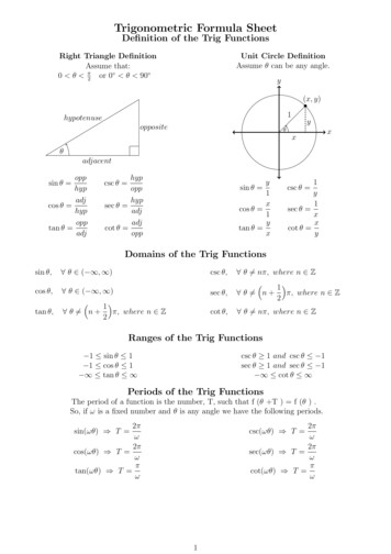
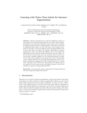
DEWS DEFINITION AND CLASSIFICATIONDRY EYEEff ect of theEnvironmentMilieu InterieurLow blink ratebehavior, VTU,microscopyWide lid aperturegaze positionAgingLow androgen poolSystemic diuretics, andsome ry EyePrimaryIntrinsicNon-SjogrenDry EyeSecondaryLacrimalDeficiencyMilieu ExterieurLow relative humidityHigh wind Gland DuctObstructionReflex BlockMeibomian OilDeficiencyExtrinsicVitamin ADeficiencyDisordersof LidApertureTopical DrugsPreservativesLow BlinkRateDrug ActionAccutaneContact LensWearOcular SurfaceDiseaseeg, AllergySystemicDrugsFigure 1.Major etiological causes of dry eye.The left hand box illustrates the influence of environment on the risk of an individual to develop dry eye. The term “environment” is usedbroadly, to include bodily states habitually experienced by an individual, whether it reflects their “milieu interieur” or is the result of exposureto external conditions which represent the “milieu exterieur.” This background may influence the onset and type of dry eye disease in anindividual, which may be aqueous-deficient or evaporative in nature.Aqueous-deficient dry eye has two major groupings, Sjogren syndrome dry eye and non-Sjogren syndrome dry eye.Evaporative dry eye may be intrinsic, where the regulation of evaporative loss from the tear film is directly affected, eg, by meibomian lipiddeficiency, poor lid congruity and lid dynamics, low blink rate, and the effects of drug action, such as that of systemic retinoids. Extrinsicevaporative dry eye embraces those etiologies that increase evaporation by their pathological effects on the ocular surface. Causes includevitamin A deficiency, the action of toxic topical agents such as preservatives, contact lens wear and a range of ocular surface diseases,including allergic eye disease. Further details are given in the text.at the 14th Congress of the European Society of Ophthalmology.25 After further clinical experience, an updated version was published in 2005, which presented three separateschemes: one based on etiopathogenesis; one based on theglands and tissues targeted in dry eye; and one based ondisease severity.26The committee felt that the concept of three differentschemes serving different purposes was attractive, but itwas noted that evidence-based referencing was limited.For this reason, the scheme as a whole was not adopted,but many conceptual aspects were incorporated into thecommittee’s final schemes.The Delphi Panel was a consensus group that met toreview the classification of dry eye.27 The panel proposedchanging the name of dry eye disease to dysfunctional tear syndrome, suggesting that the name more accurately reflectedpathophysiological events in dry eye. However, althoughthe committee felt that the term embraced the essentialfeatures of the disease, they concluded that retention of thename dry eye had much to recommend it and that its usewas embedded in the literature. The committee also rejecteda subdivision based on the presence or absence of lid disease, because it is frequently difficult to identify the relativecontribution of lid disease to a particular case of dry eye.The majority of the Definition and Classification Subcommittee was in favor of adopting a severity grading basedon the report of the Delphi Panel, recognizing it as a comprehensive approach that could form the basis of therapyaccording to severity of the disease. As noted above, theTriple Classification also presented a severity grading.B. Etiopathogenic Classification of Dry Eye DiseaseThe etiopathogenic classification developed by theSubcommittee is an updated version of that presented inthe NEI/Industry Workshop Report and reflects a morecontemporary understanding of dry eye disease (Figure 1).THE OCULAR SURFACE / APRIL 2007, VOL. 5, NO. 2 / www.theocularsurface.com77
DEWS DEFINITION AND CLASSIFICATIONAs in the 1995 report, the term dry eye is regarded as synonymous with the term keratoconjunctivitis sicca (KCS).The classification has the following features:The left hand box in Figure 1 illustrates the influence ofenvironment on an individual’s risk of developing dry eye.The term environment is used broadly to include physiological variation between individuals (their milieu interieur), aswell as the ambient conditions that they encounter (theirmilieu exterieur).The milieu interieur implies physiological conditionsparticular to an individual that could influence their riskof dry eye. For instance, a normal subject may have a lownatural blink rate, or the blink rate may be slowed for behavioral or psychological reasons.28 Slowing of the blinkrate increases the blink interval and increases the periodof evaporative loss between each blink.29Similarly, the natural height of the palpebral aperture inthe primary position varies between individuals and betweenethnic groups.30 The aperture is also wider in upgaze thandowngaze.31 Evaporative loss per eye increases with increasing palpebral width and is, therefore, increased in upgaze.32Extensive evidence supports a role for the sex hormonesin the etiology of dry eye33 with the generalization that lowlevels of androgens and high estrogen levels are risk factorsfor dry eye. Biologically active, androgens promote lacrimaland meibomian gland function.33 Androgen deficiency isassociated with dry eye34 and may be prevented by topicalor systemic androgen therapy.35-38 Dry eye occurs in patientsexposed to anti-androgens in the treatment of prostaticcancer,39,40 and women with complete androgen insensitivity syndrome show an increase in the signs and symptomsof dry eye, associated with evidence of meibomian glandand goblet cell dysfunction.41-43 A significantly depletedandrogen pool in “non-autoimmune” dry eye associatedwith meibomian gland dysfunction (MGD) has been reported.44 Also, as noted elsewhere in this issue,45 femalesex and postmenopausal estrogen therapy are importantrisk factors for dry eye,46,47 and women with prematureovarian failure suffer from the symptoms and signs of dryeye, although their tear production is not affected.48Lacrimal tear secretion is reduced by a number ofsystemic drugs, and these effects may be looked upon asdisturbances of the milieu interieur. Their details are discussed later in this report. Aging is associated with physiological changes that may predispose to dry eye, includingdecreased tear volume and flow, increased osmolarity,49decreased tear film stability,50 and alterations in the composition of the meibomian lipids.51The milieu exterieur involves the occupational andexternal environments, which may represent risk factorsfor the development of dry eye. Evaporative water lossfrom the eye is increased in conditions of low relativehumidity, occurring either as part of natural variation atdifferent geographic locations or in special circumstancescreated by air-conditioning, air travel, or other artificialenvironments.52 Similarly, tear evaporation is increased byexposure to high wind velocity, and this mechanism has78been incorporated into some of the newer experimentalmodels of dry eye.Occupational factors may cause a slow blink rate, representing a risk for dry eye in those working with video display terminals.53 Other activities associated with decreasedblinking and an increase in palpebral width, including thatassociated with upgaze, have been reported to carry a riskfor the development of dry eye symptoms.The major classes of dry eye, as in the 1995 workshop,1are still held to be aqueous tear-deficient dry eye (ADDE)and evaporative dry eye (EDE). The category ADDE referschiefly to a failure of lacrimal secretion, and this approach isretained. However, it should be recognized that a failure ofwater secretion by the conjunctiva could also contribute toaqueous tear deficiency. The class EDE has been subdividedto distinguish those causes that are dependent on intrinsicconditions of the lids and ocular surface and those that arisefrom extrinsic influences.Dry eye can be initiated in any of these classes, but theyare not mutually exclusive. It is recognized that disease initiated in one major subgroup may coexist with or even leadto events that cause dry eye by another major mechanism.This is part of a vicious circle of interactions that can amplifythe severity of dry eye. An example might be that all formsof dry eye cause goblet cell loss and that this, in turn, willcontribute to loss of tear film stability, to surface damageand evaporative water loss, and to symptoms resulting froma loss of lubrication and surface inflammatory events.The major classes and subclasses of dry eye are described below.1. Aqueous Tear-Deficient Dry Eye (Tear DeficientDry Eye; Lacrimal Tear Deficiency)Aqueous tear-deficient dry eye implies that dry eye isdue to a failure of lacrimal tear secretion. In any form ofdry eye due to lacrimal acinar destruction or dysfunction,dryness results from reduced lacrimal tear secretion andvolume.54,55 This causes tear hyperosmolarity, because,although the water evaporates from the ocular surface atnormal rates, it is from a reduced aqueous tear pool. Tearfilm hyperosmolarity causes hyperosmolarity of the ocularsurface epithelial cells and stimulates a cascade of inflammatory events involving MAP kinases and NFkB signallingpathways56,57 and the generation of inflammatory cytokines(interleukin (IL)-1A; -1B; tumor necrosis factor (TNF)-A)and matrix metalloproteinases (MMP-9).58 When lacrimaldysfunction is due to lacrimal gland infiltration and inflammation, inflammatory mediators generated in the gland areassumed to find their way into the tears and be deliveredto the ocular surface. However, when such mediators aredetected in the tears, it is not usually possible to knowwhether they derive from the lacrimal gland itself or fromthe ocular surface (conjunctiva and cornea).It is uncertain whether evaporation is reduced59 or increased59-64 in ADDE. It is possible that this is determinedby the stage of the disease. Some studies suggest that thereservoir of lid oil is larger in non-Sjogren syndrome dryTHE OCULAR SURFACE / APRIL 2007, VOL. 5, NO. 2 / www.theocularsurface.com
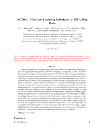
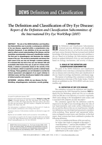
DEWS DEFINITION AND CLASSIFICATIONantibody) directed againstmuscarinic receptors within the glands.71-73I. Ocular symptoms: a positive response to at least one of the following questions:There are two forms1. H ave you had daily, persistent, troublesome dry eyes for more than 3 months?of SS, and classification2. Do you have a recurrent sensation of sand or gravel in the eyes?criteria have recently been3. Do you use tear substitutes more than 3 times a day?harmonized in a EuropeanII. Oral symptoms: a positive response to at least one of the following questions:American collaboration.741. H ave you had a daily feeling of dry mouth for more than 3 months?Primary SS consists of the2. H ave you had recurrently or persistently swollen salivary glands as an adult?occurrence of ADDE in3. Do you frequently drink liquids to aid in swallowing dry food?combination with sympIII. Ocular signs: that is, objective evidence of ocular involvement defined as a positive resulttoms of dry mouth, in thefor at least one of the following two tests:presence of autoantibod1. Schirmer I test, performed without anesthesia (b5 mm in 5 minutes)ies, evidence of reduced2. Rose bengal score or other ocular dye score (r4 according to van Bijsterveld’s scoringsalivary secretion and withsystem)a positive focus score onIV. Histopathology: In minor salivary glands (obtained through normal-appearing mucosa) focalminor salivary gland bilymphocytic sialoadenitis, evaluated by an expert histopathologist, with a focus score r1,opsy.75,76 Details of the cridefined as a number of lymphocytic foci (which are adjacent to normal-appearing mucousteria are presented in Tableacini and contain more than 50 lymphocytes) per 4 mm2 of glandular tissue181. Secondary SS consists ofV. Salivary gland involvement: objective evidence of salivary gland involvement defined by athe features of primary SSpositive result for at least one of the following diagnostic tests:together with the features1. U nstimulated whole salivary flow (b1.5 ml in 15 minutes)of an overt autoimmune2. Parotid sialography showing the presence of diffuse sialectasias (punctate, cavitary ordestructive pattern), without evidence of obstruction in the major ducts19connective disease, such as3. Salivary scintigraphy showing delayed uptake, reduced concentration and/or delayedrheumatoid arthritis, whichexcretion of tracer20is the most common, orVI. Autoantibodies: presence in the serum of the following autoantibodies:systemic lupus erythema1. Antibodies to Ro(SSA) or La(SSB) antigens, or bothtosis, polyarteritis nodosa,Wegener’s granulomatosis,Reprinted with permission from: Vitali C, Bombardieri S, Jonnson R, et al. Classification criteria for Sjogren’ssystemic sclerosis, primarysyndrome: a revised version of the European criteria proposed by the American-European Consensus Group.Ann Rheum Dis 2002;1:554-8.biliary sclerosis, or mixedconnective tissue disease.Diagnostic criteria for eachof these connective tissue disorders have been published.77eye (NSSDE)65 and that the tear film lipid layer is thicker,66The precise triggers leading to autoimmune acinarbut dynamic studies of the tear film lipid layer in ADDEdamage are not known in full, but risk factors includehave shown that spreading of the lipid layer is delayed ingenetic profile,78 androgen status79 (a low androgen poolthe interblink.67,68 Additionally, in severe ADDE, spreadfavoring an inflammatory environment within the targeting may be undetectable by interferometry, suggesting atissues), and exposure to environmental agents, rangingmajor defect in the tear film lipid layer. Delayed or absentfrom viral infections affecting the lacrimal gland to pollutedspreading of the tear film could lead to an increase in waterenvironments. A nutritional deficiency in omega-3- andloss from the eye.other unsaturated fatty acids and unsupplemented intakeADDE has two major subclasses, SS dry eye (SSDE)of vitamin C has also been reported in patients with SS.80and non-SS dry eye.It is generally accepted that environmental factors leadingto increased evaporative water loss from the eye (eg, lowa. Sjogr en Syndr ome Dr y Eyehumidity, high wind velocity, and increased exposure of theSjogren syndrome is an exocrinopathy in which theocular surface) may act as a trigger by invoking inflammalacrimal and salivary glands are targeted by an autoimmunetory events at the ocular surface through a hyperosmolarprocess; other organs are also affected. The lacrimal andmechanism (see Section V).salivary glands are infiltrated by activated T-cells, whichThe ocular dryness in SSDE is due to lacrimal hyposecause acinar and ductular cell death and hyposecretioncretion and the accompanying characteristic inflammatoryof the tears or saliva. Inflammatory activation within thechanges in the lacrimal gland, together with the presenceglands leads to the expression of autoantigens at the surfaceof inflammatory mediators in the tears and within theof epithelial cells (eg, fodrin, Ro and La)69 and the retentionconjunctiva.81 It is not known whether the conjunctivalof tissue-specific CD4 and CD8 T-cells.70 Hyposecretion ischanges are due to an autoimmune targeting of this tissueamplified by a potentially reversible neurosecretory block,or whether they are due to the effect of inflammatory mediadue to the effects of locally released inflammatory cytokinestors released from the lacrimal glands into the tears.or to the presence of circulating antibodies (eg, anti-M3Table 1. Revised international classification criteria for ocular manifestations of SjogrensyndromeTHE OCULAR SURFACE / APRIL 2007, VOL. 5, NO. 2 / www.theocularsurface.com79
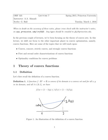
DEWS DEFINITION AND CLASSIFICATIONThe frequency of MGD is higher in patients with SSthan in the normal population; thus, a defective tear filmlipid layer may contribute to dry eye by leading to excessevaporation.82b. Non-Sjogren Syndrome Dry EyeNon-Sjogren syndrome dry eye is a form of ADDE dueto lacrimal dysfunction, where the systemic autoimmunefeatures characteristic of SSDE have been excluded. Themost common form is age-related dry eye, to which theterm KCS has sometimes been applied in the past. However,as noted earlier, the term KCS is now used to describe anyform of dry eye. In the 1995 Dry Eye Workshop report, itwas referred to as primary lacrimal disease,1 but this term hasnot been generally adopted. The different forms of NSSDEare briefly discussed below (Table 2).1) Primary Lacrimal Gland DeficienciesAge-Related Dry Eye (ARDE): There is some uncertaintyas to whether tear dynamics are affected by age in thenormal population.83 Mathers et al showed significant agerelated correlations for tear evaporation, volume, flow, andosmolarity,49 but no such relationship was noted by Craigand Tomlinson84 or in other reports of tear turnover,85tear evaporation86,87 and lipid layer.88 ARDE is a primarydisease.With increasing age in the normal human population,there is an increase in ductal pathology that could promotelacrimal gland dysfunction by its obstructive effect.89,89aThese alterations include periductal fibrosis, interacinarfibrosis, paraductal blood vessel loss and acinar cellatrophy.89,89a Damato et al found lymphocytic glandularinfiltrates in 70% of lacrimal glands studied and considered this to be the basis of the fibrosis. Appearances werelikened to the less severe grades of Sjogren syndrome. Theypostulated a sequence of periductal fibrosis, interacinarfibrosis and, finally, acinar atrophy. It has been suggestedthat the low-grade dacryoadenitis could be caused bysystemic infection or conjunctivitis89 or, alternatively, thatsubclinical conjunctivitis might be responsible for stenosisof the excretory ducts.89aC ongenital Alacrima: Congenital alacrima is a rare causeof dry eye in youth.90 It is also part of certain syndromes,91including the autosomal recessive, triple A syndrome (Allgrove syndrome), in which congenital alacrima is associatedwith achalasia of the cardia, Addison’s disease, central neurodegeneration, and autonomic dysfunction. It is caused bymutations in the gene encoding the protein ALADIN, whichplays a role in RNA and/or protein trafficking between thenucleus and cytoplasm.92,93Familial Dysautonomia: Lacrimal dysfunction is a majorfeature of the autosomal recessive disorder, familial dysautonomia (Riley Day syndrome), in which a generalizedinsensitivity to pain is accompanied by a marked lack ofboth emotional and reflex tearing, within a multisystemdisorder. There is a developmental and progressive neuronalabnormality of the cervical sympathetic and parasympa80Table 2 . Conditions associated with non-Sjogrensyndrome dry eyePrimary lacrimal gland deficienciesAge-related dry eyeCongenital alacrimaF amilial dysautonomiaSecondary lacrimal gland deficienciesLacrimal gland infiltrationSarcoidosisLymphomaAIDSGraft vs host diseaseLacrimal gland ablationLacrimal gland denervationObstruction of the lacrimal gland ductsTrachomaCicatricial pemphigoid and mucous membrane pemphigoidErythema multiformeChemical and thermal burnsReflex hyposecretionReflex sensory blockContact lens wearDiabetesNeurotrophic keratitisReflex motor blockVII cranial nerve damageMultiple neuromatosisExposure to systemic drugsthetic innervations of the lacrimal gland and a defectivesensory innervation of the ocular surface, which affects bothsmall myelinated (AD) and unmyelinated (C) trigeminalneurons.94,95 The chief mutation affects the gene encodingan IKB kinase-associated protein.2) Secondary Lacrimal Gland DeficienciesLacrimal gland infiltration: Lacrimal secretion may failbecause of inflammatory infiltration of the gland, as in:Sarcoidosis: Infiltration of the lacrimal gland by sarcoidgranulomata may cause dry eye.96Lymphoma: Infiltration of the lacrimal gland by lymphomatous cells causes dry eye.97AIDS: Dry eye may be caused by lacrimal gland infiltration by T-cells. However, in AIDS-related dry eye, unlikethe situation in SSDE, there is a predominance of CD8suppressor cells, rathe
tion presented at the 19 9 5 National Eye Institute (N E I)/In-dustry Dry Eye Workshop, which was: Dry eye is a disorder of the tear lm due to tear de ciency or excessive evaporation, which causes damage to the interpalpebral ocular surface and is associated with symptoms of ocular discomfort.1 The committee agreed that the de nition could be