Transcription
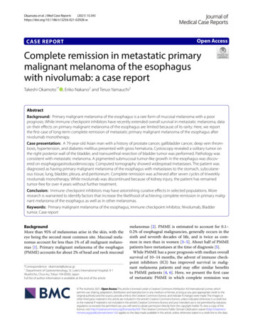
Open Access CaseReportDOI: 10.7759/cureus.25614Wandering Mucosal Melanoma Presenting asOccult Gastrointestinal Blood Loss AnemiaAimen Farooq 1 , Hamaad Rahman 2 , Baha Aldeen Bani Fawwaz 1 , Abu Hurairah 3Review began 05/23/2022Review ended 05/30/20221. Internal Medicine, AdventHealth Orlando, Orlando, USA 2. College of Osteopathic Medicine, Kansas City Universityof Medicine and Biosciences, Kansas, USA 3. Gastroenterology and Hepatology, AdventHealth Orlando, Orlando, USAPublished 06/02/2022 Copyright 2022Farooq et al. This is an open access articledistributed under the terms of the CreativeCommons Attribution License CC-BY 4.0.,Corresponding author: Aimen Farooq, aimen.farooq.md@adventhealth.comwhich permits unrestricted use, distribution,and reproduction in any medium, providedthe original author and source are credited.AbstractMalignant melanoma is a highly aggressive cancer arising from the skin, retina, and mucosal lining of therespiratory, gastrointestinal (GI), or genitourinary tracts, all of which contain melanocytes. Mucosal orextracutaneous melanomas (ECMs) are rare accounting for 1% of all melanomas. We herein report a case of ametastatic mucosal melanoma presenting as occult blood loss anemia.A 58-year-old male presented with generalized weakness, anorexia, weight loss, and intermittent melenafor one year. On exam, he was tachycardic, borderline hypotensive, and pale without epigastric tenderness.Labs showed severe anemia [hemoglobin, Hgb 3.8 mg/dL, mean corpuscular volume (MCV) 72 fl] for which hereceived two units of red cells. Endoscopy revealed an 8 mm non-bleeding, gastric ulcer with a raised borderand a clean base on the wall of the gastric body. Histologic analysis was consistent with malignantmelanoma displaying strong positivity for S-100, Melan A, and HMB 45 stains. The CT of theabdomen revealed multifocal metastatic disease with subcutaneous, intramuscular, and perinephricimplants with suspicion of small bowel carcinomatosis. The patient underwent an excisional biopsy for theabdominal wall mass and surgical pathology confirmed melanoma. The patient is planned to be started onimmunotherapy for advanced disease.Most melanomas found in the GI tract are metastatic. Mucosal melanoma presenting as a gastric ulcer isextremely rare. As a result, metastasis from other sites must be ruled out before making a diagnosis ofprimary gastric melanoma (PGM). In our case, a widespread disease with unknown primary elucidated thediagnosis but post-operative inspection failed to find any potential lesion on the skin, genitals, or otherorgans, suggesting the possible diagnosis of metastatic gastric melanoma. However, follow-up is stillrequired to confirm the diagnosis according to the established criteria. Pathologic diagnosis of melanomarequires the identification of melanin in the cytoplasm and immunohistochemistry with specific markerssuch as S-100, Melan A, and HMB-45. Although the pathologic diagnosis of PGM is similar to cutaneousmelanoma, preoperative diagnosis is difficult due to the extremely low incidence, lack of obvious melaninpigmentation, similar microscopic patterns as more common gastric cancers, and lack of awareness amongphysicians and pathologists.The prognosis of mucosal melanoma is poor, with a five-year survival rate of 25% versus 80% for cutaneousmelanoma. Advanced age, surgically unresectable disease, and lymph node involvement are all poorprognostic markers. There is no standard protocol for treatment. Surgery is the only curative treatment forthe resectable disease. Adjuvant chemotherapy, radiation, and immunotherapy have an established role incutaneous melanoma but there is only limited data on adjuvant systemic therapy with mucosal melanoma.Further research is imperative to establish proper management guidelines for this rare disease entity.Categories: Pathology, Gastroenterology, OncologyKeywords: iron deficiency anemia (ida), chronic gastrointestinal bleeding, endoscopic diagnosis, cancerimmunotherapy, mucosal malignant melanoma, primary gastric malignant melanomaIntroductionMalignant melanoma is a highly aggressive cancer arising from the skin, retina, and mucosal lining of therespiratory, gastrointestinal (GI), or genitourinary tracts, all of which contain melanocytes [1]. Mucosal orextracutaneous melanomas (ECMs) are rare accounting for 0.3% of all cancer diagnoses and 1% of allmelanomas [2-3]. The most common sites for mucosal melanomas are head and neck in men andvulvovaginal in women [4-5]. Although cutaneous melanomas frequently metastasize to the GI tract,primary GI melanomas, especially gastric melanoma, are extremely rare [6-7]. Mucosal melanomas areinteresting due to their aggressive nature and poor prognosis when compared to their cutaneouscounterparts. We herein report a case of mucosal melanoma with probable gastric origin presenting asoccult blood loss anemia.Case PresentationHow to cite this articleFarooq A, Rahman H, Bani Fawwaz B, et al. (June 02, 2022) Wandering Mucosal Melanoma Presenting as Occult Gastrointestinal Blood LossAnemia. Cureus 14(6): e25614. DOI 10.7759/cureus.25614
A 58-year-old male with a past medical history of hypertension, type 2 diabetes, and hypothyroidismpresented to the emergency department (ED) with complaints of weakness, fatigue, and shortness of breathover the past few months. He endorsed intermittent black, tarry stools, dizziness, loss of appetite, andweight for the past year. He reports noticing enlarging masses in the right upper quadrant of his abdomenand right axillary area over the past few weeks which concerned him since his friend had similar symptomsand had been diagnosed with lymphoma in the past. He denied pertinent surgical or family history. Henever had a colonoscopy or esophagogastroduodenoscopy (EGD) in the past. Upon arrival, the patient wastachycardic (heart rate, HR 110 beats per minute) otherwise hemodynamically stable. Physical exam wasnotable for a 5-cm firm, non-fluctuating, and non-tender subcutaneous mass in the right upper quadrant ofthe abdomen, as well as a 5-cm palpable, firm, mobile, non-tender, and non-fluctuating lymph node in theright axillary region.Laboratory testing (Table 1) was consistent with severe iron deficiency anemia and reactive thrombocytosisfor which he received two units of packed red blood cells (pRBCs).Laboratory testObserved valueReference rangeWhite blood cells8.66 10*3/uL3.4 - 9.6 10*3/uLHgb3.8 g/dL13.2 - 16.6 g/dLHematocrit14.4 %38.3% - 48.6%MCV72 fL80 - 100 fLPlatelets499 10*3/uL135 - 317 10*3/uLIron13 ug/dL45 - 182 ug/dLFerritin5 ng/mL24 - 336 ng/mLTIBC532 ug/dL221 - 481 ug/dLIron saturation %2%30% - 44%Transferrin380 mg/dL200 - 360 mg/dLTABLE 1: Pertinent laboratory work-up results.Hgb, hemoglobin; MCV, mean corpuscular volume; TIBC, total iron binding capacityAn EGD demonstrated an 8-mm, non-bleeding, clean base gastric ulcer with raised borders (Forrest ClassIII) on the anterior wall of the gastric body (Figure 1).2022 Farooq et al. Cureus 14(6): e25614. DOI 10.7759/cureus.256142 of 7
FIGURE 1: Endoscopic image of an 8-mm non-bleeding, cratered ulcerwith a clean base on the anterior wall of the gastric body (Forrest ClassIII).Histopathological analysis showed an aggregate of malignant tumor cells in a nodular pattern (Figure 2)which displayed strong positivity with S-100 protein, HMB-45, and Melan-A, consistent with malignantmelanoma (Figure 3a-b).2022 Farooq et al. Cureus 14(6): e25614. DOI 10.7759/cureus.256143 of 7
FIGURE 2: Endoscopic biopsy of the stomach ulcer: pathology showingnodular melanoma cells in a nodular pattern (black arrow) andsurrounding normal gastric epithelium.FIGURE 3: Immunohistochemical staining showing A) positive Melan Ain melanoma cells and negative in stomach epithelium and B) positiveHMB 45 in melanoma cells and negative in stomach epithelium.A contrast CT scan of the abdomen which was performed for staging revealed multiple subcutaneous andintramuscular implants within the abdomen and pelvis including a dominant lesion in the right upperabdominal wall measuring 4.3 cm x 4.3 cm, bilateral perinephric tumor implants, periportal andgastrohepatic ligament adenopathy, multiple areas of thickening and nodularity in the small bowelsuspicious for carcinomatosis, and mild mesenteric edema. A CT angiogram of the chest showed rightparatracheal lymphadenopathy and a 6.6-mm medial right apical pulmonary nodule (Figure 4).2022 Farooq et al. Cureus 14(6): e25614. DOI 10.7759/cureus.256144 of 7
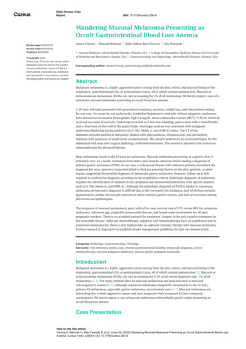
FIGURE 4: Axial CT image showing dominant lesion (arrow) in the rightupper abdominal wall.The patient underwent an excisional biopsy for the abdominal wall mass, consistent with metastaticmelanoma, positive for S-100 protein and Melan-A, and negative for cytokeratin AE1/AE3. Detailedinspection failed to find any potential lesions on the skin, eyes, or genitals. The diagnosis of metastaticmucosal melanoma was established, and the patient was discharged with plans to start combinationimmunotherapy with Nivolumab and Ipilimumab. Currently, the patient is continuing treatment withimmunotherapy.DiscussionMelanomas arise from the malignant transformation of melanocytes, commonly in cutaneous tissues andinfrequently in mucosal tissues including the GI, respiratory, and genitourinary tracts. Approximately 1.4%of all melanomas arise from mucosal tissues and have shown higher rates among women compared tomen [8-9]. Mucosal melanomas are more aggressive and carry a worse prognosis than their cutaneouscounterparts, making accurate and early diagnosis critically important [1].Due to the rare nature of mucosal melanomas, information regarding their pathogenesis is limited.Cutaneous melanomas usually develop because of mutations in the BRAF gene, whereas studies have shownthat 16%-39% of mucosal melanomas develop due to mutations in the KIT gene. Unlike the role ofultraviolet light exposure in cutaneous melanomas, there are no identifiable risk factors for mucosalmelanomas.Mucosal melanomas of the GI tract can either be primary or secondary/metastatic (Figure 5). Primary gastricmelanomas (PGMs) make up less than 1% of all ECMs [6-7]. Therefore, before diagnosing a primary lesion ofthe GI tract, it is imperative to perform a thorough cutaneous and ophthalmologic examination to ruleout possible primary cutaneous or ocular lesions.2022 Farooq et al. Cureus 14(6): e25614. DOI 10.7759/cureus.256145 of 7
FIGURE 5: Schematic representation of different types of melanomasfound in the stomach.Given the rarity of the disease, there are no agreed-upon diagnostic criteria for PGMs, rendering it hard todistinguish metastatic gastric melanoma from an advanced PGM with metastatic disease, as in our case.Blecker et al. suggested the following criteria for diagnosing PGM: 1. absence of invasive or insitu cutaneous melanocytic lesions and 2. in situ changes in the GI epithelium [10]. An alternative approachby Song et al. suggested the following: 1. a single lesion of melanoma in the stomach proven by pathology;2. absence of cutaneous lesions anywhere else in the body; 3. no history of melanoma; and 4. disease-freesurvival of at least 12 months after curative surgery [11].The proposed diagnostic criteria suggest a lack of metastatic lesions as a criterion for diagnosis, however, aPGM diagnosed in the later stages could very well present with a gastric lesion as well as concomitantmetastatic lesions. In a systematic review by Mellotte et al., 10 out of 34 patients with PGM were found tohave distant solid metastasis in organs including the liver, brain, and lungs at initialpresentation [9]. Additionally, the 12-month survival rate following treatment has been shown to varysignificantly, making it an unreliable criterion for diagnosis.In our case, a thorough full-body cutaneous examination and ophthalmologic inspection did not reveal anyother potential primary lesions. Furthermore, a colonoscopy was also performed which did not reveal anypotential primary lesions. The patient did not have any history of melanoma. Although the patient did havesignificant metastasis throughout this body, it is quite possible that this has spread from the primary gastriclesion.Similarly, there are no clear staging guidelines for PGMs. The American Committee on Cancer (AJCC)guidelines for head and neck mucosal melanomas are normally improvised for the staging of GI mucosalmelanomas [1]. Patients with PGM can experience several non-specific symptoms including abdominal pain,weight loss, occult upper GI bleed, and anemia. Because of a lack of specificity in clinical presentation,physicians often suspect other more common gastric cancers before considering PGM [1, 9, 11].The CT scan is an acceptable choice for imaging and commonly reveals thickening of the gastric wall. EGDwith a biopsy of the lesion plays a pivotal role in diagnosis. The majority of documented cases of PGM hadlarge ulcerated or polypoidal masses of varying size in the stomach. Oftentimes lesions will be pigmented,however, lesions can also be amelanotic, so immunohistochemistry staining becomes critical for establishingthe diagnosis [11].In localized cases, total resection of the lesion with negative margins has proven to be curative. In metastaticcases, other treatment modalities should be considered as surgical resection is often not a viable option.Chemotherapy is not an effective treatment option against melanoma due to its resistance and low responserate [9]. Novel therapies focusing on molecular targets, such as anti-PD-1 agents and cytotoxic T-lymphocyteantigen-4 checkpoint inhibitors, have shown to be the most effective treatment [12]. Currently, the Food andDrug Administration (FDA) has approved ipilimumab, a cytotoxic T-lymphocyte antigen-4 checkpointinhibitor, and nivolumab, an anti-PD-1 agent, for treating mucosal melanomas. Recent studies suggest thatthese two agents used in combination showed greater efficacy than either agent alone [13].Overall PGM has a poor prognosis, with a one-year survival rate of less than 60% and a five-year survivalrate of less than 3% [9, 11]. The most important prognostic factor is the presence of metastatic lesions, aspatients with metastasis had significantly lower survival rates [9]. This further emphasizes the importance of2022 Farooq et al. Cureus 14(6): e25614. DOI 10.7759/cureus.256146 of 7
raising awareness regarding the need for early detection of the disease which can significantly improveoutcomes by stopping the spread of the disease.ConclusionsMalignant melanoma, although thought to be a primary cutaneous disease, can arise from mucosal tissues.The rarity of gastric melanomas, non-specific presenting symptoms, amelanotic lesions on the endoscopicexam, and the lack of clear guidelines for diagnosing a PGM, make diagnosis extremely challenging.Cutaneous and ocular lesions must be ruled out before the diagnosis of PGM is established. Early detectionof the disease is imperative as metastasis is the most important prognostic factor. It is crucial to alwaysconsider immunohistochemistry staining. Surgery is the first-line treatment for localized disease, whereasimmunotherapy has proven effective for metastatic disease. Further research is needed to establishdiagnostic and management guidelines for this rare disease entity.Additional InformationDisclosuresHuman subjects: Consent was obtained or waived by all participants in this study. Conflicts of interest: Incompliance with the ICMJE uniform disclosure form, all authors declare the following: Payment/servicesinfo: All authors have declared that no financial support was received from any organization for thesubmitted work. Financial relationships: All authors have declared that they have no financialrelationships at present or within the previous three years with any organizations that might have aninterest in the submitted work. Other relationships: All authors have declared that there are no otherrelationships or activities that could appear to have influenced the submitted lovic M, Vlajkovic S, Jovanovic P, Stefanovic V: Primary mucosal melanomas: a comprehensive review .Int J Clin Exp Pathol. 2012, 5:739-753.Lourenço SV, Fernandes JD, Hsieh R, Coutinho-Camillo CM, Bologna S, Sangueza M, Nico MM: Head andneck mucosal melanoma: a review. Am J Dermatopathol. 2014, 36:578-587.10.1097/DAD.0000000000000035Spencer KR, Mehnert JM: Mucosal melanoma: epidemiology, biology and treatment . Cancer Treat Res.2016, 167:295-320. 10.1007/978-3-319-22539-5 13Patel SG, Prasad ML, Escrig M, et al.: Primary mucosal malignant melanoma of the head and neck . HeadNeck. 2002, 24:247-257. 10.1002/hed.10019Chang AE, Karnell LH, Menck HR: The National Cancer Data Base report on cutaneous and noncutaneousmelanoma: a summary of 84,836 cases from the past decade. The American College of Surgeons Commissionon Cancer and the American Cancer Society. Cancer. 1998, 83:1664-1678. ncr23%3E3.0.co;2-gLam KY, Law S, Wong J: Malignant melanoma of the oesophagus: clinicopathological features, lack of p53expression and steroid receptors and a review of the literature. Eur J Surg Oncol. 1999, 25:168-172.10.1053/ejso.1998.0621Lens M, Bataille V, Krivokapic Z: Melanoma of the small intestine . Lancet. 2009, 10:516-521.10.1016/S1470-2045(09)70036-1McLaughlin CC, Wu XC, Jemal A, Martin HJ, Roche LM, Chen VW: Incidence of noncutaneous melanomas inthe U.S. Cancer. 2005, 103:1000-1007. 10.1002/cncr.20866Mellotte GS, Sabu D, O'Reilly M, McDermott R, O'Connor A, Ryan BM: The challenge of primary gastricmelanoma: a systematic review. Melanoma Manag. 2020, 7:MMT51. 10.2217/mmt-2020-0009Blecker D, Abraham S, Furth EE, Kochman ML: Melanoma in the gastrointestinal tract . Am J Gastroenterol.1999, 94:3427-3433. 10.1111/j.1572-0241.1999.01604.xSong W, Liu F, Wang S, Shi H, He W, He Y: Primary gastric malignant melanoma: challenge in preoperativediagnosis. Int J Clin Exp Pathol. 2014, 7:6826-6831.Shoushtari AN, Munhoz RR, Kuk D, et al.: The efficacy of anti-PD-1 agents in acral and mucosal melanoma .Cancer. 2016, 122:3354-3362. 10.1002/cncr.30259D'Angelo SP, Larkin J, Sosman JA, et al.: Efficacy and safety of Nivolumab alone or in combination withIpilimumab in patients with mucosal melanoma: a pooled analysis. J Clin Oncol. 2017, 35:226-235.10.1200/JCO.2016.67.92582022 Farooq et al. Cureus 14(6): e25614. DOI 10.7759/cureus.256147 of 7
Endoscopy revealed an 8 mm non-bleeding, gastric ulcer with a raised border and a clean base on the wall of the gastric body. Histologic analysis was consistent with malignant . HMB-45, and Melan-A, consistent with malignant melanoma (Figure 3a-b). 2022 Farooq et al. Cureus 14(6): e25614. DOI 10.7759/cureus.25614 3 of 7.