Transcription
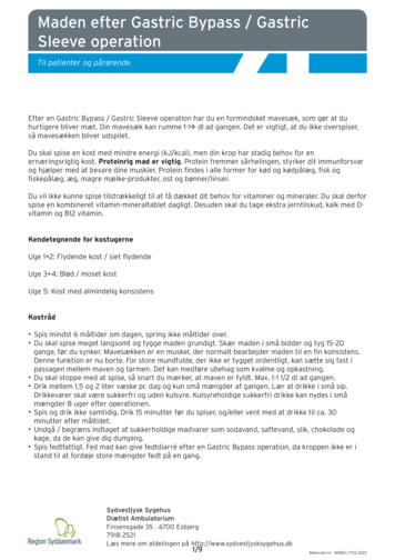
Article published online: 2020-07-03106THIEMEInterpretationReview Article of Benign Gastric Mucosal Lesions Using Narrow Band ImagingPaghadhar et al.Interpretation of Benign Gastric Mucosal LesionsUsing Narrow-Band ImagingSameer Paghadhar1Mayank Jain2B. Mahadevan11 Department of Gastroenterology, Gleneagles Global Health City,Chennai, Tamil Nadu, India2 Department of Gastroenterology, Arihant Hospital and ResearchCentre, Indore, Madhya Pradesh, India3 Department of Gastroenterology, Sri Ramachandra MedicalCollege, Porur, Chennai, Tamil Nadu, IndiaJayanthi Venkataraman3Address for correspondence Mayank Jain, MD, DNB, Departmentof Gastroenterology, Arihant Hospital and Research Centre 283-AGumasta Nagar, Indore 452009, Madhya Pradesh, India(e-mail: mayank4670@rediffmail.com).J Digest Endosc 2020;11:106–111AbstractKeywords endoscopy gastric imaging mucosaThe major drawback of conventional white light endoscopy (WLE) is that it lacks accuracy in diagnosis and differentiation of various benign and premalignant mucosal gastrointestinal lesions. To overcome this, image-enhanced endoscopy techniques, whichprovide high-definition images with good resolution and contrast enhancement, havebeen developed. One such technique is narrow-band imaging (NBI). NBI functions byfiltering the illumination light. The red component of the standard red, green, andblue filters is rejected and the selected bandwidth of the blue and green light is transmitted. The present review highlights the role of NBI in diagnosis of benign gastriclesions like atrophic gastritis, Helicobacter pylori–related gastritis, intestinal metaplasia,and other rarer conditions. NBI is a simple procedure which does not require any additional equipment and does not have a long learning curve. Use of NBI in daily practiceis likely to improve detection of mucosal abnormalities.IntroductionSince the invention of flexible fiber-optic endoscope in 1957,several modifications and improved techniques have beendeveloped to enhance the diagnostic yield of the endoscopyprocedure. The major drawback of conventional white lightendoscopy (WLE) is that it lacks accuracy in diagnosis anddifferentiation of various benign and premalignant mucosal gastrointestinal lesions.1 To overcome this, image-enhanced endoscopy techniques providing high-definitionimages with good resolution and contrast enhancementhave been developed. One such technique is narrow-bandimaging (NBI).2NBI functions by filtering the illumination light. The redcomponent of the standard red, green, and blue filters isrejected and the selected bandwidth of the blue and greenlights is transmitted ( Fig. 1). The mucosa is illuminated selectively with narrow-band wavelengths of blue (415 15 nm)and green (540 15 nm).3 The hemoglobin absorbs light atboth these wavelengths and mucosa reflects it. The bloodvessels thereby appear dark brown against a light backgroundDOI https://doi.org/10.1055/s-0040-1713553ISSN 0976-5042.providing the necessary contrast between blood vesselsand mucosa. While doing endoscopy procedure, real-timechange from white light to NBI is possible by using a “switch”whereby wavelength of specific bandwidths 415 15 nmand 540 15 nm is only transmitted by a filter that existsat the distal tip of the endoscope. This narrow band lightilluminated mucosa is reflected and the light reaches thecouple charged device (CCD) which produces electronic signals based on amount and wavelengths reflected. The signalfrom the CCD gets processed by the video processor whichresynthesizes the final output image. Colors in final imageare allocated by processor according to the human visualperception.Normal Appearance of Stomach on NBIOn NBI, the normal gastric mucosa reflects different morphological architecture of surface (S) and microvascular pattern(V) in the corpus and antrum referred to as SV pattern.4 Yaodescribed the surface and vascularpattern of gastric mucosausing microanatomical components of mucosa.5 The major 2020 Society of GastrointestinalEndoscopy of India
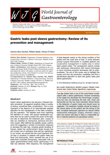
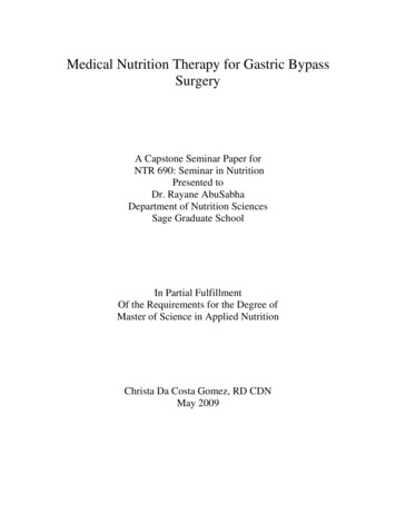
Interpretation of Benign Gastric Mucosal Lesions Using Narrow Band ImagingPaghadhar et al.Fig. 1 Overview of NBI system and its principle. NBI, narrow-band imaging.Fig. 2 (A) Normal corpus mucosa of stomach: (left) as seen by NBIwithout magnification, (right) NBI with magnification: regular roundto oval pits with honeycomb like SECN pattern (blue arrow) and spider like collecting venules (green arrow). (B) Normal antral mucosa ofstomach: (left) as seen by NBI without magnification, (right) NBI withmagnification: regular polygonal pits (red arrow) with coil or springlike SECN pattern (blue arrow) and absence of collecting venules.NBI, narrow-band imaging; SECN, subepithelial capillary network.structural or surface components, that is, S pattern refers tothe marginal crypt epithelium, the crypt opening or pit, andthe intervening portion between crypts. In V, that is, vascularpattern, vessels are described as the subepithelial capillarynetwork and the collecting venule. When there is difficultyin categorizing a vessel as capillary or venule, this is referredto microvessel.In corpus, the marginal crypt epithelium is seen as whitish circle surrounding the crypt opening.5-7 The latter is seenas a round to oval brown dot in center of subepithelial capillary network which appears as dark brown anastomosingcapillaries giving a honeycomb appearance. The interveningpart appears light pink. The capillaries drain into deeperplaced collecting venules that are perceived as cyancoloredspider-like/starfish-like thick vessels interspersed regularlyin mucosa4-8 ( Fig. 2A).In the antrum, there are ridges that are separated by sulci.Each coil or wave-shaped dark brown colored subepithelialcapillary network is located at apical part of the ridge and areseparated by linear or reticular crypt opening7-9 ( Fig. 2B).The capillary vessels often anastomose to each other andappear as open loops. The collecting venules are not normally seen in antrum as they are anatomically located in thedeeper plane compared with those in the corpus.Body of the stomach is characterized by regular arrangement of connecting venules and mucosa showing regular,small pits with dark areas encircling light areas. On the otherhand, antrum has well-defined ridge pattern without connecting venules and regular circular areas in mucosa withlight areas surrounding dark areas. These differences areattributable to differences in vascular pattern and presenceof connecting venules at deeper level in antrum.10NBI Appearances of Common Benign Gastric LesionsAtrophic Gastritis/Gastric AtrophyIn atrophic gastritis, secondary to chronic inflammationof the gastric mucosa as with Helicobacter pylori infection(85%), the rate of cell loss may exceed the ability of the stemcells to replace lost cells of surface mucosa and glands resulting in thinning of the mucosa. With white light endoscopy,atrophic gastritis is seen as atrophic mucosal folds that arepale with a shiny surface; submucosal vessels are prominent.These appearances are neither sensitive nor specific for atrophic gastritis.11With NBI, there is loss of pits and subepithelial capillarynetwork with irregular arrangement of prominent collectingvenules ( Fig. 3). The sensitivity and specificity of these findings approaches up to 90 and 96%, respectively.12,13 AtrophicJournal of Digestive Endoscopy Vol. 11No. 2/2020107
108Interpretation of Benign Gastric Mucosal Lesions Using Narrow Band Imaginggastritis may vary from mild to severe. Complete loss of pitsmay be seen in severe atrophy. There is not much data on roleof NBI in detecting mild atrophy.Intestinal MetaplasiaWhite light endoscopy of the gastric mucosa correlates poorlywith histological findings.14-17 Intestinal metaplasia refers toreplacement of foveolar and glandular epithelium in oxynticor antral mucosa by intestinal epithelium and is consideredas the “break point” in the gastric carcinogenesis cascade.On WLE, intestinal metaplasia appears as shallowdepressed and reddish area, slightly raised or whitish flatarea or flat lesion with color, similar to the background withminimal morphological changes.18,19 NBI has an additionalbenefit of differentiating intestinal metaplasia from normalmucosa by outlining the color differences. A meta-analysisof four studies reported sensitivity and specificity of 86 and77%, respectively, for diagnoses of intestinal metaplasia (IM)by NBI.20Bansal et al observed that the presence of a ridge or villouspattern by NBI (pseudopylorization of oxyntic mucosa) has ahigh specificity and sensitivity (80 and 100%, respectively)for identifying intestinal metaplasia.10 Uedo et al described anovel finding of fine blue–white lines at the crest of epithelialsurface described as the “light blue crests” on NBI–magnifyingendoscopy (ME) with a high sensitivity (89%), specificity(93%), and accuracy (91%) for the diagnosis of IM.21 In absenceof magnification, the light blue crest is seen as white-bluePaghadhar et al.(cyan color) patches on NBI. It is defined as a fine blue–whiteline on the crest of the epithelial surface/gyri ( Fig. 4).A prospective blinded study showed that WLE with fiverandom biopsies as per the Sydney system was insufficientfor detection of gastric intestinal metaplasia. This low yieldis likely because metaplastic lesions are often focal and arelikely to be missed on random biopsy sampling. Authorstherefore recommend NBI-targeted biopsies plus five mapping biopsies as per the updated Sydney system. Mappingbiopsies alone without NBI has a poor yield.22Savarino et al reported that NBI detects gastric IM withan accuracy of 93%, a sensitivity of 80%, a specificity of 96%,a positive predictive value of 84%, and a negative predictivevalue of 95%.23 A randomized crossover study by Dutta et al24showed superiority of NBI over WLE in diagnosing atrophicgastritis, as well as IM. The authors noted that NBI identifiedadditional lesions not detected on WLE.Pimentel-Nunes et al,25 in 2012, proposed a simple andreproducible classification system for the diagnosis of IMand dysplasia. The authors noted that regular vessels withcircular mucosa was associated with normal histology (accuracy 85%) and tubulovillous mucosa was associated with IM(accuracy 84%, 95% confidence interval [CI]: 77–91%). Lightblue crest had moderate reliability (k 0.62) and high specificity (87%) for IM.Apart from light blue crest, other findings have beendescribed in IM. White opaque substance (WOS) was firstreported by Yao et al. It is a substance present in the superficial part of gastric neoplasias that obscures the subepithelialmicrovascular architecture. WOS is an optical phenomenoncaused by accumulated lipid droplets.26-28The marginal turbid band (MTB) is another findingnoted in IM. It is defined as an enclosing, white turbid bandon the epithelial surface/gyri.21 Recently, another finding,namely, white villiform type mucosa which suggests atrophy and intestinal metaplasia in the gastric antrum has beendescribed29Helicobacter pylori InfectionFig. 3 Chronic atrophic gastritis (CAG): atrophied folds with palemucosa in CAG. NBI showing obscured surface and vessel patternwith irregular SECN and visible submucosal vessels. NBI, narrow-bandimaging; SECN, subepithelial capillary network.NBI is a potential tool to diagnose H. pylori related gastritis ( Fig. 5). In gastric corpus, nonvisualization of normalcollecting venules irrespective of central opening and subepithelial capillary changes suggests H. pylori–associatedFig. 4 Intestinal metaplasia (IM): (left) antrum showing slightlyraised pale lesion (blue arrow); (right) NBI-ME showing ridge patternwith LBC. ME, magnifying endoscopy; NBI, narrow-band imaging.Fig. 5 H. pylori related chronic non atrophic gastritis: (left) on WLE,changes are not well appreciated; (right) NBI: showing variable vascular density, pit enlargement, and absence of CV. H, helicobacter;NBI, narrow-band imaging; WLE, white light endoscopy.Journal of Digestive Endoscopy Vol. 11No. 2/2020
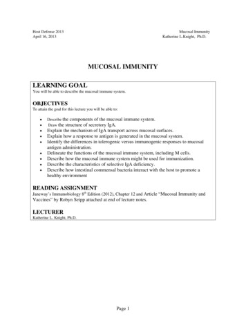
Interpretation of Benign Gastric Mucosal Lesions Using Narrow Band Imaginggastritis (100% sensitivity, 92% specificity, and positive predictive value (PPV) of 100%).30-32 Tahara et al using NBI ofnonneoplastic mucosa of gastric corpus, classified four typesto predict H. pylori infection and also described histologicalseverity of gastritis and gastric atrophy.33 Normal patternwas defined by small, round pits surrounded by SECN. Type-1pattern showed slightly enlarged, round pits with unclear orirregular SECN. Type-2 pattern showed obviously enlarged,oval or prolonged pits with increased density of irregularvessels and type-3 pattern revealed well-demarcated, oval ortubulovillous pits with clearly visible coiled or wavy SECN.33Pimentel-Nunes et al documented that regular vascularand surface pattern with variable vascular density favoredthe presence of H. pylori infection.24 Bansal et al showedthat the sensitivity and specificity of a regular mucosal andvascular pattern for the diagnosis of normal mucosa/mildgastritis were 89 and 78%, while the sensitivity and specificity of an irregular pattern with decreased density of vesselsfor the diagnosis of H. pylori was 75 and 88%.10 Banerjee et alprospectively compared NBI with WLE in 74 patients andshowed that NBI can be a potential tool for real time diagnosis of H. pylori infection based on the presence of obscurepit pattern.34 Yagi et al compared the diagnostic value ofconventional endoscopy and magnifying NBI for predictionof H. pylori status in patients after endoscopic resection ofgastric cancer. The inter observer agreement was moderate(k 0.56) for conventional endoscopy and substantial (k 0.77) for magnifying NBI. The sensitivity and specificity were79 and 52% for conventional endoscopy and 91 and 83% formagnifying NBI endoscopy, respectively.35NBI has been used to investigate the changes of gastricmucosal patterns before and 12 weeks after H. pylori eradication. Patients who were successfully treated (confirmedwith 13C Urea breath test) showed a change back to small ovalor pinhole-like round pits, as well as a reduction in the density of fine irregular vessels. In absence of severe atrophy andintestinal metaplasia, the sensitivity and specificity of NBIfor predicting the H. pylori eradication was 100%. However,H. pylori eradication did not change NBI pattern in those withpreexisting severe gastric atrophy and intestinal metaplasia.36Despite the changes associated with H. pylori infectionbeing commonly seen, tests for H. pylori infection like rapidurease test, and histopathology may still be required fordetection of active infection where clinically indicated. Moreevidence is required on the role of NBI in distinguishing current ongoing infection from past infection especially in thesetting of atrophic gastritis and intestinal metaplasia.NBI Appearance of Other Benign Gastric LesionsFundic gland polyps ( Fig. 6A) are usually small (1–5 mm)and multiple and are most commonly located in fundus andbody of the stomach. On WLE, these polyps are sessile, shiny,and translucent with normal background mucosa and onNBI, as regular round mucosal pit pattern and regular honeycomb or dense vascular pattern on a background normalgastric body mucosa.37,38Paghadhar et al.Fig. 6(A) FGP-on WLE and NBI showing round pits with honeycombSECN and CVs with demarcation line. (B) Hyperplastic polyp showingpits were dilated with coil-like enlarged vessels and absent CVs. DLwas present. NBI, narrow-band imaging; WLE, white light endoscopy;CV, collecting venule; FGP, fundic gland polyp; LBC, light blue crest;SECN, subepithelial capillary network; DL, demarcation line.Hyperplastic polyp ( Fig. 6B) are usually 2 cm in size,solitary (66%), and commonly located in antrum. They areoften associated with chronic H. pylori infection against abackground of atrophic mucosa. On NBI, these polyps mayhave tubular mucosal pattern, of several shapes, with thickbut regular vessels or dense vascular pattern.37,38Portal hypertensive gastropathy is commonly seen in fundus and body and rarely in antrum of stomach. The changesare usually submucosal; superficial mucosal biopsies are frequently false negative.39 Characteristics WLE appearance ismosaic or snake skin like pattern or a diffuse, erythematous,and reticular cobblestone pattern of gastric mucosa consisting of small polygonal areas, with superimposed red punctatelesions, 2 mm in diameter and a depressed white border.40-42On NBI, red mosaic-like mucosa of portal hypertensive gastropathy is seen as extended and swollen gastric pits withvarying degrees of dilated and convoluted capillaries surrounding the gastric pits, collecting venules are obscured.43Gastric antral vascular ectasia (GAVE) is common inelderly ( 70 years) women (80%); 30% cases are associatedwith portal hypertension. On WLE, GAVE appears as tortuouscolumns of ectatic vessels simulating a “watermelon” or isseen as a diffuse pattern. These areas of erythema are commonly arranged in a linear manner along folds in the antrumand less commonly arranged as diffuse erythema in the antrum.40,41,44,45 Hayashi et al described NBI appearance of GAVEas partial and marked dilatation of the capillaries surrounding the gastric pits and capillaries located below the gastricJournal of Digestive Endoscopy Vol. 11No. 2/2020109
110Interpretation of Benign Gastric Mucosal Lesions Using Narrow Band Imagingpits.43 Chen et al described GAVE on magnifying NBI as ringtype of red spots, which has dilated, tortuous telangiectaticcapillaries at the intervening part, providing a sensitivity andnegative predictive value of 100 and 100%, respectively.46Clinical Applications of NBI in PresentScenarioNBI is useful today to differentiate low-grade adenoma andhigh-grade adenoma/early cancer47 to determine chronicgastritis48 in diagnosis of papillary adenocarcinoma usingvessel within an epithelial circle pattern49 and in diagnosis ofhistological differentiation.50,51 Majority of the studies on useof NBI in stomach lesions have been done in Japan. The datafrom Japan need to be validated in Indian setting and currently histopathology remains the gold standard for diagnosis for majority of these benign lesions. NBI helps in targetedbiopsy which definitely improves yield of histopathologytesting. Prospective, pan Indian data are required to ascertainthe status of NBI in Indian setting.9101112131415ConclusionNBI alone or with magnification endoscopy helps in bettercharacterization of benign gastric lesions. The procedureis safe and can be done during a regular endoscopy, at the“switch of a button.” It is ideal for identifying intestinal metaplasia and diagnosing H. pylori positivity with high accuracy.161718Conflict of InterestNone.19References1 Ezoe Y, Muto M, Horimatsu T, et al. Magnifying narrow-bandimaging versus magnifying white-light imaging for the differential diagnosis of gastric small depressive lesions: a prospective study. Gastrointest Endosc 2010;71(3):477–4842 Sano Y. New diagnostic method based on color imaging usingnarrow-band imaging (NBI) system for gastrointestinal tract.Gastrointest Endosc 2001;53:1253 Gono K, Obi T, Yamaguchi M, et al. Appearance of enhancedtissue features in narrow-band endoscopic imaging. J BiomedOpt 2004;9(3):568–5774 Yao K, Oishi T. Microgastroscopic findings of mucosal microvascular architecture as visualized by magnifying endoscopy.Dig Endosc 2001;13(suppl 1) :S27–S335 Microanatomies as visualized using magnifying endoscopywith narrow band imaging in the stomach: which microanatomical structures can we visualize in the glandular epithelium using narrow band imaging, and how is this achieved?In: Yao K, ed. Zoom Gastroscopy: Magnifying Endoscopy in theStomach. Tokyo, Japan: Springer; 2013 57–696 Yao K, Anagnostopoulos GK, Ragunath K. Magnifying endoscopy for diagnosing and delineating early gastric cancer.Endoscopy 2009;41(5):462–4677 Yao K, Clinical applications of magnifying endoscopy with narrow-band imaging (M-NBI) of the stomach. In: Yao K, ed. ZoomGastroscopy: Magnifying Endoscopy in the Stomach. Tokyo,Japan: Springer; 2013 83–878 Yagi K, Nakamura A, Sekine A. Characteristic endoscopicand magnified endoscopic findings in the normal stomachJournal of Digestive Endoscopy Vol. 11No. 2/202020212223242526Paghadhar et al.without Helicobacter pylori infection. J Gastroenterol Hepatol2002;17(1):39–45Yao K. Gastric microvascular architecture as visualized by magnifying endoscopy: body and antral mucosa without pathologicchange demonstrate two different patterns of microvasculararchitecture. Gastrointest Endosc 2004;59(4):596–597, authorreply 597Bansal A, Ulusarac O, Mathur S, Sharma P. Correlation betweennarrow band imaging and nonneoplastic gastric pathology: apilot feasibility trial. Gastrointest Endosc 2008;67(2):210–216Eshmuratov A, Nah JC, Kim N, et al. The correlation of endoscopic and histological diagnosis of gastric atrophy. Dig Dis Sci2010;55(5):1364–1375Banerjee R, Reddy N, A Primer on Narrow Band Imaging. India:Paras Publishing,; 2009Anagnostopoulos GK, Yao K, Kaye P, et al. High-resolutionmagnification endoscopy can reliably identify normal gastricmucosa, Helicobacter pylori-associated gastritis, and gastricatrophy. Endoscopy 2007;39(3):202–207Sauerbruch T, Schreiber MA, Schüssler P, Permanetter W.Endoscopy in the diagnosis of gastritis. Diagnostic valueof endoscopic criteria in relation to histological diagnosis.Endoscopy 1984;16(3):101–104Redéen S, Petersson F, Jönsson KA, Borch K. Relationship ofgastroscopic features to histological findings in gastritis andHelicobacter pylori infection in a general population sample.Endoscopy 2003;35(11):946–950Lin BR, Shun CT, Wang TH, Lin JT. Endoscopic diagnosis ofintestinal metaplasia of stomach - accuracy judged by histology. Hepatogastroenterology 1999;46(25):162–166Meshkinpour H, Orlando RA, Arguello JF, DeMicco MP.Significance of endoscopically visible blood vessels as an indexof atrophic gastritis. Am J Gastroenterol 1979;71(4):376–379Nagata N, Shimbo T, Akiyama J, et al. Predictability of gastricintestinal metaplasia by mottled patchy erythema seen onendoscopy. Gastroenterol Res 2011;4(5):203–209Kaminishi M, Yamaguchi H, Nomura S, et al. Endoscopic classification of chronic gastritis based on a pilot study by theResearch Society for Gastritis. Dig Endosc 2002;14:138–151Kikuste I, Marques-Pereira R, Monteiro-Soares M, et al.Systematic review of the diagnosis of gastric premalignantconditions and neoplasia with high-resolution endoscopictechnologies. Scand J Gastroenterol 2013;48(10):1108–1117Uedo N, Ishihara R, Iishi H, et al. A new method of diagnosinggastric intestinal metaplasia: narrow-band imaging with magnifying endoscopy. Endoscopy 2006;38(8):819–824Buxbaum JL, Hormozdi D, Dinis-Ribeiro M, et al. Narrow-bandimaging versus white light versus mapping biopsy for gastricintestinal metaplasia: a prospective blinded trial. GastrointestEndosc 2017;86(5):857–865Savarino E, Corbo M, Dulbecco P, et al. Narrow-band imaging with magnifying endoscopy is accurate for detecting gastric intestinal metaplasia. World J Gastroenterol2013;19(17):2668–2675Dutta AK, Sajith KG, Pulimood AB, Chacko A. Narrow bandimaging versus white light gastroscopy in detecting potentially premalignant gastric lesions: a randomized prospectivecrossover study. Indian J Gastroenterol 2013;32(1):37–42Pimentel-Nunes P, Dinis-Ribeiro M, Soares JB, et al. A multicenter validation of an endoscopic classification with narrowband imaging for gastric precancerous and cancerous lesions.Endoscopy 2012;44(3):236–246Yao K, Iwashita A, Tanabe H, et al. White opaque substancewithin superficial elevated gastric neoplasia as visualized bymagnification endoscopy with narrow-band imaging: a newoptical sign for differentiating between adenoma and carcinoma. Gastrointest Endosc 2008;68(3):574–580
Interpretation of Benign Gastric Mucosal Lesions Using Narrow Band Imaging27 Yao K, Iwashita A, Nambu M, et al. Nature of white opaquesubstance in gastric epithelial neoplasia as visualized by magnifying endoscopy with narrow-band imaging. Dig Endosc2012;24(6):419–42528 Ueo T, Yonemasu H, Yada N, et al. White opaque substancerepresents an intracytoplasmic accumulation of lipid droplets: immunohistochemical and immunoelectron microscopicinvestigation of 26 cases. Dig Endosc 2013;25(2):147–15529 Yamasaki Y, Uedo N, Kanzaki H, et al. Investigation of mucosal pattern of gastric antrum using magnifying narrow-bandimaging in patients with chronic atrophic fundic gastritis. AnnGastroenterol 2017;30(3):302–30830 Yagi K, Nakamura A, Sekine A. Characteristic endoscopic andmagnified endoscopic findings in the normal stomach without helicobacter pylori infection. J Gastroenterol Hepatol2002;17(1):39–4531 Yagi K, Nakamura A, Sekine A. Comparison between magnifying endoscopy and histological, culture and urease testfindings from the gastric mucosa of the corpus. Endoscopy2002;34(5):376–38132 Nakagawa S, Kato M, Shimizu Y, et al. Relationship betweenhistopathologic gastritis and mucosal microvascularity:observations with magnifying endoscopy. Gastrointest Endosc2003;58(1):71–7533 Tahara T, Shibata T, Nakamura M, et al. Gastric mucosal patternby using magnifying narrow-band imaging endoscopy clearlydistinguishes histological and serological severity of chronicgastritis. Gastrointest Endosc 2009;70(2):246–25334 Banerjee R, Ramji C, Mansard MJ, et al. High resolution narrow band imaging (NBI) can identify normal gastric mucosaandH. pylori (HP) associated gastritis. Gastrointest Endosc2007;65:AB17835 Yagi K, Saka A, Nozawa Y, Nakamura A. Prediction ofHelicobacter pylori status by conventional endoscopy, narrowband imaging magnifying endoscopy in stomach after endoscopic resection of gastric cancer. Helicobacter 2014;19(2):111–11536 Okubo M, Tahara T, Shibata T, et al. Changes in gastric mucosalpatterns seen by magnifying NBI during H. pylori eradication.J Gastroenterol 2011;46(2):175–18237 Castro R, Pimentel-Nunes P, Dinis-Ribeiro M. Evaluation andmanagement of gastric epithelial polyps. Best Pract Res ClinGastroenterol 2017;31(4):381–38738 Omori T, Kamiya Y, Tahara T, et al. Correlation between magnifying narrow band imaging and histopathology in gastricprotruding/or polypoid lesions: a pilot feasibility trial. BMCGastroenterol 2012;12(1):1739 Ripoll C, Garcia-Tsao G. Management of gastropathy and gastric vascular ectasia in portal hypertension. Clin Liver Dis2010;14(2):281–295Paghadhar et al.40 Kamath PS, Shah VH, Portal hypertension and bleeding esophageal varices. In: Boyer TD, Manns MP, Sanyal AJ, eds. Zakimand Boyer’s Hepatology: A Textbook of Liver Disease. 6th ed.Philadelphia, PA: Elsevier Saunders,; 2012 296–32641 Feldman M, Lee EL, Gastritis. In: Feldman M, Friedman LS,Brandt LJ, eds. Sleisenger and Fordtran’s Gastrointestinal andLiver Disease: Pathophysiology, Diagnosis, Management. 10thed. Philadelphia, PA: Elsevier Saunders,; 2010 868–88342 Vigneri S, Termini R, Piraino A, Scialabba A, Pisciotta G, FontanaN. The stomach in liver cirrhosis. Endoscopic, morphological, and clinical correlations. Gastroenterology 1991;101(2):472–47843 Hayashi S, Saeki S. Endoscopic microvascular architecture ofthe portal hypertensive gastric mucosa on narrow band imaging. Dig Endosc 2007;19:116–12344 Patwardhan VR, Cardenas A. Review article: the managementof portal hypertensive gastropathy and gastric antral vascular ectasia in cirrhosis. Aliment Pharmacol Ther 2014;40(4):354–36245 Kamath PS, Lacerda M, Ahlquist DA, McKusick MA, Andrews JC,Nagorney DA. Gastric mucosal responses to intrahepatic portosystemic shunting in patients with cirrhosis. Gastroenterology2000;118(5):905–91146 Chen PH, Hou MC, Hsin IF, Wang YP. Magnifying endoscopywith narrow band imaging is superior to histology for thediagnosis of gastric antral vascular ectasia in cirrhotic patients.Gastrointest Endosc 2016;83(5S) :x47 Yao K. How is the VS (vessel plus surface) classification systemapplicable to magnifying narrow-band imaging examinationsof gastric neoplasias initially diagnosed as low-grade adenomas? Gastric Cancer 2012;15(2):118–12048 Kanzaki H, Uedo N, Ishihara R, et al. Comprehensive investigation of areae gastricae pattern in gastric corpus using magnifying narrow band imaging endoscopy in patients with chronicatrophic fundic gastritis. Helicobacter 2012;17(3):224–23149 Kanemitsu T, Yao K, Nagahama T, et al. The vessels within epithelial circle (VEC) pattern as visualized by magnifying endoscopy with narrow-band imaging (ME-NBI) is a useful markerfor the diagnosis of papillary adenocarcinoma: a case-controlled study. Gastric Cancer 2014;17(3):469–47750 Nakayoshi T, Tajiri H, Matsuda K, Kaise M, Ikegami M, SasakiH. Magnifying endoscopy combined with narrow band imaging system for early gastric cancer: correlation of vascularpattern with histopathology (including video) Endoscopy2004;36(12):1080–108451 Kanesaka T, Sekikawa A, Tsumura T, et al. Absent microsurfacepattern is characteristic of early gastric cancer of undifferentiated type: magnifying endoscopy with narrow-band imaging.Gastrointest Endosc 2014;80(6):1194–1198.e1Journal of Digestive Endoscopy Vol. 11No. 2/2020111
108 Interpretation of Benign Gastric Mucosal Lesions Using Narrow Band Imaging Paghadhar et al. Journal of Digestive EndoscopyVol. 11 No. 2/2020