Transcription
Journal of Cancer 2021, Vol. 12IvyspringInternational PublisherResearch Paper253Journal of Cancer2021; 12(1): 253-263. doi: 10.7150/jca.49709NKCC1 promotes proliferation, invasion and migrationin human gastric cancer cells via activation of theMAPK-JNK/EMT signaling pathwayJun-fu Wang#, Kun Zhao#, Ye-yang Chen, Yue Qiu, Jin-hui Zhu, Bo-pei Li, Zheng Wang and Jun-qiang Chen Department of Gastrointestinal Surgery, The First Affiliated Hospital of Guangxi Medical University, 6 Shuangyong Road, Nanning 530021, Guangxi Zhuang AutonomousRegion, China.#These authors contributed equally to this work. Corresponding author: Jun‑qiang Chen, Department of Gastrointestinal Surgery, The First Affiliated Hospital of Guangxi Medical University, 6 Shuangyong Road,Nanning, Guangxi Zhuang Autonomous Region 530021, P.R. China; E‑mail: gxhans@163.com. The author(s). This is an open access article distributed under the terms of the Creative Commons Attribution License (https://creativecommons.org/licenses/by/4.0/).See http://ivyspring.com/terms for full terms and conditions.Received: 2020.06.19; Accepted: 2020.10.06; Published: 2021.01.01AbstractAims: This study aimed to explore the function of NKCC1 in the proliferation, migration and invasion ofGastric cancer (GC) cells.Materials and Methods: GC data extracted from the database was analyzed using molecular bioinformatics.The expression levels of NKCC1 in tissue samples from GC patients and GC cell lines were determined byWestern blotting, qRT-PCR, and immunohistochemistry. Immunofluorescence was used to detect proteinlocalization. The GC cell lines were transfected with NKCC1-shRNA or expression plasmid, and in vitroproliferation, invasion and migration were analyzed by the CCK8, wound healing and transwell tests.Results: The NKCC1 mRNA level was significantly increased in GC tissues than that in normal gastric tissues(P 0.0195). This phenomenon was further confirmed by the analysis of the TCGA-GTEx database thatincludes 408 gastric cancer tissues and 211 normal gastric tissues (P 0.01). Furthermore, the increased levelof NKCC1 was significantly correlated with Tumor size (P 0.039), lymphatic node metastasis (P 0.035) andtumor stage (P 0.034). In vitro experiments confirmed that NKCC1 expression was higher in GC cellscompared to that in GES-1 cells, and was mainly localized to the cytoplasm and membrane. NKCC1 silencinginhibited GC cell proliferation, invasion, migration and EMT, whereas its overexpression had the oppositeeffects. Furthermore, NKCC1 overexpression upregulated and activated JNK, and the targeted inhibition ofJNK by SP600125 abrogated the pro-metastatic effects of NKCC1.Conclusions: NKCC1 promotes migration and invasion of GC cells by MAPK-JNK/EMT pathway and can bea potential therapeutic target.Key words: Na /K /2CI- cotransporter 1 (NKCC1); gastric cancer; migration; invasion; c cancer (GC) originates from the gastricepithelial cells, and is one of the most commonlydiagnosed malignancies and the third leading causeof cancer-related deaths worldwide [1-3]. Theoccurrence and development of GC are controlled bymultiple genes and factors. Furthermore, due to lackof sensitive non-invasive diagnostic indicators, mostpatients are diagnosed in the advanced stages. Inaddition, the post-surgery five-year survival rate ofgastric cancer is only 20% [4] owing to the high ratesof local and distal metastasis and postoperativerecurrence [5-6]. Therefore, it is vital to elucidate themechanisms underlying GC metastasis in order toidentify novel early diagnostic markers andtherapeutic targets, and improve patient prognosis.Na /K /2CI- cotransporter 1 (NKCC1) is amember of the SLC12 family of cationic chlorideco-transporter membrane proteins, including NKCC1and NKCC2 isotypes [7-8]. NKCC1 transports Na, K,and Cl ions across epithelial and non-epithelial cells,http://www.jcancer.org
Journal of Cancer 2021, Vol. 12thereby regulating cell proliferation, differentiationand metastasis [9-11]. It is present in the stomach,esophagus, colorectal, liver, pancreas, lung, and othertissues and is aberrantly expressed in many tumors.High levels of NKCC1 in glioma tissues affect theshape, polarity, and adhesion of tumor cells [10]. Inaddition, NKCC1 promotes the growth of GC cells [9],although its potential role in GC metastasis has notbeen elucidated.Epithelial-mesenchymal transition (EMT) refersto the transformation of polar epithelial cells intometastatic mesenchymal cells. This process endowstumor cells with the ability to metastasize and invadedistant tissues [12]. It is initiated with the degradationor redistribution of connexins and dissociation of theβ-catenin/E-cadherin complex, resulting in the loss ofcell stability, polarity and intercellular adhesion, andcytoskeletal rearrangement from keratin to vimentin[13-15]. The tumor cells acquire characteristics ofembryonic mesenchymal cells and become moreinvasive following EMT, resulting in greaterinfiltration into adjacent tissues, blood, or lymphaticvessels and eventually distant metastasis [16-18]. EMTis regulated by multiple pathways, including NF-kB,Wnt, TGF-β, PI3k/Ak, Ras/MAPK, Rho/Rac, andothers [19-21]. It is believed that a greaterunderstanding of the underlying molecularmechanisms of EMT in GC can help identify noveltherapeutic targets.We found that NKCC1 was overexpressed in GCtissues. It promoted EMT in GC cells, and upregulatedMMP2 and MMP9 through the MAPK-JNK signalingpathway. NKCC1 is a potential treatment target forGC.Materials and methodsPatients and sample collectionThe study was approved by the EthicsCommittee of the First Affiliated Hospital of GuangxiMedical University. All the patients were diagnosedwith gastric cancer by electronic gastroscope biopsy,excluding other systematic tumors or tumormetastasis to the stomach, and the patients did notreceive chemotherapy or radiotherapy beforeoperation. All primary tumor and paired normalgastric epithelial tissues were obtained from 95pathologically confirmed GC patients who underwentsurgical resection at our center between May 2010 andJanuary 2019. The tissue samples were flash frozenand stored at -80 C for further analysis. Informedconsent was acquired from all patients.Database information acquisitionThe Cancer Genome Atlas project database wasanalysis by GEPIA2 software (http://gepia.cancer-254pku.cn/) to determine expression levels of NKCC1 inGC. The RNA-seq expression data of 408 gastriccancer tissue specimens and 211 normal gastric tissuesin the database (Log FC 1, p-value 0.01) were alsoanalyzed.Cell cultureHuman GC cell lines (HGC-27, BGC-823,SGC-7901, MKN-28, AGS, and MGC-803) wereobtained from the Cell Bank of the Chinese Academyof Sciences (Shanghai, China). Normal gastricepithelial cell GES-1 was obtained from Shanghai FuXiang Biotechnology Co. Ltd. The GES-1 and AGScells were proliferated in DMEM (Gibco-BRI, USA)supplemented with 10% FBS (Gibco-BRI, USA), whilethe others cells were grown in complete RPMI1640medium. All cells were cultured under 5% CO2 at37 C.qRT-PCRTotal RNA was extracted using the NucleoZOLRNA Isolation Kit (Genecompany, Germany) andreverse transcribed using the PrimerScript RT Kit(Takara, China) as per the manufacturers’instructions. The relative amounts of NKCC1 andGAPDH (internal control) mRNAs were measured byqRT-PCR in the ABI (7500) RT-PCR System (AppliedBiosystems, USA) using the SYBR Green QRT-PCRKit (Takara, China). The primers were used: NKCC1forward 5′-TAAAGGAGTCGTGAAGTTTGGC-3′ andreverse5′-CTTGACCCACAATCCATGACA-3′;GAPDH forward 5′-TGACTTCAACAGCGACACCCA-3′ and reverse 5′-CACCCTGTTGCTGTAGCCAAA-3′. All samples were tested in triplicate.Western blottingProteins were extracted from cells and tissuesusing RIPA lysis buffer (Thermo Fisher Scientific)supplemented with protease inhibitors (Solarbio) anddetermined using the Bradford protein assay kit(Beyotime). Cell lysates containing equal amount ofproteins (30 µg) were separated via SDS/PAGE gelsand transferred to 0.22 µm PVDF membranes (MerckMillipore Ltd). Blots were blocked with 5% milk for30min at 37 C, and incubated with primaryantibodies, including E-cadherin (CST. No, 3195S,1:1000), Vimentin (CST. No, 5741S, 1:1000), snail (CST,no, 3879S, 1:1000), MMP2 (CST. No, 40994S, 1:1000),MMP9 (CST. no, 13667S, 1:1000), NKCC1 (Proteintech,no,13884-1-AP 1:10000), JNK (Santa Cruz, no, 7345,1:200), p-JNK (Santa Cruz, no, 6254, 1:200), ERK(ABclonal, no, A19630, 1:1000), p-ERK (ABclonal, no,APO485, 1:1000), P38 (ABclonal, no, A4771, 1:1000),p-P38 (ABclonal, no,AP05261:1000), and GAPDH(Cell Signaling Technology, 1:10000). The blots wereprobed with secondary antibody (Cell Signalinghttp://www.jcancer.org
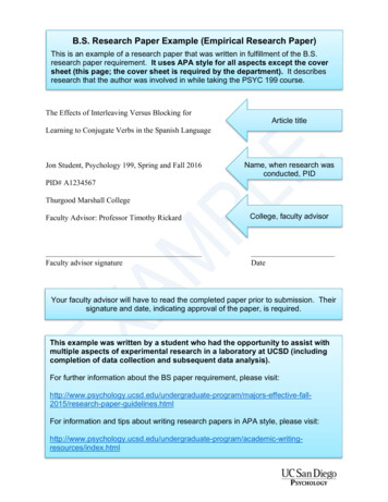
Journal of Cancer 2021, Vol. 12Technology 1:5000 dilution) for 30min at 37 C. Thepositive bands were detected by ECL kit (Pierce).Constructs and vectorsNKCC1-shRNA (sh1: 5’-CCAGCACTACTATTATGATAC-3’, sh2: 5’-GGTGATTTCGTCATAGGAACA-3’, sh3: dbyGenecopoeia, Inc. (Guangzhou, China). The GC cellswere transfected with suitable constructs usingLipofectamine 3000 (Thermo Fisher Scientific, USA) asper the manufacturer’s instructions. After culturingfor 48 h, stable transfectants were screened usingpolybrene, and the expression levels of target proteinand mRNA were analyzed.Cell proliferation assayCCK-8 assay (CCK-8 SAB biotech. College Park,MD, USA) was used for cell proliferation detection.Following the manufacturer’s protocol. The Cellswere seeded into 6-well plates at a density of 1.0 105cells per well and incubated for 24 h in mediumsupplemented with 5% FBS, and incubated at 37 Cwith 5% CO2. At 24 h after transfection, cells weredigested with trypsin and seeded in triplicate into96-well plates (3 104 cells/ well). Each well wasincubated with 10 μl/well of Cell Counting Kit-8solution for 2 h daily for 5 days. Optical density at 450nm was measured on a microplate reader. Threeindependent experiments were performed.ImmunofluorescenceCells were fixed with 4% polymerizedformaldehyde (Solarbio) for 15 min and thenpermeabilized with 0.1% Triton-X100 (Solarbio) foranother 15 min. The samples were incubatedovernight with anti-E-cadherin and anti-vimentinantibodies (Protein Tech, 1:50) at 4 C. Afterincubating with the secondary antibody (1:200) in 5%BSA (Solarbio) for 1 h at 37 C, the cells werecounterstained with DAPI for 25 min. The cells wereobserved using a fluorescence microscope andphotographed (200 magnification).Wound-healing assayCells (5 105) were seeded in a 6-well plates andcultured cells reached 85% confluence. The monolayerwas scratched longitudinally across the plates with asterile p200 pipette tip to simulate a wound. Thecellular debris was removed by PBS. Cell migrationwas observed 0, 12, and 24 h after wounding.Cell invasion and migration assaysThe suitably transfected GC cells were plated inthe top chambers of Corning Incorporated Cell255Culture Inserts (#3422; Franklin Lakes, New Jersey,USA) or Corning Incorporated BioCoat Matrigel coated Invasion Chamber (#3422) in 2% FBS, and thelower chambers were placed with media containing5% FBS as the chemoattractant. The cells wereincubated at 37 C in a humidified atmospherecontaining 5% CO2 for 24 h or 48h. The migratedcancer cell on the lower surface were fixed with 95%alcohol, and stained using 0.1% crystal violet(Solarbio). The number of invaded cells was countedand photographed (100 magnification) under aphase contrast microscope (four random fields perwell).Statistical analysisExperiments data are summarized as mean SDof three times repeation. GraphPad Prism 5(GraphPad Software, Inc, USA) was used to statisticalanalysis. The differences between groups werecompared using two-tail Student’s t test and variance.P values were less than 0.05 were consideredstatistically significant.ResultsNKCC1 is overexpressed in primary GC tissuesNKCC1 was upregulated in the 408 GC tissuescompared to the 211 normal gastric epithelial tissues(P 0.01; Figure 1A) in TCGA datasets as per GEPIA(http://gepia.cancer-pku.cn/). Consistent with this,NKCC1 mRNA levels were significantly higher in thegastric tumors relative to the paired adjacent normaltissues in our cohort (Fig. 1B), and 87.5% of the GCtissues overexpressed NKCC1 protein compared tothe paired adjacent normal samples (Fig. 1C, D).Representativeimmunohistochemistryimagesshowed in situ NKCC1 expression in the tumor (Fig.1E). As shown in Table 1, the clinical relevance of highNKCC1 expression was significantly associated withGC Tumor size (P 0.039), TNM stage (P 0.034) andlymphatic node metastasis (P 0.035) but did notshow any significant correlation to age, gender, tumorlocalization and differentiation. Altogether, NKCC1 issignificantly upregulated in GC tissues and can be apotential oncogenic factor.Knockdown of NKCC1 inhibited theproliferation, migration and invasion of GCcell linesTo elucidate the potential biological function ofNKCC1 in GC development, the expression ofNKCC1 in GC cell lines (HGC-27, BGC-823,SGC-7901, MGC-803, AGS, MKN-28) was firstevaluated by Western-blot and qRT-PCR. NKCC1protein and mRNA were markedly upregulated in theGC cells compared with GES-1 cells (Fig. 2A, B). Inhttp://www.jcancer.org
Journal of Cancer 2021, Vol. 12256addition, NKCC1 was mainly localized in the vesiclesand plasma membrane in AGS and MGC-803 cells(Fig. 2C). This is particularly important because theexact function of NKCC1 varies depending on itsintra- or extracellular localization. To furtherdemonstrate the role of NKCC1 in GC progression,NKCC1 was knocked down in AGS and MKN-28lines (Fig. 2D–G). As Figure 2H and I illustrate, stableknockdown of NKCC1 obviously suppressed growthof GC cells in vitro in the CCK-8 assays. As Figure 2Jand K illustrate, stable knockdown of NKCC1decreased migration of GC cells in the wound healingassay. Furthermore, the transwell assay showed anobvious reduction in the motility and invasiveness ofthe NKCC1-knockdown GC cells compared to thecontrol cells (Fig. 2L M). These results implied thatknocking down NKCC1 inhibited the proliferation,invasiveness and metastasis of GC cells.Table 1. Correlation between NKCC1 expression and patientsclinicopathological characteristics in 95 GC patientsParametersTotal case95Age (years) 6846 6849GenderMale72Female23Tumor size 6.5m40 6.5 cm55Lymphatic node metastasisNegative32Positive63Invasion depth (T)T1-212T3-483NN1-241N3-454MM091M14TNM stageI–II42III–IV53P valueNKCC1High expression(N 50)Low expression(N 0.034*NOTE: The values are statistically significant (*** P 0.001; ** P 0.01; * P 0.05);The 8thTNM Classification of Malignant Tumors proposed by the AJCC/UICC.The mean age at diagnosis is 68 years in patients with GC. Samples are divided intotwo groups based on the mean age.The mean tumor size was 6.5 cm. They were divided into two groups based on themean tumor size.Overexpression of NKCC1 enhances theproliferation, migration and invasion of GC celllinesNKCC1 was overexpressed in MGC-803 andSGC-7901 cells (Fig. 3A–D). In contrast, stableoverexpressed of NKCC1 significantly accelerated cellgrowth (Fig. E-F). and markedly increased migrationof GC cells in the wound healing assay (Fig. 3G, H),and the transwell assay indicated a significantincrease in the migration and invasiveness of theoverexpressed NKCC1 GC cells compared to thecontrol cells (Fig. 3I, J). Taken together, intracellularNKCC1 enhances the proliferation, invasiveness andmetastasis of GC cells.NKCC1 induced EMT and increased MMP2/9expression in GC cellEMT and extracellular matrix remodeling arecritical for cancer cell migration and invasion and arethus key to metastasis [22]. Therefore, we analyzedthe levels of EMT-related proteins in theNKCC1-knocked down and overexpressing GC cells.As shown in Figure 4A, knocking down NKCC1downregulatedthemesenchymalmarkerEMT-related transcription factors Snail and Vimentin,and ECM proteins MMP-2 and MMP-9, andupregulated epithelial E-cadherin. In contrast,overexpression of NKCC1 upregulated Vimentin,Snail, MMP-2 and MMP-9 but downregulatedE-cadherin (Fig. 4B). Thus, NKCC1 promotes EMT ofGC cells.NKCC1 induced EMT of GC cells by activatingthe MAPK-JNK pathwayThe MAPK-JNK/p38 pathway has beenimplicated in EMT, and the JNK pathway in particularis essential for the progression and maintenance ofEMT-related phenotypes and cellular changes [23-24].Therefore, we analyzed the levels of the MAPK-JNKpathway proteins in GC cells following NKCC1knockdown or overexpression. The p-JNK/JNK ratiowas significantly reduced after silencing NKCC1 (Fig.4E), while no significant change was observed in thephosphorylation of P38 and ERK. As expected,p-JNK/JNK increased markedly in the NKCC1overexpressing cells whereas p-ERK and p-P38 werenot affected (Fig. 4F). To determine whether theMAPK-JNK signaling pathway mediates themetastatic effects of NKCC1, the JNK inhibitorSP600125 (Selleckchem, Inc.) was dissolved in DMSO,we treated the NKCC1-overexpressing MGC-803 andAGS cells with 10 μM SP600125 and found that theinvasion and migration of the cells were significantlyinhibited (Fig. 5A-D). Immunofluorescent assay alsoconfirmed the changes of EMT markers (vimentin andE-cadherin) after SP600125 treatment in MGC-803 andAGS cells (Fig. 5E-F). Consistent with this, SP600125downregulated the mesenchymal markers andEMT-related transcription factors in the MGC-803 andAGS cells overexpressing NKCC1 and upregulatedE-cadherin (Fig. 5G-H). Overall, NKCC1 regulateshttp://www.jcancer.org
Journal of Cancer 2021, Vol. 12EMT by the MAPK- JNK pathway to promote GCinvasion and migration.DiscussionNKCC1 is an active Na /K / 2Clcotransporter, which regulates the changes in cellvolume by controlling the intracellular content ofwater and sodium. Ion transporters and ion channelshave recently gained attention for their potentialtumorigenic roles. They regulate the intracellularcontent of water and ions and therefore control cellvolume. The CI- channel (CLIC1) enhanced theinvasion and migration of gastric and liver cancercells [25-26], and K /Ca2 channels are frequentlyoverexpressed in colon cancer [27-28]. Ectopicexpression of NKCCI induced the proliferation and257phenotypic transformation of mouse fibroblasts [29].In addition, NKCC1 has been reported to play animportant role in many different tumorigenesis, it isaberrantly expressed in gastric cancer [9], esophagealcancer [30], meningioma [25], liver cancer [11], andglioma [10] and functions as an oncogene [9-11, 30].We found that NKCC1 was highly expressed in avariety of tumors through analyzing The CancerGenome Atlas project database using GEPIA software(http://gepia.cancer-pku.cn/). NKCC1 was alsohighly expressed in GC tissues by analyzing the RNAseq expression data of 408 GC tissue specimens and211 normal gastric tissues in the database. Thesubsequent experiments will be carried out for furtherverification.Figure 1. Expression of NKCC1 in Gastric cancer tissues and normal Gastric tissues. A. NKCC1 mRNA levels in 408 GC tissues and 211 para-tumor tissues inTCGA database. B. NKCC1 mRNA levels in 38 GC tissues and paired adjacent tissues. C-D. Western blotting showing NKCC1 protein expression levels in GC tissues andpara-tumor tissues with GAPDH as the internal reference control and the quantification of the three independent repeated experiment were shown (n 16, p 0.019). E.Representative immunohistochemistry images showing in situ NKCC1 expression in the tumor, magnification - 200X, Scale bars: 50 µm, magnification - 400X, Scale bars: 20 µm.http://www.jcancer.org
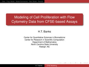
Journal of Cancer 2021, Vol. 12258Figure 2. Knockdown of NKCC1 inhibited the proliferation, migration and invasion of GC cells. A-B. NKCC1 protein and mRNA levels were observed in the GCcell lines and GES-1 cells. C. Representative immunofluorescence images showing localization of NKCC1 on the plasma membrane of the GC cells (magnification - 200X, Scalebars: 50 µm). D-G. NKCC1 mRNA and protein levels in GC cells transfected with shNKCC1 and scrambled control. H-I. The proliferation capacities were detected by CCK8assays in GC cells transfected with shNKCC1 and scrambled control. J-K. Representative images of wound healing assay showing the in vitro migration of control and shNKCC1GC cells (magnification - 40X, Scale bars: 200 µm). L-M. Transwell assay showing in vitro migration and invasion of control and shNKCC1 GC cells (magnification - 100X, Scalebars: 100 µm). * P 0.05; ** P 0.01; *** P 0.001.http://www.jcancer.org
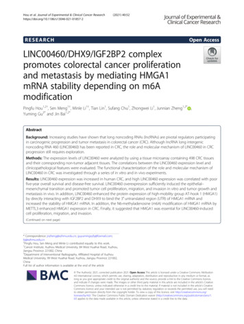
Journal of Cancer 2021, Vol. 12259Figure 3. Overexpress of NKCC1 enhances the proliferation, invasion and migration of GC cells. A-D. NKCC1 protein and mRNA levels in GC cells transfectedwith NKCC1 expression plasmid. E-F. The proliferation capacities were detected by CCK8 assays in GC cells transfected with the control or the NKCC1-overexpressingplasmid. G-H. Wound healing assay showing the in vitro migration of control and NKCC1-overexpressing GC cells (magnification - 40X, Scale bars: 200 µm). I-J. Transwell assayshowing in vitro invasion and migration of control and NKCC1-overexpressing GC cells (magnification - 100X, Scale bars: 100 µm). ** P 0.01; *** P 0.001.http://www.jcancer.org
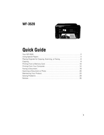
Journal of Cancer 2021, Vol. 12260Figure 4. NKCC1-induced EMT activates the MAPK-JNK pathway in GC cells. Western blotting showing expression levels of EMT makers in A. NKCC1-knockdownGC cells and B. NKCC1-overexpressing GC cells. C-D. Representative immunofluorescence images showing in situ expression of E-cadherin and vimentin in GC cells treatedas above (magnification - 200X, Scale bars: 50 µm). E-F. Immunoblots showing expression levels of JNK/p-JNK, ERK/p-ERK and p38/p-p38 in E. NKCC1-knockdown GC cells andF. NKCC1-overexpressing GC cells.Previous studies show that NKCC1 promotes theinvasion and migration of meningioma [31], glioma[10, 32], and liver cancer [11] cells and proliferation ofesophageal cancer [30] and liver cancer [11] cells. Inaddition, a previous study reported that blockingNKCC1 in GC cells can inhibit cell cycle progressionby disrupting the intracellular Cl- concentration [33].Significantly higher levels of NKCC1 were detected inGC tissues patients compared to the pairedpara-tumor tissues, which was closely related to theTumor size, lymph node metastasis and TNM stage,suggesting that upregulated expression of NKCC1 inGC might facilitate the tumor growth and metastaticphenotype. This was consistent with the data fromTGCA datasets, further confirming that NKCC1 ishighly expressed in GC tissues. These findingsunderscore a potentially important role of NKCC1 asan underlying biological mechanism in theprogression of GC. To clarify the biological function ofNKCC1 in regulating GC cell growth, motility andinvasiveness, a series of in vitro assays wereconducted. The results showed that knocking downNKCC1 in the GC cell lines inhibited EMT anddownregulated the related factors, resulting in lowerproliferation, invasion and migration in vitro. In invitro experiments, the expression of NKCC1 in gastricepithelial cells (GES-1) and six gastric cancer cell lineswas first detected, which was relatively high in AGSand MKN-28 cells, but relatively low in SGC-7901 andMGC-803 cells. The expression of proteins in differentlocations of the cell plays a different role.Immunofluorescence experimental results indicatethat NKCC1 is mainly expressed in the cytoplasm andcell membrane of gastric cancer cells as a plasmamembrane protein. Next, we changed the expressionof NKCC1 in gastric cancer cells. In contrast, NKCC1http://www.jcancer.org
Journal of Cancer 2021, Vol. 12overexpression promoted EMT and increased theproliferation, invasion and migration of GC cells. Inaddition, it is reported that the matrix metalloproteinases (MMPs) enhanced tumor cell motility andinvasion by degrading the extracellular matrix [34-35].Silencing NKCC1 decreased MMP 2/9 levels, while261its overexpression produced the opposite effects. Theabove data suggested that NKCC1 may be a newindicator for the growth and metastasis of gastriccancer, and may act as an oncogene in the progressionof gastric cancer as well as plays an important role inthe proliferation, invasion and migration of GC cells.Figure 5. The MAPK-JNK pathway mediates the pro-metastatic effects of NKCC1 in GC cells. A-B. Transwell assay showing invasion and migration of MGC-803and AGS cells treated as above (magnification - 100X, Scale bars: 100 µm). The experiments were repeated 3 times, and the results were summarized as mean SD (**, P 0.01).C-D. Wound healing assay showing in vitro migration of NKCC1-overexpressing MGC-803 and AGS cells treated with JNK inhibitor SP600125 (magnification - 40X, Scale bars:200 µm). E-F. Representative immunofluorescence images showing in situ expression of vimentin and E-cadherin in MGC-803 and AGS cells treated as above (magnification 200X, Scale bars: 50 µm). G-H. Immunoblots showing expression levels of p-JNK, ERK, E-cadherin snail, vimentin, MMP2 and MMP9 in MGC-803 and AGS cells treated as above.http://www.jcancer.org
Journal of Cancer 2021, Vol. 12EMT plays an important role in the infiltration,invasion, and distant metastasis of various tumors.EMT not only initiates tumor invasion and migration,but is also an indicator of the distant metastasis abilityof tumor cells [36-37]. Studies show that EMTpromotes GC progression via the MAPK-JNKsignaling pathway, and activating this pathwayinduces tumor EMT [38-40]. Consistent with this,knockdown of NKCC1 inhibited EMT-relatedproteins including Snail, vimentin, MMP2 and n had the opposite effects. Severalstudies have shown that JNK promoted EMT andenhanced the invasion and migration of GC cells [24,38-40], which was consistent with our findings.Furthermore, NKCC1 silencing also inhibited JNKactivation without affecting P-38 and ERK. Notsurprisingly, therefore, targeted inhibition of JNK inthe GC cells overexpressing NKCC1 reversed EMT.Immunofluorescence staining demonstrated thatE-cadherin expression was up-regulated andvimentin expression was down-regulated after GCcells overexpressing NKCC1 was treated with the JNKinhibitor SP600125. Hence, NKCC1 may regulate theoccurrence of EMT through MAPK-JNK signalpathway. After SP600125, a JNK inhibitor was addedto gastric cancer cells with overexpressed NKCC1, theEMT-related proteins were changed. The inactivationof MAPK-JNK reversed the overexpression of NKCC1and induced EMT. The invasion and migration abilityof GC cells with overexpressed NKCC1 was inhibited,indicating that NKCC1 could mediate EMT byactivating the MAPK-JNK signal pathway. As a result,it was concluded that MAPK-JNK signal pathwayplayed an important role in the progression of GCmediated by NKCC1.In summary, NKCC1 acts as an oncogene in GCand promotes invasion and migration throughactivating the MAPK-JNK/EMT pathway. This studyproves NKCC1 to be a potential therapeutic target inGC. NKCC1 was highly expressed in human GCsamples, and may be a marker for regulating theprognosis of gastric cancer. NKCC1 could regulate theproliferation, invasion and migration of gastriccancer cells in vitro. It was further confirmed thatNKCC1 could induce EMT to promote the invasionand migration of GC cells through MAPK-JNKpathway. Our study showed that NKCC1 played animportant role in the proliferation and metastasis ofgastric cancer and may become a candidate markerfor clinical diagnosis and treatment of GC.AbbreviationsNKCC1: Na /K /2CI- cotransporter 1; sition;qRT-PCR:real-timepolymerase chain reaction.quantitativeAcknowledgmentsThe authors thank the Key Research andDevelopment program of Science and TechnologyDepartment of Guangxi (grant no. 2017AB45153), theScientific Research and Technology-departmentProgram of Guangxi (grant no. 1598011-4) andInnovation Project of Guangxi Graduate Education(grant no. YCBZ2019043).Authors’ ContributionsAll authors are the guarantors of integrity of theentire study. Jun-fu Wang and Kun Zhao contributedto the study, collected the data, analyzed the data andwrote this manuscript. Ye-yang Chen, Yue Qiu andJin-hui Zhu contributed to the Immunohistochemistryexperiment. Conduted the western blot analyses.Bo-pei Li and Zheng Wang contributed to theReal-time PCR. Jun-qiang Chen contributed todesigned and check this manuscript.Ethical StatementThe authors are accountable for all aspects of thework in ensuring that questions related to theaccuracy or integrity of any part of the work areappropriately investigated and resolved.Author biographyDr. Jun-qiang Chen is the director of thedepartment of the First Affiliated Hospital of GuangxiMedical University, director of GastrointestinalSurgery, doctor of Clinical Medicine, Professor, chiefphysician, and a doctoral tutor. He has profoundknowledge in basic research and clinical work ofgastric cancer, has a solid clinical foundation and richscientific research experience, is proficient in cellbiology, molecular biology and animal experimenttechnology, and has been published more than 50articles related to basic and clinical research of Gastriccancer as the first or corresponding author, and hasundertaken and completed a number of researchprojects related to Gastric cancer. Among them,“Clinical and Basic Research Related to Gastric CancerProgress” won the second prize of Guangxi Scienceand Technology Progress Award in 2018, “Synthesis,Evaluation and Promotion of Medical Evidence forOncology Surgery and Related Diseases” won thethird prize of Guangxi Science and TechnologyProgress Award in 2017.FundingThe present study was supported by the KeyResearch and Development program of Science andTechnology De
qRT-PCR in the ABI (7500) RT-PCR System (Applied Biosystems, USA) using the SYBR Green QRT-PCR Kit (Takara, China). The primers were used: NKCC1 . (GraphPad Software, Inc, USA) was used to statistical analysis. The differences between groups were compared using two-tail Student's t test and variance.