Transcription
Hou et al. Journal of Experimental & Clinical Cancer (2021) 40:52RESEARCHOpen AccessLINC00460/DHX9/IGF2BP2 complexpromotes colorectal cancer proliferationand metastasis by mediating HMGA1mRNA stability depending on m6AmodificationPingfu Hou1,2†, Sen Meng1†, Minle Li1†, Tian Lin1, Sufang Chu1, Zhongwei Li1, Junnian Zheng1,2* ,Yuming Gu3* and Jin Bai1,2*AbstractBackground: Increasing studies have shown that long noncoding RNAs (lncRNAs) are pivotal regulators participatingin carcinogenic progression and tumor metastasis in colorectal cancer (CRC). Although lncRNA long intergenicnoncoding RNA 460 (LINC00460) has been reported in CRC, the role and molecular mechanism of LINC00460 in CRCprogression still requires exploration.Methods: The expression levels of LINC00460 were analyzed by using a tissue microarray containing 498 CRC tissuesand their corresponding non-tumor adjacent tissues. The correlations between the LINC00460 expression level andclinicopathological features were evaluated. The functional characterization of the role and molecular mechanism ofLINC00460 in CRC was investigated through a series of in vitro and in vivo experiments.Results: LINC00460 expression was increased in human CRC, and high LINC00460 expression was correlated with poorfive-year overall survival and disease-free survival. LINC00460 overexpression sufficiently induced the epithelial–mesenchymal transition and promoted tumor cell proliferation, migration, and invasion in vitro and tumor growth andmetastasis in vivo. In addition, LINC00460 enhanced the protein expression of high-mobility group AT-hook 1 (HMGA1)by directly interacting with IGF2BP2 and DHX9 to bind the 3′ untranslated region (UTR) of HMGA1 mRNA andincreased the stability of HMGA1 mRNA. In addition, the N6-methyladenosine (m6A) modification of HMGA1 mRNA byMETTL3 enhanced HMGA1 expression in CRC. Finally, it suggested that HMGA1 was essential for LINC00460-inducedcell proliferation, migration, and invasion.(Continued on next page)* Correspondence: jnzheng@xzhmu.edu.cn; guyumingxyfy@foxmail.com;bj@xzhmu.edu.cn†Pingfu Hou, Sen Meng and Minle Li contributed equally to this work.1Cancer Institute, Xuzhou Medical University, 84 West Huaihai Road, Xuzhou,Jiangsu Province 221002, China3Department of Interventional Radiography, Affiliated Hospital of XuzhouMedical University, 99 West Huaihai Road, Xuzhou, Jiangsu Province 221002,ChinaFull list of author information is available at the end of the article The Author(s). 2021, corrected publication 2021. Open Access This article is licensed under a Creative Commons Attribution4.0 International License, which permits use, sharing, adaptation, distribution and reproduction in any medium or format, aslong as you give appropriate credit to the original author(s) and the source, provide a link to the Creative Commons licence,and indicate if changes were made. The images or other third party material in this article are included in the article's CreativeCommons licence, unless indicated otherwise in a credit line to the material. If material is not included in the article's CreativeCommons licence and your intended use is not permitted by statutory regulation or exceeds the permitted use, you will needto obtain permission directly from the copyright holder. To view a copy of this licence, visit http://creativecommons.org/licenses/by/4.0/. The Creative Commons Public Domain Dedication waiver ) applies to the data made available in this article, unless otherwise stated in a credit line to the data.
Hou et al. Journal of Experimental & Clinical Cancer Research(2021) 40:52Page 2 of 18(Continued from previous page)Conclusions: LINC00460 may be a novel oncogene of CRC through interacting with IGF2BP2 and DHX9 and bind tothe m6A modified HMGA1 mRNA to enhance the HMGA1 mRNA stability. LINC00460 can serve as a promisingpredictive biomarker for the diagnosis and prognosis among patients with CRC.Keywords: LINC00460, Colorectal cancer, IGF2BP2, DHX9, HMGA1, m6ABackgroundLong noncoding RNAs (lncRNAs) are a class of large transcripts with more than 200 nucleotides having no or limited protein-coding capacity, lncRNAs are pervasivelytranscribed from the human genome [1, 2]. With the unprecedented progress in understanding the function oflncRNAs, increasing evidence has shown that lncRNAsplay a role in the physiological and pathological processesof various diseases, especially malignancies [3]. Althoughmany findings should be further validated, lncRNAs cancarry out diverse functions in carcinogenesis, metastasis,and poor prognosis in cancer [4, 5]. For example, the upregulated expression of CCAT2 in colorectal cancer(CRC) is correlated with migration and metastasis [6].MALAT1 was identified as a prognostic biomarker thatpromotes cell proliferation and metastasis in non-smallcell lung cancer (NSCLC) and other cancers [7]. ManylncRNAs and their underlying mechanisms in CRC havebeen previously reported [8, 9]. However, the specificmechanisms of lncRNAs in CRC development and metastasis should be further elucidated.CRC is one of the most lethal malignancies worldwideand is also the leading cause of cancer incidence rateand mortality in China [10]. Most patients are diagnosedat the advanced stage and suffer from poor prognosis[11]; furthermore, malignant proliferation and extensivemetastasis have already occurred in the advanced stageof CRC upon diagnosis [12]. Reports indicate that 50%of patients with CRC die from developing distant metastasis [13], and those with liver metastasis feature a shortfive-year survival rate of less than 10% [14]. The mechanism of colorectal carcinogenesis and metastasis is considered a multifactorial and multistep process that mayinvolve several biological pathways and genetic alterations. Therefore, novel biomarkers that can promote orinhibit the development and progression of CRC shouldbe sought.LncRNAs are increasingly being discovered, and theymay affect the complicated metastasis progress. In thepresent study, we assayed clinical specimens and adjacentnormal tissues from patients with CRC to screen differentially expressed lncRNAs. We identified a novel lncRNA,called long intergenic noncoding RNA 460 (LINC00460),which is positively correlated with CRC proliferation andmetastasis. LINC00460 is located on chromosome13q33.2 and is transcribed into a 935 bp transcript; thislncRNA has recently been reported in several cancers.Most recently, several studies have shown LINC00460 wasoverexpressed in multiple cancers, such as gastric cancer[15], lung cancer [16], head and neck squamous cell carcinoma (HNSCC) [17], hepatocellular carcinoma [18] andcolon cancer [19, 20]. Studies showed that LINC00460mainly located in cytoplasm and could bind to miRNAs toaffect CRC progression [19, 20]. A recent study showedLINC00460 interacted with PRDX1 and facilitated PRDX1entry into the nucleus to promote HNSCC malignantproperties [17]. However, its biological role and specificmechanism in CRC malignant proliferation and extensivemetastasis should be further uncovered.Most recently, Marco Mineo et al. have demonstratedthat lncRNA HIF1A-AS2 binding proteins insulin-likegrowth factor 2 mRNA-binding protein 2 (IGF2BP2) andATP-dependent RNA helicase A (DHX9) are engaged indownstream mRNA stabilization [21]. The High Mobility Group A1 (HMGA1) is a member of non-histonechromatin protein, and regulates multiple biology events,such as embryogenesis, proliferation, transformation, invasion, and metastasis [22]. Recently, studies haveshowed that HMGA1 can be regulated by IGF2BP2through regulating HMGA1 mRNA stability [21, 23].IGF2BP2 is also reported as a N6-methyladenosine(m6A) reader enhancing mRNA stability through recognizing m6A modification sites [24]. Methyltransferaselike 3 (METTL3) is a catalytic enzyme that promotesm6A modification of mRNAs [25]. The m6A modification in mRNAs have been shown regulating mRNA stability [26]. However, the m6A modification in regulatingHMGA1 has not been reported.In the present study, we showed that LINC00460 ishighly expressed in CRC and indicates poor prognosis.We also revealed that LINC00460 regulated the CRCgrowth and metastasis in vitro and in vivo. LINC00460interacted with IGF2BP2 and (DHX9) to promote themRNA stability of HMGA1 and further leading to biological response to CRC malignant proliferation and extensive metastasis. In addition, the (m6A) modificationof HMGA1 mRNA by METTL3 enhanced its expressionin CRC and LINC00460 regulate HMGA1 expressiondepending on METTL3. It suggested LINC00460/DHX9/IGF2BP2 complex may regulate HMGA1 expression through recognizing the m6A modification sites ofHMGA1 to enhance its mRNA stability.
Hou et al. Journal of Experimental & Clinical Cancer Research(2021) 40:52MethodsPatients and sample collectionFor this study, the tissue microarrays (TMAs) slides included examination of 498 pairs of tissue specimens including CRC tissues and the matched correspondingadjacent normal colorectal tissues from CRC patient cohorts that were enrolled at Affiliated Hospital of XuzhouMedical University from 2010 to 2015 in China. Clinicaland pathological information was obtained from themedical records of the Affiliated Hospital of XuzhouMedical University. Survival time was calculated basedon the date of surgery to the date of death or to the lastfollow-up. The patient studies were conducted in accordance with Declaration of Helsinki. The use of thesespecimens and data for research purposes were grantedapproval by the Ethics Committee of the Affiliated Hospital of Xuzhou Medical University.Page 3 of 18Amico Ultra centrifugal filters (Millipore 100KDMWCO). The concentrated virus was used to infectHCT116 and SW480 cells and stable LINC00460 overexpression or knockdown cells were generated by lentivirus infection. The relative LNC00460 and METTL3shRNA sequences were described as below:ShLINC00460#1-For: CCTTTTTG;ShLINC00460#2-For: CCTTTTTG;shMETTL3#1-For: TCGAGAACAATGGATTGTTCCTTGGCTTTTTG.Cell culture and cell treatmentThe CRC cell lines and HEK293T cell were obtainedfrom the cell bank of Chinese academy of sciences.SW480, SW620, HCT116, DLD1, LOVO, HT29, FHCand HEK293T cells were cultured in 1640 and DMEMMedium supplemented with 10% fetal bovine serum,100 U/ml penicillin, 100 μg/ml streptomycin, and incubated in a 37 C humidified incubator with 5% CO2. 1%O2 was generated by flushing a 94% N2/5% CO2 mixtureinto the incubator.The small interfering RNAs (siRNA, 50 nM) againsthuman IGF2BP2 and DHX9 were transfected into theCRC cells SilenFect reagent (Thermo Fisher ScientificInc., USA), while non-specific siRNA was used as negative controls. All siRNAs were purchased from Genepharma Technology (Shanghai, China). The sequencesof the siRNAs were listed in GGAGGCTT.LncRNA microarray analysisTotal RNA was extracted from five paired CRC tissuesand NCTs using TRIzol Reagent (Invitrogen, USA).RNA quantity and quality were measured by NanoDropND-1000. RNA integrity was assessed by standard denaturing agarose gel electrophoresis. LncRNA microarray was performed by (Aksomics, Shanghai, China).Arraystar Human LncRNA Microarray V3.0 is used fordetecting the global profiling of human LncRNAs. Agilent Feature Extraction software (version 11.0.1.1) wasused to analyze acquired array images. LncRNAs weredeemed differentially expressed if their fold change between the CRC and NCT (normal colon tissue) groupsexceed 2.0 and their P-values were less than 0.05.RNA extract, reverse transcription-PCR and qRT-PCRRNA was extracted using TRIzol (Invitrogen) and cDNAwas synthesized using the HiScript 1st Strand cDNASynthesis Kit (Vazyme Biotech, Nanjing, China). Realtime PCR was carried out on ABI-7500 using UltraSYBROne Step RT-qPCR Kit (CWBIO, Beijing, China). Theprimers using for quantitative RT-PCR analysis werelisted in Additional file 1: Table S1:Establishment of stable cell linesThe cDNA of LINC00460 was inserted into pCDHCMV-MCS-EF1-GreenPuro lentivirus vector at ECOR1/BamH1 sites. LINC00460 shRNA sequences were clonedto the vector pLko.1 at Age1/ECOR1 sites. The METTL3 shRNA vectors were purchased from GENE company(Shanghai, China). The overexpression and knockdownlentiviruses were generated by co-transfecting 293 T cellswith the other two packing vectors pMD2G and psPAX.The supernatants of 293 T cultured medium were collected 48 h later, filtered through 0.45-mm filters (Millipore, Temecula, CA, USA) and concentrated usingWestern blot and antibodiesCells were harvested, total protein from cells was extracted by using RIPA lysis buffer and was qualified byusing a BCA detecting kit (Keygen, Nanjing, China). Proteins samples were subjected to 7.5% or 10% SDS-PAGEand transferred onto a PVDF membrane, and then incubated with specific antibody at 4 C overnight, respectively. The next day, the membranes were incubated withsecondary antibodies included HRP-goat anti-mouse,HRP-goat anti-rabbit (ABclonal) at room temperaturefor 1 h. Protein bands were detected on Tanon 5200
Hou et al. Journal of Experimental & Clinical Cancer Research(2021) 40:52automatic chemiluminescence imaging analysis systemusing ECL reagent (Tanon, Shanghai, China). Antibodyagainst GAPDH was used as control (sc-32,233, SantaCruz, Dallas, TX, USA). HMGA1 (A1635, ABclonal,Wuhan, China), IGF2BP2 (11601–1-AP, Proteintech,USA), DHX9 (17721–1-AP, Proteintech, USA), Ecadherin (610,181,BD Biosciences, Bedford, Massachusetts, USA), N-cadherin (610,920,BD Biosciences, Bedford, Massachusetts, USA),β-catenin (610,154, BDBiosciences, Bedford, Massachusetts, USA), were usedfor Western blot assays.In situ hybridization (ISH) and immunohistochemistry(IHC)The in situ hybridization of LINC00460 was performedon formalinfixed paraffin-embedded sections using specific oligonucleotide probe (Hs-LINC00460, RNAscope 2.5, ACD, US), following the manufacturer instructions.Positive control probe (Hs-PPIB) and Negative controlprobe (DapB) were included for each hybridization procedure and analyzed using an OLYMPUS microscopewith OLYMPUS OlyVIA V2.9 software.IHC assays was implemented following a standardstreptavidin-peroxidase (SP) method as previously reported [27] and heat induced epitope retrieval (HIER)was performed with the retrieval buffer, citrate, pH 6.0,prior to commencing with IHC staining protocol. Forprimary antibody incubation, anti-HMGA1 (A1635,ABclonal, Wuhan, China) antibody at 1:100 dilution.The slide without primary antibody incubation served asnegative control.RNA fluorescence in situ hybridization (FISH)Cells were seeded on glass bottom cell culture dish (801,001, NEST) for about 24 h, then cells were fixed with10% neutral buffer formalin, incubation with hydrogenperoxide (2,005,617, RNAscope ) and protease III (2,004,920, RNAscope ) for about 10 min separately. After cellpreparation and pretreatment, following the manufacturer instructions and used specific oligonucleotideprobe (RNAscope 2.5, ACD, US) to visualize the singleRNA in each cell. The nuclei were stained with DAPIfor 15 min. Finally, images were taken under a confocalmicroscope.Cellular proliferation, colony formation assaysCCK-8 assay was applied to measure the cell proliferation according to the Cell Counting Kit-8 manufacturerprotocol (Dojindo, Japan). Colony formation assay wasperformed in 60 mm dishes one thousand cells seededand two weeks cultured. Colonies were counted manually after staining with 0.1% crystal violet.Page 4 of 18Real time cellular analysis (RTCA)The Real Time Cell Analyzer (xCelligence, RTCA, USA)were used for examined the effect of LINC00460 on cellproliferation. The xCELLigence system was used according to the instructions of the supplier (ACEA Biosciences). The system measured impedance differences toderive cell index values at time points and it may be setby the operator.The xCELLigence system consists of four main components: the RTCA analyzer, the RTCA DP station, theRTCA computer with integrated software and disposableE-plate 16. Firstly, the optimal seeding concentration forproliferation assay was determined. After seeding thenumber of 5000 cells in 200 ml medium to each well inE-plate 16, the attachment and proliferation of the cellswere monitored every 15 min. All experiments were carried out for 96 h .Cell migration, invasion and wound healing assaysCells were starved in serum-free media for 24 h thenadded to the top chamber of 24-well transwell chambersplates (8.0 μm, Corning, NY, USA.). Specially, for the invasion assay, cells were seeded into the top chambercoated with Matrigel (BD Biosciences). Completemedium was added to the bottom chambers to stimulatemigration or invasion. After incubation for 24–48 h, thecells those adhered to the lower surface of the membrane were stained with 0.1% Crystal Violet, and thenthe percentages of migrated or invasion cells were calculated. Five randomly selected fields per filter werecounted.In wound healing assay, cells were seeded at a densityof 1 106 cell/well onto six-well plates and cultured toabout 80% confluence. Then, a sterile 10-μl pipette tipwas used to form artificial scratches for each well. Thesuspended cells were washed away with PBS, and thenthe cells were cultured in medium with 1% FBS. Cell migration distance was photographed at 0 h and 24 h underan inverted light microscope.RNA pull-down assay and mass spectrometryRNA pull-down assay was carried out as describedbriefly: in vitro biotin-labeled RNAs (LINC00460 andthe antisense RNA) were transcribed with Biotin RNALabeling Mix (Promega Corporation, US) and T7 RNApolymerase (Thermo Fisher Scientific, US) treated withRNase inhibitor, and purified with Clean-up kit (Promega Corporation, US). The biotinylated LINC00460probes were dissolved in binding and washing buffer andincubated with Streptavidin Agarose Resin (ThermoFisher Scientific, US). Then cell lysates of HCT116 andSW480 were incubated with probes-coated streptavidinbeads, and pull-down proteins were run on SDS-PAGEgels, and then gels were stained by Coomassie Blue
Hou et al. Journal of Experimental & Clinical Cancer Research(2021) 40:52staining, and differential bands were cut off for Massspectrometry (Shanghai Applied Protein TechnologyCo., Ltd. China).RNA immunoprecipitationThe RIP experiment was carried out with the EZ-MagnaRIP Kit (Millipore) according to the manufacturer’sprotocol using 5 mg of antibody. HCT116 and SW480cells were lysed in complete RIP lysis buffer, and the cellextract was incubated with protein A/G agarose beadsconjugated with specific antibodies or control IgG for 2h at 4 C. Beads were washed and incubated with Proteinase K to remove proteins. Finally, purified RNA wassubjected to quantitative RT-PCR analysis. AntibodyIGF2BP2 (11601–1-AP, Proteintech, China), DHX9(17721–1-AP, Proteintech, China).MeRIPMeRIP, we used Anti-N6-methyladenosine (m6A) antibody (ab151230) to pull down m6A modified HMGA1.Total RNA was extracted from cells using Trizol(Thermo-Fisher). 100 μg RNA was added to 500 μlMeRIP buffer (150 mM NaCl, 10 mM Tris-HCl, pH 7.5,0.1% NP-40) and incubated briefly with 1 μl rabbit IgGand then the IgG was removed by protein A/G beads.The pre-cleaned lysates were transferred to new tubesand incubated with rabbit IgG or m6A antibody for 2 hat 4 C with rotation. Finally, m6A bound RNA was extracted with Trizol and the RNA level of HMGA1 wasmeasured by qRT-PCR.Animal workThe female BALB/c nude mice (6–8 weeks old) were purchased from Beijing Vital River Laboratory Animal Technology Co., Ltd. (Beijing, China). All animal experimentswere approved by the Animal Care and Use Committee atXuzhou Medical University. Groups of HCT116-LucshCtrl, HCT116-Luc-shLINC00460, and HCT116-LucshLINC00460 HMGA1 cells (5 106) were injected subcutaneously into the flanks of mice correspondingly. Tumors volume (V) was monitored every 2 days bymeasuring the long axis (L) and the short axis (W) ofxenograft tumor and calculated with the following formula: V (L W2)/2.Bioinformatics of gene expression databaseThe correlation of LINC00460 expression and CRC patient survival were analyzed using the datasets generatedwith Affymetrix HGU133 Plus 2.0 microarrays. Data aredeposited at the Gene Expression Omnibus (GSE40967).GSE40967 contains 566 CRC tissues.Page 5 of 18Statistical analysisStatistical analyses were carried out using SPSS 20.0software (SPSS Inc., Chicago, IL, USA) and GraphPadPrism 8 The association between LINC00460 and theclinicopathologic parameters of the CRC patients wereevaluated by a Chi-square test. The Kaplan–Meiermethod and log-rank test were used to evaluate the correlation between LINC0046 expression and CRC patientsurvival. The unpaired t test was used to determine thestatistical significance of differences between groups.Data were presented as mean SD. p 0.05 was considered statistically significant.ResultsOverexpression of LINC00460 occurs in CRC andcorrelates with poor prognosisLncRNA expression profiles were analyzed in six pairedCRC tissues and normal colorectal tissues (NCTs) toscreen differentially expressed lncRNAs by usinglncRNA microaaray. Among the lncRNAs tested in ourmicroarray, 462 were significantly upregulated, and 817were significantly downregulated in CRC relative toNCT (fold change 2, p 0.05, Additional file 2: Dataset1). Among the 50 lncRNAs showing the greatestchanges, some known oncogenic lncRNAs in CRC, suchas FEZF1-AS1, CCAT1, and H19, were found. The novellncRNA LINC00460 was one of the most expressedlncRNAs (Fig. 1a). Thus, we focused on LINC00460 forfurther studies. Expressions of LINC00460 were validated in a small CRC cohort, revealing that LINC00460was upregulated in CRC tissues relative to adjacentNCTs, as shown in RT-PCR (p 0.0048, Fig. 1b) and gelelectrophoresis results (Additional file 3: Figure S1).LINC00460 expression was examined via in situhybridization (ISH, RNAscope ) in an expanded CRC cohort. The ISH arrays showed that LINC00460 was overexpressed in CRC tissues compared with the adjacentNCTs (p 0.001, Fig. 1c–d). ISH analysis showed thatLINC00460 expression was significantly upregulated insamples with advanced stage III/IV compared with thatin stage I/II tumors (Table 1). Survival analysis showedthat the high LINC00460 expression was significantlycorrelated with poor overall survival (OS) (p 0.0001,Fig. 1e) and disease-free survival (DFS) (p 0.0005, Fig.1f). The Gene Expression Omnibus (GEO) data indicated that the high LINC00460 expression of CRC patients was correlated with poor OS and DFS (Fig. 1g–h).The GEO data also showed that LINC00460 expressionwas elevated in CRC in the advanced tumor stage (Fig.1i). Finally, univariate analysis showed that LINC00460was significantly associated with poor OS (hazard ratio[HR], 2.526; 95% confidence interval [CI], 1.696–3.761;p 0.001) and DFS (HR, 3.437; 95% CI, 1.323–8.927; p 0.001) (Additional file 4: Table S1). Multivariate analysis
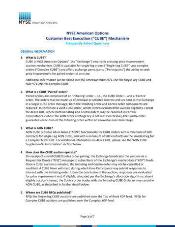
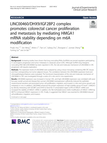
Hou et al. Journal of Experimental & Clinical Cancer Research(2021) 40:52Page 6 of 18ABRP11 807H22.5XLOC 010998XLOC 004546AC017096.1RP11 219E7.4CDKN2B AS1LOC389023SATB2 AS1LOC389332BC133032AC009133.21BC048192XLOC 001595RP4 697P8.2XLOC 009302CTD 2168K21.1RP4 677H15.6RP11 333A23.4AC092652.1RP11 342A1.1PWRN1AL928768.3AX747981H19RP11 596D21.1AK022914CTD 2026G22.1AP000525.8SH3PXD2A AS1CLK1LINC00460LINC00524RP11 267A15.1RP5 1011O1.2FEZF1 AS1RP11 138J23.1PSMD1AC123023.1RP11 474D1.3C17orf76 AS1AC019100.3RP11 141J13.5MIR17HGVAMP7RP1 170O19.17CCAT1LOC170425AC002331.1NCTsPaired LINC00460 RNA Level inCRC Tissure (Normal/Cancer)10 1P 0.00481001010.10.01Normal(n 57)Cancer(n 57)CRC lE4P 0.001210-1100200300400-2Low 17450High 324n 498P 0.0001HR 2.52495%CI: 1.825 to 3.490Disease Free Survival110Low expression of LINC00460High expression of LINC004601003FOver SurvivalCum.SurvivalCase 464Cum.SurvivalΔIn Situ Hybridization ofLINC00460 Staining(C-N)DLow expression of LINC00460High expression of LINC00460100Low 13590High 20580n 340P 0.0005HR 4.06495%CI: 1.948 to 8.4817000020406008020Over Survival(GSE40967)HDisease Free Survival(GSE40967)Low 281n 562P 0.0240High 281Time(months)Fig. 1 (See legend on next page.)Low expression of LINC00460High expression of LINC00460Cum.SurvivalCum.SurvivalLow expression of LINC00460High expression of LINC00460Low 279High 278n 557P 0.0001Time(months)ILINC00460 Expression (log2)G4060Time(months)Time(months)TNM Stage(GSE40967)P 0.025386420I/IIIII/IV80
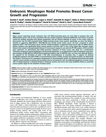
Hou et al. Journal of Experimental & Clinical Cancer Research(2021) 40:52Page 7 of 18(See figure on previous page.)Fig. 1 Overexpression of LINC00460 is observed in CRC and correlates with poor prognosis. a Heatmap of top 50 dysregulated lncRNAs identifiedfrom microarray analysis. NCTs: Normal colon tissues. b LINC00460 expression was quantified in colorectal cancer tissues and their adjacent noncancerous tissues in a small cohort (n 57, p 0.0048). b LINC00460 expression was validated in 8 pairs of CRC patient samples by PCR and gelelectrophoresis, N, normal tissue; T, tumor. c representative images of LINC00460 expression in colorectal cancer and adjacent normal tissuesusing ISH analysis in large cohort (n 498). d Staining intensities of LINC00460 in colorectal cancer tissues compared with paired adjacent noncancerous tissue. N, paired adjacent non-cancerous tissues. C, colorectal cancer tissues (n 464, p 0.001). e, f Kaplan–Meier survival curvesdepicting overall survival (n 498, p 0.0001) or disease-specific survival of patients with CRC (n 340, p 0.001) stratified by LINC00460expression levels in CRC tissues. g, h, i The GEO date of LINC00460 expression was in CRC correlated with poor survival in over survival, diseasefree survival, and advanced tumor stage (GSE40967)demonstrated that LINC00460 expression was also anindependent prognostic marker for OS (HR, 2.299; 95%CI, 1.539–3.435; p 0.001) and DFS (HR, 2.751; 95% CI,1.052–7.198; p 0.009) (Additional file 5: Table S2).LINC00460 promotes CRC cell proliferationThe endogenous expression of LINC00460 in colorectalcell lines, including DLD1, HCT116, SW480, LOVO,SW620, HT29, and the human normal colorectalTable 1 Relationship between LINC00460 expression andclinicopathological features of CRC patientsVariablesAllpatientsLINC00460 expressionLow (%)High (%)All cases498174 (35)324 (65) 60 years19481 (42)113 (58) 60 years30493 (31)211 (69)AgePvalue*0.011Gender0.700Males289103 (36)186 (64)Females20971 (34)138 (66)TNM stage0.005I/II275109 (40)166 (60)III/IV22365 (29)158 (71)Lymph node metastasis0.044N0302116 (38)186 (62)N1/N2/N319658 (30)138 (70)M0461167 (36)294 (64)M1377 (19)30 (81)Metastasis0.034Tumor diameter0.044 4.5 cm25177 (31)174 (69) 4.5 cm24797 (39)150 (61)Poor427149 (35)278 (65)Moderate/High7125 (35)46 (65)Differentiation0.959Depth of invasion0.104T1/T210343 (42)60 (58)T3/T4395131 (33)264 (67)P-value* measured by Pearson’s Chi-Squared testepithelial cell FHC were measured by RT-PCR and gelelectrophoresis. The results showed that CRC cellsexpressed higher LINC00460 compared with the normalcolonic cell (Fig. 2a–b). Subsequently, stable knockdownor overexpression cell lines were established usingshRNAs or LINC00460 overexpression lentivirus to investigate the biological roles of LINC00460 in CRC (Fig. 2cd). Then, Cell Counting Kit-8 (CCK8) assays and a RealTime Cell Analyzer (RTCA, xCELLigence) were used toexamine the effect of LINC00460 on cell proliferation.CCK8 assay showed that LINC00460 overexpression increased HCT116 and SW480 cell proliferation (Fig. 2e–g).Conversely, HCT116 and SW480 cells depleted ofLINC00460 displayed reduced rates of cell growth compared with the corresponding controls (Fig. 2f–h). TheRTCA assays also LINC00460 positively regulated theproliferation of CRC cells (Fig. 2i-j, Additional file 6: Figure S2A-B). Colony formation assays suggested thatLINC00460 promotes tumor cell growth in CRC cells(Fig. 2k-n). These results have revealed that LINC00460 isan important regulator of CRC cell proliferation.LINC00460 promotes CRC cell migration and invasionTo estimate the in the migration and invasion abilities ofLINC00460 in CRC cells, we used transwell and woundhealing assays to analyze the effects of LINC00460 onCRC cell migration and invasion. The results revealedthat LINC00460 overexpression increased migration andinvasion abilities, whereas LINC00460 knockdown decreased the abilities in HCT116 and SW480 cells(Fig. 3a-d). Wound healing assays revealed thatLINC00460 positively regulated the wound healing speedin HCT116 and SW480 cells (Additional file 7: FigureS3A-D).Given that Epithelial Mesenchymal Transition (EMT)is a well-known driving force for cell migration and invasion, we investigated the effects of LINC00460 onEMT. We observed that LINC00460 overexpression reduced the mRNA expression of E-cadherin and increased the mRNA expression of N-cadherin, whereasLINC00460 knockdown increased the mRNA level of Ecadherin and decreased N-cadherin mRNA expression(Fig. 3e-h). Consistent with mRNA changes, the proteinlevels of E-cadherin and N-cadherin were also altered
Hou et al. Journal of Experimental & Clinical Cancer hCtrlshLINC00460#1shLINC00460#2Cell 460500Colony NumbersHCT1162436-0.5L6072shCtrl84shLINC00460#1 shLINC00460#2
lent Feature Extraction software (version 11.0.1.1) was used to analyze acquired array images. LncRNAs were deemed differentially expressed if their fold change be-tween the CRC and NCT (normal colon tissue) groups exceed 2.0 and their P-values were less than 0.05. RNA extract, reverse transcription-PCR and qRT-PCR