Transcription
Haji et al. Respiratory Research(2020) SEARCHOpen AccessMitochondrial dysfunction in airways andquadriceps muscle of patients with chronicobstructive pulmonary diseaseGulam Haji1,2†, Coen H. Wiegman1*† , Charalambos Michaeloudes1, Mehul S. Patel2, Katrina Curtis2,Pankaj Bhavsar1, Michael I. Polkey2, Ian M. Adcock1, Kian Fan Chung1,2 and on behalf of the COPDMAP consortiumAbstractBackground: Mitochondrial damage and dysfunction have been reported in airway and quadriceps muscle cells ofpatients with chronic obstructive pulmonary disease (COPD). We determined the concomitance of mitochondrialdysfunction in these cells in COPD.Methods: Bronchial biopsies were obtained from never- and ex-smoker volunteers and COPD patients (GOLDGrade 2) and quadriceps muscle biopsies from the same volunteers in addition to COPD patients at GOLD Grade 3/4 for measurement of mitochondrial function.Results: Decreased mitochondrial membrane potential (ΔΨm), increased mitochondrial reactive oxygen species(mtROS) and decreased superoxide dismutase 2 (SOD2) levels were observed in mitochondria isolated frombronchial biopsies from Grade 2 patients compared to healthy never- and ex-smokers. There was a significantcorrelation between ΔΨm and FEV1 (% predicted), transfer factor of the lung for carbon monoxide (TLCOC %predicted), 6-min walk test and maximum oxygen consumption. In addition, ΔΨm was also associated withdecreased expression levels of electron transport chain (ETC) complex proteins I and II. In quadriceps muscle ofGrade 2 COPD patients, a significant increase in total ROS and mtROS was observed without changes in ΔΨm,SOD2 or ETC complex protein expression. However, quadriceps muscle of GOLD Grade 3/4 COPD patients showedan increased mtROS and decreased SOD2 and ETC complex proteins I, II, III and V expression.Conclusions: Mitochondrial dysfunction in the airways, but not in quadriceps muscle, is associated with airflowobstruction and exercise capacity in Grade 2 COPD. Oxidative stress-induced mitochondrial dysfunction in thequadriceps may result from similar disease processes occurring in the lungs.Keywords: COPD, Airways, Quadriceps, Oxidative stress, Mitochondria* Correspondence: c.wiegman@imperial.ac.uk†Gulam Haji and Coen H. Wiegman contributed equally to this work.1Airways Disease, National Heart and Lung Institute, Imperial CollegeLondon, Dovehouse Street, London SW3 6LY, UKFull list of author information is available at the end of the article The Author(s). 2020 Open Access This article is licensed under a Creative Commons Attribution 4.0 International License,which permits use, sharing, adaptation, distribution and reproduction in any medium or format, as long as you giveappropriate credit to the original author(s) and the source, provide a link to the Creative Commons licence, and indicate ifchanges were made. The images or other third party material in this article are included in the article's Creative Commonslicence, unless indicated otherwise in a credit line to the material. If material is not included in the article's Creative Commonslicence and your intended use is not permitted by statutory regulation or exceeds the permitted use, you will need to obtainpermission directly from the copyright holder. To view a copy of this licence, visit http://creativecommons.org/licenses/by/4.0/.The Creative Commons Public Domain Dedication waiver ) applies to thedata made available in this article, unless otherwise stated in a credit line to the data.
Haji et al. Respiratory Research(2020) 21:262BackgroundChronic obstructive pulmonary disease (COPD) is characterised by a persistent, progressive airflow obstructionand increased chronic inflammation in the airways andlungs. The pathological features include emphysema,mucus hypersecretion, small airways inflammation andfibrosis, and increased airway smooth muscle (ASM)mass due to ASM cell proliferation, which lead to airflow obstruction. Patients with more advanced COPDexperience frequent exacerbations caused by virusesand/or bacteria, associated with a faster decline in lungfunction and greater mortality [1, 2]. Oxidative stress derived both exogenously through cigarette smoking andendogenously through inflammatory processes is an important mechanism driving the damage and inflammation in COPD [1]. Markers of oxidative stress such ashydrogen peroxide and lipid peroxidation products suchas 8-isoprostanes and malonaldehyde are increased,while antioxidants such as glutathione are reduced [2].Mitochondria are the most important source of intracellular reactive oxygen species (ROS), generated duringthe synthesis of ATP through oxidative phosphorylation.Mitochondrial reactive oxygen species (mtROS) aremaintained as a balance between production and scavenging processes from the antioxidant defence system. Elevated levels of mtROS can initiate processes thatinfluence mitochondrial function and integrity.Indeed, failure to scavenge mtROS leads to mitochondrial dysfunction as shown in airway epithelial cells exposed to cigarette smoke extract (CSE) leading todecreased ATP levels and mitochondrial membrane potential and impaired mitophagy [3–5]. In addition, CSEexposure leads to mitochondrial fragmentation, branching and reduced cristae linked to increased levels of IL1β, IL-6, and CXCL8 that are associated with features ofsenescence [6, 7]. Similar changes of mitochondrial damage with fragmentation has been observed in airwayepithelial cells obtained from patients with COPD,and altered mitochondrial function has been shown inairway smooth muscle cells cultured from biopsiesobtained from patients with COPD as assessed by reduced membrane potential, respiration rates and ATPproduction [8].One extra-pulmonary manifestation of COPD is locomotor skeletal muscle dysfunction, specifically atrophyand weakness of these muscles, and reduced muscle endurance described in the quadriceps muscle even in milddisease [9]. The impairment in muscle endurance observed in COPD is related to a reduced muscle oxidativecapacity, together with reductions in both mitochondrialdensity and enzyme activities [10–12]. A recent studysupported the concept that the low muscle oxidativecapacity in COPD cannot be explained by physical inactivity alone and is likely driven by COPDPage 2 of 11pathophysiological processes in the lungs of patientswith COPD [13], bringing in the concept that circulatingmediators derived from the COPD lung could causemitochondrial dysfunction in skeletal muscle particularlythe quadriceps muscle.We conducted this study in order to examine mitochondrial function from mitochondria isolated frombronchial biopsies and quadriceps muscle of patientswith COPD compared to those obtained from smokerswithout COPD and to assess whether there were parallelchanges in both compartments.MethodsStudy design and proceduresNon-smoking healthy and COPD participants were recruited from the Royal Brompton Hospital and enrolledthrough the Medical Research Council COPD MAPConsortium. COPD participants were enrolled with severity levels defined according to the Global initiative forchronic Obstructive Lung Disease (GOLD) guidelines.Participants with an exacerbation or symptoms of a respiratory tract infection in the preceding month were excluded. All participants were assessed at an initial visit,then undertook an incremental maximal cycle-ergometercardiopulmonary exercise test (CPET) with cardiorespiratory measurements, a six-minute walk test and quadricepsstrength measurements over two visits. For safety considerations, COPD participants at GOLD Grade 3/4 did notundergo bronchoscopy and only underwent a muscle biopsy. All participants gave informed consent to this studythat was approved by the Research Ethics Committee (11/LO/1852 and 12/LO/0088).Fiberoptic bronchoscopyFiberoptic bronchoscopy was undertaken in participantsunder sedation, with midazolam and topical anaesthesiato the airways with lidocaine. Standard sized biopsy forceps were used to take four to six endobronchial biopsies from the right middle lobe and the right lower lobebronchi. Biopsies were collected in cold PBS and transported on ice to the laboratory for protein fractionisolation.Quadriceps muscle biopsyA percutaneous needle biopsy of the Vastus lateralis wasobtained from the dominant side at least 1 week afterbut within 1 month of phenotypic characterisation.Under sterile conditions (2% chlorhexidine/70% isopropyl), 5 ml of 2% lignocaine was injected into the skin andsubcutaneous tissue. A sub centimetre incision wasmade, and a Bergstrom needle inserted through the subcutaneous tissues and the fascia of the muscle. Severaltissue biopsies (4–6, approximately 0.5 cm2) were collected at the same pass. Biopsies were collected in cold
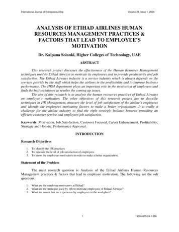
Haji et al. Respiratory Research(2020) 21:262PBS and transported on ice to the laboratory for proteinfraction isolation.Page 3 of 11Superoxide dismutase 2 (SOD2) levels in mitochondrial fractions were determined by sandwich ELISA(R&D systems) according to manufacturer’s instructionsand are expressed per mg protein of each sample.Mitochondrial functional analysisProtein fraction isolation from bronchial and quadricepsbiopsies were performed immediately after obtaining thebiopsy. Intact mitochondria and cytoplasmic proteinfractions were isolated using a Mitochondrial IsolationKit for Tissue (Thermo-Scientific) and a Dounce TissueGrinder set (SIGMA). Briefly, to disrupt cell-to-cell contact bronchial and quadriceps biopsies were douncedwith douncer A by 5 strokes and 10 strokes, respectively.This was followed by douncing with 20 and 40 strokesfor bronchial and quadriceps biopsies, respectively, withdouncer B to disrupt the cells. Cellular contents wereseparated into three different fractions by two centrifugation steps. The large nuclear membrane fraction waspelleted by centrifugation at 2500 rpm for 10 min at4 C. The supernatant containing the cytoplasm fractionwith mitochondria was transferred to new tubes andcentrifuged subsequently at 10,500 rpm for 15 min at4 C. The mitochondrial pellets were dissolved in Hanks’Buffered Salt Solution (HBSS) containing MgCl2 andCaCl2 (Gibco). After isolation the protein content of allthe fractions was determined by Bradford assay (Biorad)against a 2 mg/ml bovine serum albumin standard.Mitochondrial membrane potential (ΔΨm) was measured in intact isolated mitochondria using the cationicdye benzimidazolylcarbocyanine iodide (JC-1, Invitrogen). Intact mitochondria were incubated with JC-1 (3 μM) for 30 min at37 C and 5% CO2. JC-1 monomers emit green fluorescence but when they enter live mitochondria, they formJ-aggregates which emit red fluorescence. The ratio between red and green fluorescence was used as an indexof ΔΨm and is expressed per mg protein of each sample.Mitochondrial reactive oxygen species (ROS) weremeasured with MitoSOX Red (Invitrogen), a redoxsensitive fluorescent probe that is selectively targeted tothe mitochondria. Intact isolated mitochondria were incubated with 5 μM MitoSOX and the red fluorescencewas determined at 510/580 nm using a fluorescenceplate reader. Whole cellular ROS was detected in thecytoplasm fractions using the conversion of the cellpermeable non-fluorescent 2′-7′-dichlorofluorescin diacetate (DCF) probe into the highly fluorescent 2′-7′dichlorofluorescin when de-esterified by oxidation products. The cytoplasmic fractions were incubated with 0.1mM DCF for 30 min at 37 C and 5%CO2. Fluorescencewas measured at 485/528 nm on a SynergyTM multidetection microplate reader (Biotek Instruments, USA).Data is expressed as relative fluorescence units (RFU)per mg protein.Mitochondrial electron transport chain (ETC) complexproteins I, II, III, IV and VContent of isolated protein fractions were determined byBradford assay (Biorad). Mitochondrial protein fractions(20 μg per lane) were separated on 10% NuPAGE gels(Invitrogen) and proteins were detected as previously described [14]. Bronchial and quadriceps muscle mitochondrial electron chain transport (ETC) complexproteins were detected using the MitoProfile TotalOXPHOS Human WB Antibody Cocktail (MitoScience).Porin (VDAC1) protein expression was used as a loadingcontrol (Abcam).Statistical analysisData involving patient details and assessments areexpressed as mean SEM. Differences in continuousvariables (Table 1) were assessed parametrically usingANOVA and t-tests after normality of the dataset wastested using the Kolmogorov-Smirnov test.Pearson and Spearman’s correlations were determinedbetween mitochondrial membrane potential (JC-1) andpatient assessments (FEV1, TLCOc, 6 MW and VO2)and between JC-1 and the electron transport chain(ETC) complex protein expression levels in bronchialand quadriceps biopsies, respectively. Biopsy analysiswas assessed non-parametrically by Kruskal WallisANOVA and Mann-Whitney test. P value of 0.05 wastaken as significant.ResultsSubject characteristicsThere was no difference in lung function and exercise performance between the healthy never-smokers and exsmokers with an FEV1 80% predicted (Table 1). As expected, patients with GOLD Grade 3/4 disease demonstrated poorer exercise performance and higher symptomburden than those with GOLD Grade 2 disease andnever-smokers or ex-smokers, together with a reducedpeak oxygen consumption (VO2 Peak) and six-minutewalk distance. GOLD Grade 2 patients also demonstrateda lower VO2 Peak and six-minute walk distance whencompared to ex-smokers and never-smokers.Airway mitochondrial function, ROS and ETC complexprotein levelsPatients with GOLD Grade 2 COPD demonstrated a significant reduction in airway mitochondrial membranepotential (ΔΨm) when compared to ex-smokers (p 0.01) or never-smokers (p 0.05) (Fig. 1a), with higher
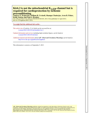
Haji et al. Respiratory Research(2020) 21:262Page 4 of 11Table 1 Subject characteristicsNever-smokersn 5Ex-smokersn 8GOLD Grade 2n 6GOLD Grade 3/4n 6Age (years)61 663 564 364 7Female: Male1:43:54:21:5Cigarette smoking(pack years)0 036 1634 1240 6Body mass index (kg/m2)24 525 429 723 3 #CAT score2 (0–6)8 (2–15)16 (4–23)24 (20–26)**#FEV1 (L)2.9 0.42.9 0.71.9 0.30.8 0.3**###FEV1 (% pred)100 9103 1171 5*30 11**##FVC (L)4.3 0.84.4 1.03.0 0.73.1 1.3FEV1/FVC (%)69 667 763 828 9**##TLCOc (% pred)90 1480 1067 8*38 13**##6-min walk (m)657 18631 55493 66**364 65**##VO2 Peak (ml/kg/min)28 325 317 3**12 4**#QMVC (Kg)34 641 1131 832 6QMVC/BMI1.5 0.11.6 0.41.1 0.3*1.4 0.2SPPB1212 (11–12)11 (10–12)11 (9–12)BMI Body Mass Index; CAT score: COPD Assessment Test score; FEV1 Forced Expiratory Volume in one sec; 6 MW: 6-Minute Walk; QMVC Quadriceps MaximalVoluntary Contraction; SPPB Short Physical Performance Battery; TLCOc Single-breath transfer factor of the lung for carbon monoxide; VO2 Oxygen consumption.Data shown as mean SD or median (range). * p 0.05 compared to never smoker or ex-smoker, **p 0.01 compared to never smoker or ex-smoker, #p 0.05compared to GOLD Grade 2, ## p 0.01 compared to GOLD Grade 2, ### p 0.01 compared to GOLD Grade 2Fig. 1 Mitochondrial function, ROS and ETC complex protein expression levels in the lung. (a) Airway mitochondrial membrane potential (ΔΨm),(b) mitochondrial reactive oxygen species (mtROS), (c) cellular ROS levels and (d) superoxide dismutase 2 (SOD2) levels in never-smoking healthy,ex-smokers and GOLD Grade 2 patients. Data is expressed as mean SEM; healthy never-smokers (n 5), ex-smokers (n 8) and GOLD Grade 2patients (n 6); *p 0.05, **p 0.01, Mann-Whitney test; RFU: Relative Fluorescence Units
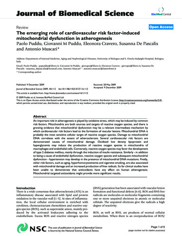
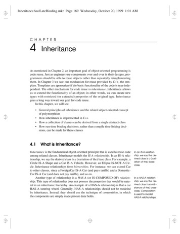
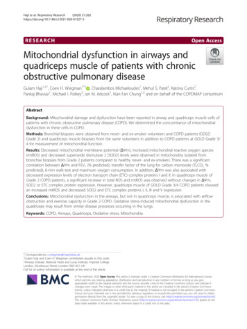
Haji et al. Respiratory Research(2020) 21:262levels of mitochondrial ROS when compared to healthyex-smokers (p 0.01) and healthy never-smokers (p 0.05) (Fig. 1b). Overall cellular ROS burden was alsohigher in the GOLD Grade 2 patients when compared tothe healthy never-smoker (p 0.01) and ex-smoker (p 0.05) groups (Fig. 1c). Superoxide dismutase 2 (SOD2)levels were decreased in GOLD Grade 2 COPD patientmitochondrial fractions compared to ex-smokers (p 0.05) and never-smokers (p 0.01) (Fig. 1d).ETC complex protein I (NADH dehydrogenase) protein expression was reduced in mitochondria isolatedfrom COPD patient biopsies compared to healthy ( 85.1%, p 0.01) and ex-smoker ( 42.2%, p 0.01)groups (Fig. 2a). ETC complex protein II (ubiquinolcytochrome c reductase complex) protein expressionwas also reduced in lung mitochondria isolated fromCOPD patient lung compared to healthy ( 45.8%, p 0.01) and ex-smoker ( 39.4%, p 0.01) groups (Fig. 2b).A small reduction in ETC complex protein V (ATPase)protein expression was observed in mitochondria isolated from the ex-smoker group lungs compared to thenever-smoker group ( 13.9%, p 0.05). No change inprotein expression of the ETC complex V protein wasobserved in the COPD group. Representative Westernblot images from 2 never-smoker, 2 ex-smokers and 2GOLD Grade 2 COPD patients are shown in Fig. 2d.There was a significant correlation between airwaymitochondrial membrane potential (ΔΨm) and FEV1%Page 5 of 11predicted (r 0.72, p 0.001), and TLCOc % predicted(r 0.46, p 0.05) (Fig. 3a & b). Six-minute walk distance was also correlated with membrane potential (r 0.52, p 0.05) as was the peak oxygen consumption VO2(r 0.65, p 0.01) (Fig. 3c & d). In addition, there was acorrelation between ΔΨm and the level of ETC complexproteins I and II measured on the Western blots frombronchial biopsy mitochondria (r 0.73; p 0.001 andr 0.79, p 0.001, respectively; Fig. 4a & b).Quadriceps muscle mitochondrial function & ROS and ETCcomplex protein levelsThere was no difference in ΔΨm in the quadricepsmuscle between the study groups (Fig. 5a). There werehigher whole cell and mitochondrial ROS levels in thequadriceps muscle of GOLD Grade 2 and GOLD Grade3/4 patients when compared to the healthy neversmokers and ex-smoker groups (Fig. 5b & c). SOD2levels were decreased in GOLD Grade 3/4 COPD patients compared to never-smokers (p 0.05) and exsmokers (p 0.05) but not to GOLD Grade 2 COPD patients. There was no significant correlation betweenΔΨm and ROS levels in quadriceps muscle with quadriceps muscle strength, FEV1 (%predicted), TLCOc (%predicted), six-minute walk distance and peak VO2 (datanot shown).There was no difference in the level of ETC complexproteins I, II, III and V in the quadriceps of never-Fig. 2 ETC complex protein expression levels in the lung. (a) Electron transport chain (ETC) complex protein I, (b) complex protein II, and (c)complex protein V expression levels. (d) Representative Western blot images from 2 never-smoker, 2 ex-smokers and 2 GOLD Grade 2 COPDpatients of mitochondrial ETC proteins. Data is expressed as mean SEM; healthy never-smokers (n 5), ex-smokers (n 5) and COPD patients(n 5). *P 0.05 and **P 0.01, Mann-Whitney test
Haji et al. Respiratory Research(2020) 21:262Page 6 of 11Fig. 3 Correlations between airway mitochondrial membrane potential and FEV1, TLCOc and VO2 Peak. (a) Correlations between airwaymitochondrial membrane potential (ΔΨm) and FEV1 (% predicted), (b) single-breath transfer factor of the lung for carbon monoxide (TLCOc %predicted), (c) six-minute walk distance (6 MW), and (d) weight-adjusted peak oxygen uptake (VO2 peak ml/min/kg). never-smokers (n 5), ex-smokers (n 8) and COPD GOLD Grade 2 (n 6)Fig. 4 Correlation between mitochondrial membrane potential and ETC protein expression levels in quadriceps muscle. (a) Correlations betweenbronchial mitochondrial membrane potential (ΔΨm) and protein expression levels of lung electron transport chain (ETC) complex proteins I and(b) lung ETC complex protein II. (c) Correlation between quadriceps mitochondrial membrane potential (ΔΨm) and muscle ETC complex protein Iand (d) muscle ETC complex protein II never-smokers (n 5), ex-smokers (n 5) and COPD GOLD Grade 2 (n 5)
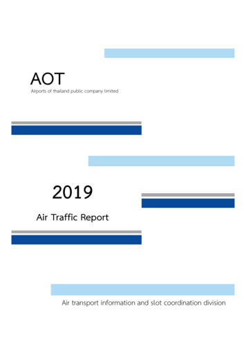
Haji et al. Respiratory Research(2020) 21:262Page 7 of 11Fig. 5 Mitochondrial function, ROS and ETC complex protein expression levels in quadricep muscle. (a) Quadriceps muscle mitochondrialmembrane potential (ΔΨm), (b) mitochondrial reactive oxygen species (mtROS) levels, (c) total cellular ROS levels and (d) superoxide dismutase 2(SOD2) levels in healthy never-smokers, ex-smokers, GOLD Grade 2 and GOLD Grade 3/4 patients. Data shown as mean SEM; never-smokers(n 5), ex-smokers (n 8), GOLD Grade 2 patients (n 6), and GOLD Grade 3/4 patients (n 6); *p 0.05, **p 0.01, Mann-Whitney testsmokers, ex-smokers and GOLD Grade 2 COPD subjects(Fig. 6 a-e). However, there was a significant reduction inthe level of these ETC complex proteins in GOLD Grade3/4 COPD subjects (Fig. 6a-e). There was a significantcorrelation between ΔΨm and the levels of mitochondrialETC complex proteins I and II in muscle (r 0.70; p 0.01; r 0.56, p 0.05, respectively; Fig. 4c &d).Mitochondrial function and ROS levels between bronchialand muscle biopsiesThere was no correlation between ΔΨm of mitochondriaof bronchial and quadriceps biopsies, but mitochondrialand cellular ROS levels did correlate between bronchialand quadriceps biopsies (Table 2).DiscussionWe have shown the presence of mitochondrial dysfunction in the mitochondrial fraction of bronchial biopsycells in terms of higher levels of mitochondrial ROS anda reduction in ΔΨm in patients with GOLD Grade 2COPD compared to healthy never-and ex-smokers. Thiswas associated with a reduction in protein expression ofelectron transfer chain (ETC) complex proteins I, II andV in the mitochondria of patients with COPD, with a reduction in ETC activity as indicated by the impairedΔΨm. These changes were associated with an increasedoxidative stress that is probably resulting from a reducedantioxidant capacity in the cells contained within the biopsy. The ΔΨm generated by the proton pumps (ETCcomplex proteins I, III and IV) drives ATP productionduring oxidative phosphorylation. By contrast, in the patients with COPD, concomitant examination of mitochondrial dysfunction of the quadriceps muscle obtainedfrom the same subjects at GOLD Grade 2, we found noevidence of mitochondrial dysfunction with respect toΔΨm. Although there was an increase in both cellularand mitochondrial ROS levels, this was not associatedwith any reduction in the mitochondrial ETC complexproteins, particularly of I and II, which were clearly reduced in the mitochondria of the bronchial biopsies. Ofgreater interest is the observation that in the quadricepsmuscle of patients with more severe COPD at GOLDGrade 3/4, there was a considerable loss of ETC complex I, II, III, and V proteins, without any perturbationof ΔΨm but with excess cellular and mitochondrialROS, similar to that observed for the quadriceps muscleof COPD patients at GOLD Grade 2.Although we did not obtain bronchial biopsies fromthe patients at GOLD Grade 3/4, our data indicate thatthe mitochondrial dysfunctionality in the lung is quitedifferent from that observed in the locomotor quadriceps muscle. There was a significant correlation between
Haji et al. Respiratory Research(2020) 21:262Page 8 of 11Fig. 6 ETC complex protein expression levels in quadriceps muscle.(a) Representative Western blot of mitochondrial electron transportchain (ETC) complex proteins from quadriceps muscle of 3 neversmokers, 3 ex-smokers, 3 GOLD Grade 2 COPD patients and 3 GOLDGrade 3/4 COPD patients. (b) Levels of ETC complex proteins V, (c)levels of ETC complex protein III, (d) levels of ETC complex protein II,(e) levels of ETC complex protein I; Healthy never-smoker (n 5),ex-smoker (n 4), COPD Grade 2 patients (n 6), COPD Grade3/4 (n 6). Data is expressed as mean SEM; *P 0.05 and**P 0.01, Mann-Whitney testthe airway ΔΨm and the physiologic measures of airflowobstruction (FEV1% predicted) and transfer factor tocarbon monoxide (TLCOC), a measure of emphysema,and measures of exercise capacity such as the maximaloxygen consumption and the distance walked during the6-min walk test. This supports the possibility that mitochondrial dysfunction in the airways may be a determinant of exercise capacity, which is also dependent on thedegree of the airflow obstruction and lung damage.Changes in mitochondrial content, mitochondrial integrity, mitochondrial fusion/fission processes and mitophagy most likely influence these correlations. Bycontrast, we found no correlation between ΔΨm andROS levels in quadriceps muscle with quadriceps musclestrength, FEV1, TLCOc, 6-min walk distance and peakVO2 consumption, supporting the notion that mitochondrial dysfunction in muscle may not be related to exercise capacity.Mitochondrial dysfunction in terms of enhanced production of mitochondrial ROS, a reduction in oxidativecapacity and an increased autophagy and apoptosis ofmitochondria has already been reported in locomotormuscle in patients with COPD [15–18]. Understandingthe contribution of physical inactivity, or of the diseaseprocess of COPD, could be important in determiningthe degree of mitochondrial dysfunction particularly inthose with advanced COPD. Recent evidence suggeststhat the low muscle oxidative capacity in COPD cannotbe explained by physical inactivity alone and is likelydriven by an interaction between epigenetic factors [19]and pathophysiological correlates of COPD [13]. Indeed,in a study of patients with COPD compared to healthyindividuals who perform a similar amount of physicalactivity, there was attenuated skeletal muscleTable 2 Correlation of mitochondrial function and oxidativestress between bronchial and quadriceps biopsiesBronchial vs QuadricepsRp valueCellular ROS0.463p 0.045Mitochondrial ROS0.4868p 0.0345ΔΨm 0.175p 0.4736 (NS)
Haji et al. Respiratory Research(2020) 21:262mitochondrial density, reduced mitochondrial respiration and increased oxidative stress in the COPD muscle[20]. Furthermore, the fact that we found mitochondrialabnormalities in the airways would also support the possibility that the widespread mitochondrial abnormalitiesmay be secondary to the COPD. This is supported by arecent study that showed that COPD is accompanied bycoordinated patterns of transcription in the quadricepsinvolving the mitochondria and extracellular matrix, thatincluded genes previously implicated in the process ofCOPD [21].We presume that the antioxidant potential of the locomotor striated muscle in COPD is adequate to tackle theexcess ROS production. Indeed, the SOD2 levels inmuscle biopsies were not affected in the GOLD Grade 2COPD patient group. However, we did observe a smalldecrease in SOD2 levels in the GOLD Grade 3/4 COPDpatient group, but this did not affect the membrane potential probably due to compensatory effect of otherantioxidant defence processes. Overall, our observationsindicate that the major site of mitochondrial injury inCOPD occurs in the airways. The increased oxidativestress is likely related to exposure to cigarette smoke.However, the number of pack-years was similar betweenthe ex-smoker and COPD groups indicating that thepresence of the COPD pathology or severity may not beentirely related to amount of smoking. Another important factor is how the lungs respond to the oxidativestress of cigarette smoke. Genetic factors, epigenetic processes and environmental factors are likely to influencethis response, but these have not been assessed in thisstudy. Mitochondrial dysfunction in other organs suchas locomotor muscle has been attributed to secondarydamage due to circulating agents emanating from thedamaged lungs. A reduction in mitochondrial ETC complex protein expression particularly of ETC complex I,the NADH coenzyme Q reductase, which is a protonpump, was seen in bronchial but not in muscle mitochondria at GOLD Grade 2 but observed in musclemitochondria at GOLD Grade 3/4. However, thesechanges in ETC complex expression were accompaniedby a reduction in ΔΨm in the mitochondria from bronchi but not from the quadriceps muscle. The explanationfor this is not provided in this study but could relate tothe differences in antioxidant defences available in thequadriceps muscle compared to the bronchial biopsycells. In addition, the metabolic consequences of loss ofETC complex proteins may also be different in these celltypes. This difference may indicate that the airways ofpatients with COPD show a different mitochondrial defect to the one observed in the skeletal muscle. Skeletalmuscle may maintain a high mitochondrial membranepotential, despite a reduction in ETC complex expression, due to an increase in the activity of thesePage 9 of 11complexes. Indeed, increased ETC complex I, III and IVactivity accompanied by elevated mitochondrial ROSlevels has been reported in the skeletal muscle of patients with moderate and severe COPD [22, 23].One limitation of this study is the use of bronchial andmuscle tissues that contain many different cell typessuch that the results cannot be related to any specificcell type. These results indicate changes at the organlevel, and we can only speculate on the cell type thatmay be driving these differences. Further research isneeded to elucidate how the different cellular components in these tissues are able to contribute to the observed effects.Isolation of mitochondria from biopsies was performedusing the douncer method which potentially can causemechanical damage to the isolated mitochondria. Disruption of tissue and cellular material might lead to lossor alteration of ETC complex proteins or induce ROSproduction [24]. However, the advantage of the douncermethod is that it is a very quick assay method. Analysisof the isolated mitochondria can thus be done veryquickly after taking the biopsy from the patient. In themitochondrial fractions of both the bronchial and quadriceps biopsies, we did
ging processes from the antioxidant defence system. Ele-vated levels of mtROS can initiate processes that influence mitochondrial function and integrity. Indeed, failure to scavenge mtROS leads to mitochon-drial dysfunction as shown in airway epithelial cells ex-posed to cigarette smoke extract (CSE) leading to