Transcription
Heo et al. BMC Cancer(2020) ESEARCH ARTICLEOpen AccessRadotinib enhances cytarabine (Ara-C)induced acute myeloid leukemia cell deathSook-Kyoung Heo1, Eui-Kyu Noh2, Ho-Min Yu1, Do Kyoung Kim1, Hye Jin Seo1, Yoo Jin Lee2, Jaekyung Cheon2,Su Jin Koh2, Young Joo Min2, Yunsuk Choi2* and Jae-Cheol Jo1,2*AbstractBackground: Acute myeloid leukemia (AML) is a heterogeneous disease that frequently relapses after standardchemotherapy. Therefore, there is a need for the development of novel chemotherapeutic agents that could treatAML effectively. Radotinib, an oral BCR-ABL tyrosine kinase inhibitor, was developed as a drug for the treatment ofchronic myeloid leukemia. Previously, we reported that radotinib exerts increased cytotoxic effects towards AMLcells. However, little is known about the effects of combining radotinib with Ara-C, a conventionalchemotherapeutic agent for AML, with respect to cell death in AML cells. Therefore, we investigated combinationeffects of radotinib and Ara-C on AML in this study.Methods: Synergistic anti-cancer effects of radotinib and Ara-C in AML cells including HL60, HEL92.1.7, THP-1 andbone marrow cells from AML patients have been examined. Diverse cell biological assays such as cell viability assay,Annexin V-positive cells, caspase-3 activity, cell cycle distribution, and related signaling pathway have beenperformed.Results: The combination of radotinib and Ara-C was found to induce AML cell apoptosis, which involved themitochondrial pathway. In brief, combined radotinib and Ara-C significantly induced Annexin V-positive cells,cytosolic cytochrome C, and the pro-apoptotic protein Bax in AML cells including HL60, HEL92.1.7, and THP-1. Inaddition, mitochondrial membrane potential and Bcl-xl protein were markedly decreased by radotinib and Ara-C.Moreover, this combination induced caspase-3 activity. Cleaved caspase-3, 7, and 9 levels were also increased bycombined radotinib and Ara-C. Additionally, radotinib and Ara-C co-treatment induced G0/G1 arrest via theinduction of CDKIs such as p21 and p27 and the inhibition of CDK2 and cyclin E. Thus, radotinib/Ara-C inducesmitochondrial-dependent apoptosis and G0/G1 arrest via the regulation of the CDKI–CDK–cyclin cascade in AMLcells. In addition, our results showed that combined treatment with radotinib and Ara-C inhibits AML cell growth,including tumor volumes and weights in vivo. Also, the combination of radotinib and Ara-C can sensitize cells tochemotherapeutic agents such as daunorubicin or idarubicin in AML cells.Conclusions: Therefore, our results can be concluded that radotinib in combination with Ara-C possesses a stronganti-AML activity.Keywords: Radotinib, Acute myeloid leukemia, Cytarabine, Ara-C, Anti-leukemic activity* Correspondence: choiysmd@gmail.com; jcjo97@hanmail.net2Department of Hematology and Oncology, Ulsan University Hospital,University of Ulsan College of Medicine, 877 Bangeojinsunhwan-doro,Dong-gu, Ulsan 44033, Republic of Korea1Biomedical Research Center, Ulsan University Hospital, University of UlsanCollege of Medicine, Ulsan 44033, Republic of Korea The Author(s). 2020 Open Access This article is licensed under a Creative Commons Attribution 4.0 International License,which permits use, sharing, adaptation, distribution and reproduction in any medium or format, as long as you giveappropriate credit to the original author(s) and the source, provide a link to the Creative Commons licence, and indicate ifchanges were made. The images or other third party material in this article are included in the article's Creative Commonslicence, unless indicated otherwise in a credit line to the material. If material is not included in the article's Creative Commonslicence and your intended use is not permitted by statutory regulation or exceeds the permitted use, you will need to obtainpermission directly from the copyright holder. To view a copy of this licence, visit http://creativecommons.org/licenses/by/4.0/.The Creative Commons Public Domain Dedication waiver ) applies to thedata made available in this article, unless otherwise stated in a credit line to the data.
Heo et al. BMC Cancer(2020) 20:1193BackgroundAcute myeloid leukemia (AML) is characterized by therapid growth of abnormal white blood cells thataccumulate in the bone marrow and/or peripheral blood[1, 2]. It is a heterogeneous disease, characterized by numerous cytogenetic and molecular alterations [3]. Therefore, AML is still one of the most difficult malignanciesto cure. Moreover, many patients with AML die fromdisease recurrence and therapy options in both therelapsed and refractory settings of this disease are restricted [4]. In addition, there are unmet needs in AMLtreatment because the current standard treatment isbased on old chemotherapeutic regimens. Therefore, thedevelopment of novel chemotherapeutic agents that cantreat AML effectively is required.Cytarabine (1-β-D-arabinofuranosylcytosine, cytosinearabinoside, Ara-C) has been used as a mainstream therapy for AML for over 40 years [5, 6]. It slows downDNA synthesis in the S-phase of the cell cycle, and itsaction is mainly related to DNA fragmentation andchain termination [7, 8]. Generally, it is prescribed aloneor in combination with other drugs. Induction therapyconsisting of Ara-C in combination with anthracyclinesleads to responses in approximately 60% of adult AMLpatients [9]. However, the development of resistance andhigh rates of relapse is a significant obstacle to the successful treatment of AML [10]. Therefore, it is essentialto develop potential therapeutic regimens that includenew drugs that can maximize the effectiveness of Ara-C.Radotinib, an oral BCR-ABL tyrosine kinase inhibitor,was developed as a drug for the management of chronicphase-chronic myeloid leukemia (CML-CP) in SouthKorea [11, 12]. It is an effective inhibitor of naive andkinase-domain mutant BCR-ABL1 [12]. More recently, aphase III clinical trial on the efficacy and safety of radotinib showed the generation of complete cytogeneticresponses and major molecular responses in patientsnewly evaluated with Philadelphia chromosome-positiveCML-CP [13]. Radotinib also shows neuroprotective effects in a Parkinson’s disease mouse model [14]. More recently, radotinib was found to activate NK cell cytotoxicityagainst various Fas-expressing solid cancer cells [15].We previously demonstrated that radotinib exhibitsincreased cytotoxicity against diverse AML cells [16]. Inaddition, it inhibits AML cell proliferation by inducingCDK inhibitors including p21 and p27 [17]. Moreover,radotinib induces apoptosis in differentiated cells fromAML blasts [16]. Furthermore, targeting c-KIT (CD117)with radotinib promotes cell death in c-KIT-positiveAML [18, 19]. However, little is known about the effectsof a combination of radotinib and Ara-C with respect tocell death and cell cycle distribution in AML cells. Here,we show that combination therapy, comprising radotiniband Ara-C, induces AML cell apoptosis, which involvesPage 2 of 15the mitochondrial- and caspase-dependent pathway.Further, we attempt to show that radotinib treatmentwith Ara-C-based regimens could be clinically evaluatedfor AML patients.MethodsReagentsRadotinib was generously gifted by Ilyang Pharmaceutical Co., Ltd., (Seoul, South Korea). Its purity was foundto be 99.9% by HPLC analysis [16]. The cell cultureplates were obtained from SPL Life Sciences (Pocheon,South Korea). All reagents were obtained from SigmaAldrich (St. Louis, MO, USA) unless otherwise indicated.The Apoptosis Detection Kit I was purchased from BDBiosciences (San Jose, CA, USA). The CellTiter 96AQueous One Solution Cell Proliferation Assay was purchased from Promega (Madison, WI, USA). NE-PERNuclear and Cytoplasmic Extraction Reagents wereobtained from Thermo Scientific (Rockford, IL, USA).All antibodies for western blot were purchased from CellSignaling Technology (Beverly, MA, USA).Patient samplesAll patients were newly diagnosed with AML (n 5) atUlsan University Hospital, Ulsan, South Korea, as described in Supplementary Table 1. Bone marrow sampleswere collected before administering the first round ofchemotherapy.Isolation of patient cells and cultureThe patient cells were isolated by the density gradientmethod, as previously described [18]. In brief, bonemarrow cells (BMCs) were isolated via density gradientcentrifugation at 400 g using Lymphoprep (Axis-Shield,Oslo, Norway). They were washed with phosphatebuffered saline (PBS) and cultured in RPMI1640 with10% FBS and 1% penicillin-streptomycin in a 5% CO2humidified atmosphere at 37 C.Cell cultureThe human AML cell lines HL60, HEL92.1.7, and THP1 in this study were grown as suspension cultures inRPMI-1640 medium with 10% FBS and a 1% penicillinstreptomycin solution (final concentration: 100 units/mland 100 μg/ml, respectively) in a 5% CO2 humidifiedatmosphere at 37 C, as previously described [16]. Inaddition, the human small cell lung cancer (SCLC) cellline H209 were cultured as described previous herein.Cell viability assayThe effect of each drug on cell growth both as a singleagent and in combination was determined by cell viability assay. Cells were seeded (density, 2 104 cells/well)in 96-well plates containing 200 μl medium per well and
Heo et al. BMC Cancer(2020) 20:1193Page 3 of 15were incubated with 5 μM radotinib and/or 50 nM AraC for 48 h at 37 C. CellTiter 96 solution (20 μl; Promega, Madison, WI, USA) was added directly to eachwell, and the plates were incubated for 4 h in a humidified atmosphere of 5% CO2 at 37 C. Absorbance wasmeasured at 490 nm using a SpectraMax iD3 MicroplateReader (Molecular Devices, San Jose, CA, USA). Resultsare expressed as percent change from baseline conditions determined using four to five culture wells for eachexperimental condition. The following equation wasused: death (% of control) 100 cell viability [(OD target group / OD of 0 μM radotinib group) 100]. In someexperiments HL60 cells were treated with various concentrations of radotinib (0, 10, 30, 40 and 50 μM) andAra-C (0, 40, 80, 120 and 160 nM) for 48 h. Additionally,cells were treated with a combined low dosage of idarubicin and daunorubicin.Preparation of cytosolic extractions for cytochrome CanalysisDetection of Annexin V-positive cellsXenograft animal model5HL60 and HEL92.1.7 cells (1 10 cells/ml) were seededin 24-well plates and treated with 5 μM radotinib and/or50 nM Ara-C for 48 h at 37 C. The cells were harvestedand washed twice with FACS buffer (PBS containing0.2% bovine serum albumin and 0.1% NaN3). Then, thecells were stained with Annexin V-FITC from the Apoptosis Detection Kit I according to the manufacturer’s instructions. Cells were analyzed using the FACSCaliburflow cytometer and CellQuest Pro software.Measurement of caspase-3 activityCells were examined using the CaspGLOW FluoresceinActive Caspase-3 Staining Kit according to the manufacturer’s instructions (Thermo Fisher Scientific, MA, USA).Cell cycle analysisHL60, HEL92.1.7 and THP-1 cells were treated with5 μM radotinib and/or 50 nM Ara-C for 48 h at 37 C.They were then washed twice with PBS and fixed with70% ethanol overnight at 20 C, followed by washingagain with PBS and incubation with 0.5 ml PI/RNasestain buffer for 15 min at room temperature. The sampleswere then analyzed using a FACSCalibur flow cytometerand CellQuest Pro software (BD Biosciences).Analysis of mitochondrial membrane potentialHL60 and HEL92.1.7 cells were incubated with 5 μMradotinib and/or 50 nM Ara-C for 48 h at 37 C, harvested, and washed twice with PBS buffer. Mitochondrialmembrane potential (MMP, ΔΨm) was evaluated bystaining the cells with DiOC6(3) for 30 min. After incubation, the cells were harvested and washed. Percentagesof DiOC6(3)-positive cells were determined using a flowcytometer and CellQuest Pro software.HEL92.1.7 cells were treated with 5 μM radotinib and/or50 nM Ara-C for 48 h at 37 C. Cells were washed withice-cold PBS, resuspended in cold lysis buffer, and incubated on ice for 30 min. Next, the cytosolic fractions ofcells were separated using the NE-PER Nuclear andCytoplasmic Extraction Reagents according to the manufacturer’s instructions (Thermo Fisher Scientific, MA,USA). The release of cytochrome C was analyzed by immunoblotting with an anti-cytochrome C mAb.Western blotting analysisCells were incubated with each drug and their combination for 48 h at 37 C. They were then washed threetimes with ice-cold PBS and harvested. Western blottingwas performed as previously described [17, 18].Specific-pathogen-free five-week-old athymic nude malemice were purchased from Koatech (Pyeongtaek, Korea)and kept in a clean environment of the Ulsan Universityof Korea (Korea, Ulsan). All mice were housed in standard conditions (12-h light/dark cycle) under constanttemperature (22–24 C) and humidity (50–60%), givenfree access to food and water, and handled in accordancewith the Institutional Animal Care and Use Committee(IACUC) of the University of Ulsan (Ulsan, Korea,Approval No. 0117–07). For anesthesia, mice wereinjected intraperitoneally with tribromoethanol (250mg/kg). Mice were sacrificed using carbon dioxide(CO2) gas per IACUC protocol.All mice were naïve to previous experimental manipulations. Each mouse was considered as one experimentalunit, and mice were housed in 3–5 mice per cage. Tominimize experimental bias, mice were randomized intoall prospective treatment cages for in vivo preclinical experiments. The inoculations of tumor cells ex vivo werealso blinded. The number of cohorts/mice used in eachexperiment is described in Supplementary Table 2. Thexenograft animal model was generated as previously described [18]. Briefly, HEL92.1.7 tumors were establishedby subcutaneous injection of 1 107 cells into the rightflank of five-week-old athymic nude male mice (n 5per group). To aid precise inoculations, mice wereanaesthetized. Once tumors were established, mice weretreated with vehicle (0.5% carboxymethylcellulose/DW),50 mg/kg po radotinib daily, 50 mg/kg ip Ara-C dailyevery 5/7 day or their combination for up to 24 day. Themaximal length and width of the tumor were measuredonce per week using digital calipers, and the tumor volume (V) was calculated using the following formula: V (length width2) 0.5. The mice were sacrificed on days30–34 following tumor cell implantation. If the size of
Heo et al. BMC Cancer(2020) 20:1193the subcutaneous tumor exceeded 1000 mm in volume,the animals were excluded from the study and the standard was established for euthanasia before a predetermined time point. All tumors met the criteria, and therewere no exclusions. The body weights of the tumorbearing mice did not change significantly during the duration of study. The tumors were excised and weighed, andeach tumor tissue was homogenized for the preparation ofcell samples for several analyses including western blottingfor specific molecular markers.TUNEL assay for measurement of DNA double-strandbreaks in tumor tissueTumors were frozen in optimal cutting temperature(OCT) compound, and stored at 80 C until use. Thefrozen tissue samples were sectioned by a microtomecryostat (CM1950, Leica Biosystems, IL, USA). Sampleswere fixed in 4% paraformaldehyde for 10 min, washedin PBS and then treated with 0.1% Triton X-100 in PBSfor 10 min. Then, tumor tissue samples were evaluatedfor apoptosis using the TUNEL Assay Kit according tothe manufacturer’s instructions (Abcam, Cambridge,United Kingdom). Cells were analyzed with a Fluorescence microscope (Olympus, NY, USA).Staining of proliferating cell nuclear antigen (PCNA)positive cells in tumor tissueUnder the above experimental conditions, tumor tissuewas stained with anti-PCNA monoclonal antibody(mAb) or isotype control mAb at 4 C for 30 min. Thesamples were then analyzed with a Fluorescence microscope (Olympus, NY, USA).Statistical analysisData are presented as means the standard error of themean (SEM) based on at least three independent experiments. All values were evaluated by a one-way analysisof variance followed by Tukey post-hoc test, as implemented by GraphPad Prism 7.0 (GraphPad Software,Inc., La Jolla, USA). Differences were considered significant when P 0.05.ResultsRadotinib enhances Ara-C-induced AML cell death in AMLcell lines and primary patient samplesHL60 cells were treated with various concentrations ofradotinib (0, 10, 30, 40 and 50 μM) and Ara-C (0, 40, 80,120 and 160 nM) for 48 h. Radotinib and Ara-C significantly inhibited the viability of the HL60 cells in a dosedependent manner (Supplementary Figure 1A and 1B).Interestingly, however, although 5 μM of radotinib and50 nM of Ara-C alone had little effect on the viability ofthese cells (over 89 and 81% cell viability, respectively),in combination these concentrations of radotinib andPage 4 of 15Ara-C produced a significant inhibitory effect of cell viability at 48 h and 72 h (42% at 48 h, 36% at 72 h; see Supplementary Figure 1C and Fig. 1a). Combination effectwas better at 48 h than 72 h. Accordingly, we used theseconditions for the remainder of the experiments.We examined the cell viability of diverse AML cells including HL60, HEL 92.1.7, THP-1, and BMCs fromAML patients (n 5). Combined treatment with radotinib and Ara-C exerted synergistic effects on AML celldeath (Fig. 1a-d). In addition, the human SCLC cell lineH209 were tested for cell viability after radotinib andAra-C treatment. Here, these agents did not have a synergistic effect on cell viability, as expected (Fig. 1e).Therefore, radotinib enhances Ara-C-induced AML, butnot MM and SCLC, cell death (Fig. 1).Combined treatment with radotinib and Ara-C hassynergistic effects on HL60 and HEL92.1.7 cell apoptosisvia activation of the mitochondrial- and caspasedependent apoptosis pathwayWe first observed that combined treatment with radotinib and Ara-C had a synergistic effect on AML celldeath. We then performed Annexin V staining to seewhat pathways regulate this cell death using HL60 andHEL 92.1.7 cells. As a result, we showed that combinedtreatment with 5 μM radotinib and 50 nM Ara-C exertsa synergistic effect on HL60 and HEL92.1.7 cell apoptosis (Fig. 2a-c).Next, we measured the effects of radotinib and Ara-Con the mitochondrial-dependent apoptotic pathway.Cells were collected and MMP was measured by flow cytometry using DiOC6(3) dye. As shown in Fig. 3a and b,the DiOC6(3)-positive cells were markedly decreased bycombined treatment with radotinib and Ara-C stimulation in AML cells including HL60 and HEL92.1.7 cells.Further, as shown in Fig. 3c, cytosolic cytochrome C wasdrastically increased in radotinib and Ara-C-treatedHEL92.1.7 cells. In addition, combined radotinib andAra-C also increased the pro-apoptotic protein Bax inAML cells such as HL60, HEL92.1.7, and THP-1. As expected, this treatment also decreased Bcl-xl expressionin diverse AML cells (Fig. 3d). To inhibit MMP disruption, bongkrekic acid (BA, 50 μM) was added to HL60cells for 2 h prior to combined treatment of radotiniband Ara-C. After 48 h, the cells were analyzed by immunoblotting using an anti-Bax mAb. The BA efficientlyblocked the radotinib/Ara-C-induced Bax expression inHL60 cells (relative band density: with 5 μM radotiniband 50 nM Ara-C, 100%; with 50 μM BA preinculation,4.6%), as shown in Fig. 3e. Thus, these data indicate thatradotinib/Ara-C-induces cell death via a mitochondrialdependent apoptosis pathway (Fig. 3).We further confirmed that caspase-3 activity was enhanced by treatment with radotinib and Ara-C in HL60
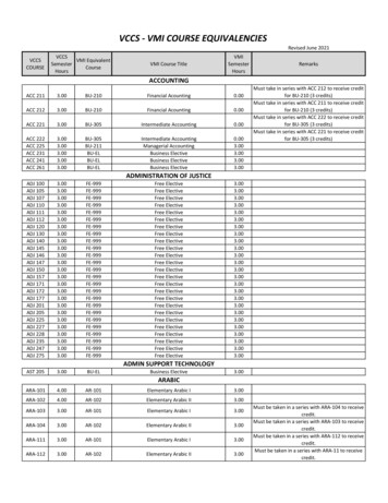
Heo et al. BMC Cancer(2020) 20:1193Page 5 of 15Fig. 1 Combination of radotinib and Ara-C enhances acute myeloid leukemia (AML) cell death. Cells were treated with 5 μM radotinib and/or 50nM Ara-C for 48 h. The cytotoxicity was then evaluated by an MTS assay. a HL60 cells. b HEL92.1.7 cells. c THP-1 cells. d Bone marrow cells (BMCs)from AML patients (n 5). e H209 cells. Representative data are shown from at least three independent experiments. These data represent themeans SEM. Significantly different from control (*) or combination of radotinib and Ara-C (#); ***, ###: P 0.001. R, radotinib; A, Ara-C; R A,combination of radotinib and Ara-Cand HEL92.1.7 cells (Fig. 4a and b). At the same time,we observed that cleaved caspase-3, 7, and 9 increasedwith radotinib/Ara-C administration in both cell types(Fig. 4c-e). To inhibit caspase activation, pan caspase inhibitor, Z-VAD-FMK (10 μM) was added to HL60 cellsfor 30 min prior to combined treatment of radotinib andAra-C. After 48 h, the cells were analyzed by capase-3activity using the CaspGLOW Fluorescein ActiveCaspase-3 Staining Kit. The Z-VAD-FMK efficientlyblocked the radotinib/Ara-C-induced capase-3 activity inHL60 cells (relative intensity: with 5 μM radotinib and50 nM Ara-C, 100%; with 10 μM Z-VAD-FMK preinculation, 4%), as shown in Fig. 4f. The cells were also collected and treated under the same conditions described
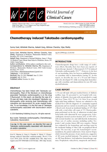
Heo et al. BMC Cancer(2020) 20:1193Page 6 of 15Fig. 2 Combination of radotinib and Ara-C increases Annexin V-positive HL60 and HEL92.1.7 cells. Cells were treated with 5 μM radotinib and/or 50nM Ara-C for 48 h. Cells were stained with annexin V-FITC followed by flow cytometric analysis. a Annexin V staining of HL60 cells. b Data show thepercentage of Annexin V-positive cells (apoptotic cells) in (a). c Data show the percentage of Annexin V-positive HEL92.1.7 cells. Representative dataare shown for at least three independent experiments. These data represent the means SEM. Significantly different from control (*) or combinationof radotinib and Ara-C (#); *: P 0.05; **: P 0.01; ***, ###: P 0.001. R, radotinib; A, Ara-C; R A, combination of radotinib and Ara-Cin Fig. 4f. The cells were analyzed by immunoblottingusing an anti-cleaved PARP-1 (cPARP-1) mAb and ananti-cleaved caspase-3 mAb. The Z-VAD-FMK efficiently reversed the radotinib/Ara-C-induced cPARP-1and cleaved caspase-3 expression in HL60 cells (relativeband density of cPARP-1: with 5 μM radotinib and 50nM Ara-C, 100%; with 10 μM Z-VAD-FMK preinculation, 6%, and relative band density of cleaved caspase-3:with 5 μM radotinib and 50 nM Ara-C, 100%; with10 μM Z-VAD-FMK preinculation, 2.7%), as shown in asshown in Fig. 4g. Therefore, these data indicate thatradotinib/Ara C induces cell death via a caspasedependent pathway. In addition, the bongkrekic acidpowerfully blocked the radotinib/Ara-C-induced cPARP1 and cleaved caspase-3 expression in HL60 cells (relative band density of cPARP-1: with 5 μM radotinib and50 nM Ara-C, 100%; with 50 μM BA preinculation, 3.7%,and relative band density of cleaved caspase-3: with5 μM radotinib and 50 nM Ara-C, 100%; with 50 μM BApreinculation, 3.2%), as shown in as shown in Fig. 4h.Therefore, these data indicate that radotinib/Ara C induces cell death via a mitochondrial-dependent pathway.Generally, it is well-known that the caspase-dependentapoptosis pathway is downstream of the mitochondrialpathway [20]. Therefore, combined treatment with radotinib and Ara-C has a synergistic effect on AML celldeath via the activation of mitochondrial-dependentapoptosis.Combined treatment with radotinib and Ara-C has asynergistic effect on G0/G1 phase arrest in HL60 andHEL92.1.7 cells via the induction of p21 and p27We also monitored the cell cycle distribution after radotinib and Ara-C treatment in HL60, HEL92.1.7 andTHP-1 cells. First, combination treatment with radotiniband Ara-C caused G0/G1 phase cell cycle arrest in AMLcells such as HL60 and HEL92.1.7 cells (Fig. 5a). Moreover, we examined the effects of combined radotinib andAra-C on cell cycle regulatory proteins including notonly CDK2 and cyclin E but also p21 and p27. As a result, the expression of CDK2 and cyclin E was remarkably decreased in the radotinib and Ara-C combinationgroup (Fig. 5b). Moreover, as shown in Fig. 5c, the expression levels of p21 and p27 in cells co-treated with
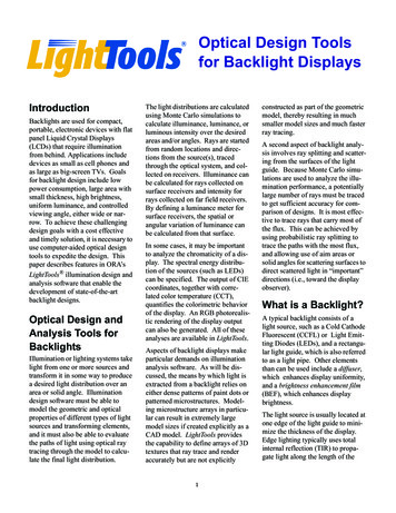
Heo et al. BMC Cancer(2020) 20:1193Fig. 3 (See legend on next page.)Page 7 of 15
Heo et al. BMC Cancer(2020) 20:1193Page 8 of 15(See figure on previous page.)Fig. 3 Radotinib/Ara-C-induced apoptosis involves the mitochondrial pathway. A combination of radotinib and Ara-C inhibited the mitochondrialmembrane potential of HL60 (a) and HEL92.1.7 cells (b). The cells were also collected and treated under the same conditions described in Fig. 2.The mitochondrial membrane potential was measured by flow cytometry using DiOC6(3) dye. c The effects of radotinib/Ara-C on cytosoliccytochrome C. HEL92.1.7 cells were incubated with 5 μM radotinib and/or 50 nM Ara-C for 48 h. The cytosolic fractions were then separated, afterwhich each sample was analyzed for the expression of cytochrome C. The membrane was stripped and re-probed with an anti-β-actin mAb toconfirm equal loading. d The effects of radotinib/Ara-C on Bax and Bcl-xl expression. Diverse AML cells including HL60, HEL92.1.7, and THP-1 cellswere treated with 5 μM radotinib and/or 50 nM Ara-C for 48 h at 37 C. Then, cells were washed twice PBS buffer and stained with an anti-Baxand anti-Bcl-xl mAb. e HL60 cells were incubated with 5 μM radotinib and 50 nM Ara-C in the presence or absence of bongkrekic acid (BA,50 μM) for 48 h and then harvested. Cells were analyzed by immunoblotting using an anti-Bax mAb. The membrane was stripped and re-probedwith an anti-β-actin mAb to confirm equal loading. These data represent the means SEM. Significantly different from control (*) or combinationof radotinib and Ara-C (#); ***, ###: P 0.001. R, radotinib; A, Ara-C; R A, combination of radotinib and Ara-C; BA, bongkrekic acidradotinib and Ara-C were higher than those in singletreatment group, as expected. These results suggest thatcombined treatment with radotinib and Ara-C increasesexpression of the inhibitory proteins p21 and p27 anddecreases the expression of CDK2 and cyclin E in bothcell types, maintaining those cells in the G0/G1 phase.Therefore, the combination of radotinib and Ara-C induces G0/G1 arrest in HL60 and HEL92.1.7 AML cellsvia regulation of the CDKI–CDK–cyclin cascade (Fig. 5).Especially, the induction of CDK inhibitors, namely p21and p27, contributed to the G0/G1 arrest of HL60 andHEL92.1.7 cells. Consequently, these results indicate thatthe combination of radotinib and Ara-C has a synergisticeffect on G0/G1-phase arrest in HL60 and HEL92.1.7cells via the induction of CDK inhibitors including p21and p27.Furthermore, we examined cell cycle markers including p21 and p27 using pan caspase inhibitor, ZVAD-FMK and MMP disruption inhibitor, andbongkrekic acid to clarify the relationship betweencell cycle and cell death. In briefly, the cells were alsocollected and treated under the same conditions described in Fig. 4g and h. The cells were analyzed byimmunoblotting using an anti-p21 mAb and an antip27 mAb. The Z-VAD-FMK significantly blocked theradotinib/Ara-C-induced p21 and p27 expression inHL60 cells (relative band density of p21: with 5 μMradotinib and 50 nM Ara-C, 100%; with 10 μM ZVAD-FMK preinculation, 2.6%, and relative banddensity of p27: with 5 μM radotinib and 50 nM AraC, 100%; with 10 μM Z-VAD-FMK preinculation,4.7%), as shown in as shown in Fig. 5d. In addition,the bongkrekic acid strongly blocked the radotinib/Ara-C-induced (relative band density of p21: with 5 μMradotinib and 50 nM Ara-C, 100%; with 50 μM BA preinculation, 11.4%, and relative band density of p27: with5 μM radotinib and 50 nM Ara-C, 100%; with 50 μM BApreinculation, 1.2%), as shown in as shown in Fig. 5e.Thus, these data showed that radotinib/Ara C-inducedcell cycle markers, p21 and p27, were closely related toAML cell death. It also indicates that mitochondrialdependent apoptosis and G0/G1 phase cell cycle arrestcontribute to radotinib/Ara-C mediated anti-AMLactivity.Combined treatment with radotinib and Ara-C inhibitsAML cell growth in vivoTo examine the effect of combined treatment withradotinib and Ara-C on AML cell death in vivo, weimplanted HEL92.1.7 cells into nude mice. Throughprevious preliminary studies, radotinib and Ara C wereadministered by concentration, and then the tumor volume was used in combination with each concentrationthat decreased by 5–8% compared to the control group(data not shown). As shown in Fig. 6a-b, combinedtreatment with radotinib and Ara-C inhibited AML cellgrowth, including tumor volume and weight in vivo. Inaddition, the body weights of the tumor-bearing micedid not change significantly during the duration of study(Fig. 6c). The expression of Bcl-xl, Cyclin E, and PCNAwith combined radotinib and Ara-C treatment wassignificantly decreased in tumor tissues isolated from themice, as shown in Fig. 6d-e. Further, the expression ofBax and p21, as a well-known CDK inhibitor, wassignificantly increased in tumor tissues with combinedtreatment comprising radotinib and Ara-C. In addition,the expression of TUNEL-positive cells was significantlyamplified, while the expression of PCNA-positive cellswas dramatically reduced in the tumor tissue with combined treatment of radotinib and Ara-C (Fig. 6f-g). Inparticular, the combination of the two drugs was shownto lead to the profound inhibition of acute myeloidleukemia tumor growth (Fig. 6).Radotinib and Ara-C sensitize AML cells tochemotherapeutic agents including daunorubicin oridarubicinFinally, we performed cell viability tests to determine theefficacy of a combination of radotinib and various anticancer drugs commonly used for AML therapy.
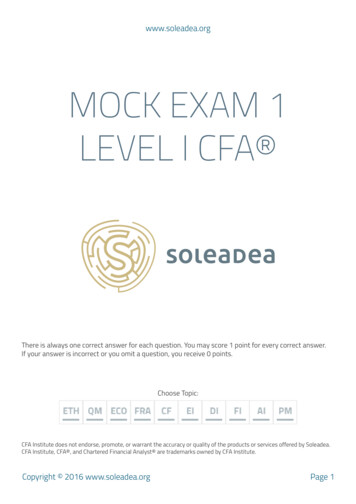
Heo et al. BMC Cancer(2020) 20:1193Fig. 4 (See legend on next page.)Page 9 of 15
Heo et al. BMC Cancer(2020) 20:1193Page 10 of 15(See figure on previous page.)Fig. 4 Radotinib/Ara-C induces caspase-dependent apoptosis and activates caspase-3 activity in HL60 and HEL92.1.7 cells. The cells were alsocollected and treated under the same conditions described in Fig. 2. a Caspase-3 a
were then analyzed using a FACSCalibur flow cytometer and CellQuest Pro software (BD Biosciences). Analysis of mitochondrial membrane potential HL60 and HEL92.1.7 cells were incubated with 5μM radotinib and/or 50nM Ara-C for 48h at 37 C, har-vested, and washed twice with PBS buffer. Mitochondrial membrane potential (MMP, ΔΨm) was evaluated by