Transcription
Improved elongation factor-1 alpha-based vectorsfor stable high-level expression of heterologousproteins in Chinese hamster ovary cellsOrlova et al.Orlova et al. BMC Biotechnology 2014, 14:56http://www.biomedcentral.com/1472-6750/14/56
Orlova et al. BMC Biotechnology 2014, ETHODOLOGY ARTICLEOpen AccessImproved elongation factor-1 alpha-based vectorsfor stable high-level expression of heterologousproteins in Chinese hamster ovary cellsNadezhda A Orlova1,2, Sergey V Kovnir1,2, Julia A Hodak1,2, Ivan I Vorobiev1,2*, Alexandre G Gabibov2,3and Konstantin G Skryabin1AbstractBackground: Establishing highly productive clonal cell lines with constant productivity over 2–3 months of continuousculture remains a tedious task requiring the screening of tens of thousands of clonal colonies. In addition, long-termcultivation of many candidate lines derived in the absence of drug selection pressure is necessary. Expression vectorsbased on the elongation factor-1 alpha (EEF1A) gene and the dihydrofolate reductase (DHFR) selection marker (withseparate promoters) can be used to obtain highly productive populations of stably transfected cells in the selectionmedium, but they have not been tested for their ability to support target gene amplification under gradually increasingmethotrexate pressure.Results: We have modified EEF1A-based vectors by linking the DHFR selection marker to the target gene in thebicistronic RNA, shortening the overall plasmid size, and adding an Epstein-Barr virus terminal repeat fragment(EBVTR) element. Presence of the EBVTR element increased the rate of stable transfection by the plasmid by 24times that of the EBVTR-minus control and improved the rate of methotrexate-driven gene amplification. The meanexpression level of the enhanced green fluorescent protein (eGFP) used herein as a model protein, increased up toeight-fold using a single round of amplification in the case of adherent colonies formation and up to 4.5-fold inthe case of suspension polyclonal cultures. Several eGFP-expressing cell populations produced using vectors withantibiotic resistance markers instead of the DHFR marker were compared with each other. Stable transfection ofChinese hamster ovary (CHO) DG44 cells by the p1.2-Hygro-eGFP plasmid (containing a hygromycin resistance marker)generated highest eGFP expression levels of up to 8.9% of the total cytoplasmic protein, with less than 5% of the cellpopulation being eGFP-negative.Conclusions: The p1.1 vector was very effective for stable transfection of CHO cells and capable of rapid MTX-driventarget gene amplification, while p1.2-Hygro achieved similar eGFP expression levels as p1.1. The set of vectors we havedeveloped should speed-up the process of generating highly productive clonal cell lines while substantially decreasingthe associated experimental effort.Keywords: CHO cells, High level expression, Stable cell line generation, Molecular cloning* Correspondence: ptichman@gmail.com1Laboratory of Mammalian Cell Bioengineering, Centre “Bioengineering”,Russian Academy of Sciences, 60-letija Oktyabrya 7, Moscow 117312, Russia2Laboratory of Biocatalysis, Institute of Bioorganic Chemistry, RussianAcademy of Sciences, Miklukho-Maklaya 16/10, Moscow 119971, RussiaFull list of author information is available at the end of the article 2014 Orlova et al.; licensee BioMed Central Ltd. This is an Open Access article distributed under the terms of the CreativeCommons Attribution License (http://creativecommons.org/licenses/by/2.0), which permits unrestricted use, distribution, andreproduction in any medium, provided the original work is properly credited. The Creative Commons Public DomainDedication waiver ) applies to the data made available in this article,unless otherwise stated.
Orlova et al. BMC Biotechnology 2014, ackgroundMost of the proteins currently employed for therapeuticuse are produced by stably transfected mammalian cells,of which the most popular is the Chinese hamster ovary(CHO) cell line. Establishing highly productive clonalcell lines that exhibit constant productivity over a 2–3month period of continuous culture remains a tedioustask, requiring tens of thousands of clonal colonies to bescreened, followed by the long-term cultivation of candidate lines in the absence of an appropriate selectionpressure. Generally, the expression levels of a target genemay be increased by its amplification in the genome [1],which is usually achieved by linking the target gene tothe murine dihydrofolate reductase (DHFR) gene withstepwise increases in the concentration of the DHFR inhibitor, methotrexate (MTX), in the selection medium.Target gene amplification is a time-consuming process,resulting in cell populations that often contain unstableclones, and in the absence of an appropriate selectionpressure, reduced production levels. The probability ofobtaining a highly productive clonal cell line can be increased significantly by utilizing plasmids based on noncoding parts of the elongation factor-1 alpha gene (EEF1A)from Chinese hamster, as described by Running Deer andAllison [2]. Expression vector pDEF38, introduced by theseauthors, differs significantly from the widely employed vectors with the core promoter of the human ortholog elongation factor 1 alpha gene (EF1a). EEF1A-based expressionvector includes 4.1 kb upstream and 4.2 kb downstreamflanking areas of the EEF1A gene, so the ORF of the of thetarget gene replaces the coding exons of the elongation factor 1 alpha protein in the natural EEF1A gene, mimickingwith all possible accuracy the structure of the natural genein the resulting expression plasmid. It was shown thatpresence of both flanking areas in the EEF1A-based vectorsresults in the 6- to 35- fold increase of the average expression level comparing to commercial vectors with CMV orEF1alpha promoters. Removal of the downstream flankingarea from the expression vector resulted in the 4-fold dropin the expression level.Original expression vector pDEF38 contained the DHFRselection marker with a separate SV40 promoter andwas not tested for its ability to support target geneamplification under gradually increasing MTX pressure. DHFR-compatible vectors, bearing the neomycinresistance gene instead of the DHFR gene, were alsodescribed in the same work. Existing EEF1A-basedvectors, despite their high promoter strength and theirlong-term production level stability, do not accommodate very large plasmid sizes. Consequently, this can result in low-level genome integration and inability tomaintain the target gene amplification step, possibly as aresult of vector fragmentation and autonomous amplification of the DHFR-coding area.Page 2 of 11Since EEF1A-based vectors are much longer than CMVbased vectors, they are expected to have lower transfection efficiency and, subsequently, lower numbers of stably transfected cells. It was shown, that the insertionthe concatemer fragment of the Epstein-Barr virus terminal repeats (EBVTR) [3,4] in the expression vectorsincrease the rate of stably transfected colonies formation by five to ten fold [5]. The molecular mechanismof this effect is poorly understood. It is known thatG-rich repeats in the EBVTR bind to the cellular proteinterminal repeat binding protein (TRBP) [3] and at leasttwo binding sites of TRBP were identified in the repetitive cellular DNA [6]. EBVTR areas are involved in theintegration of the Epstein-Barr virus into the chromosomal DNA [7]. EBV-infected cells may harbour the virusin the chromosome-integrated form, as the independently replicating episome or the mixture of both forms[8]. Region of the EBV, called oriP, maintains the episomal replication of the EBV genome, interacts with theEBV-encoded nuclear antigen-1 (EBNA-1) and allowsEBV plasmids to separate in mitosis through binding tochromosomes [9]. EBVTR concatemer used for enhancement of expression plasmids, however, containsno sequences from the oriP region and no DNA fragments with significant homology toward oriP region,so the EBNA-1 – mediated persistence of the EBVTRcontaining plasmid as the episome in the transfectedcells is highly unlikely.We hypothesized that significant improvements toEEF1A-based vectors might be achieved by: 1) insertingthe EBVTR element outside of the EEF1A flankingDNA; 2) linking the DHFR open reading frame to thetarget gene by the internal ribosome entry site (IRES)thereby preventing the possibility of separate amplification of the selection marker; 3) reducing of thelength of the backbone DNA, which is necessary formaintaining the plasmid in the bacterial host. Similarimprovements might be applied to DHFR-compatibleEEF1A-based vectors used for monocistronic expression of a target gene; in this case by placing the antibiotic resistance genes outside the context of thenon-coding parts of the elongation factor 1 alpha gene,which might decrease genetic linkage between the selection marker and the target gene.Here, we report on the functional properties of EEF1Abased plasmid vectors for bicistronic and monocistronicexpression. We also describe the corresponding methodsfor obtaining highly productive and stable cell lines thatmaintain constant productivity levels after genome amplification of the integrated plasmid, utilizing the DHFRnegative cell line CHO DG44 [10,11]. In addition, weused the enhanced green fluorescent protein (eGFP) asa model target protein, and show eGFP accumulationinside CHO cells.
Orlova et al. BMC Biotechnology 2014, ethodsMolecular cloningThe sequences of the primers used for cloning expression plasmids are shown in Additional file 1: Table S1.The backbone vector, pBL-2, was obtained by two stagesof inverted PCR using long adapter primers and thepUC18 plasmid as a template. Non-functional parts ofthe plasmid including the pLac promoter and the LacZgene were removed. Inverted PCR was performed as described previously [12]. Oligonucleotides and PCR reagents were from Evrogen, JSC (Moscow, Russia). PCRproducts were purified from 1% agarose gels by theWizard SV Gel and PCR Clean-Up System (Promega,Madison, WI). T4 polynucleotide kinase and MalI restriction endonuclease (Sibenzyme, Novosibirsk, Russia)were used. T4 DNA ligase (Fermentas, Vilnius, Lithuania)was used for inverted PCR product circularization. TheEscherichia coli TOP10 strain (Invitrogen, Carlsbad, CA)was used for cloning. Plasmids were isolated with aGeneJet Plasmid Purification Kit (Fermentas, Vilnius,Lithuania).The pAL-EBV plasmid, containing a fragment of aconcatemer of EBV terminal repeats, as described previously [5] and nearly undistinguishable from the humanherpes virus 4 strain K4123-MiEBV sequence [GenBank:KC440852.1] was made from synthetic oligonucleotides cloned into a pAL-TA (Evrogen) vector. The ORFencoding mouse DHFR was obtained by PCR usingpOptivec-TOPO linearized vector (Invitrogen) as a template. The fragment encoding the attenuated encephalomyocarditis virus (EMCV) IRES was obtained from thepOptivec-circ plasmid (self-ligated pOptivec-TOPO) byrestriction. These fragments were cloned into the pBL-2plasmid via assembly of two different intermediate constructs, PBL-2-ID and PBL-2-ID-EBV. DNA modificationenzymes for routine molecular cloning were obtainedfrom Fermentas or Sibenzyme.Construction of p1.1 vectorsFragments corresponding to the upstream and downstream flanking regions (8532–12603 and 14545–18794sequences of [GenBank:AY188393]) of the CHO elongation factor 1 gene were obtained by PCR using CHODG44 cell (Invitrogen) genomic DNA as a template. Themodular assembly cloning technique used herein is described in detail elsewhere [13].Assembled CHO genomic regions were cloned into theintermediate plasmids, PBL-2-ID and PBL-2-ID-EBV,resulting in p1.1(EBVTR-) and p1.1 expression vectors,respectively (Figure 1).Construction of p1.2 vectorsp1.2-Mono, the intermediate backbone plasmid for expression vectors bearing antibiotic resistance genes wasPage 3 of 11obtained by removal of the region containing the EMCVIRES and the DHFR ORF from the p1.1 expression vector. Plasmid pAL-3CH123, containing first three modules of the downstream flanking region of the EEF1Awas used as the source of the donor DNA insert fragment, replacing the deleted IRES and DHFR area, soboth flanking regions of the EEF1A remained unaltered(Figure 2). Antibiotic resistance genes and the SV40 promoter and terminator regions were obtained by amplification with adaptor primers, using pcDNA3.1/Hygro,pcDNA3.1( ), and self-ligated pcDNA4/HisMax-TOPO(Invitrogen) as PCR templates. Antibiotic resistancecassettes were sub-cloned into T-vectors and then transferred into the p1.2-Mono backbone by restrictionligation resulting in p1.2-Hygro, p1.2-Neo and p1.2-Zeo.A DNA fragment encoding eGFP and a consensusKozak sequence (GCCGCCATGG) [14] was obtained byPCR with adaptor primers and the pEGFP-C2 plasmid(Clontech, Mountain View, CA) as a template and thencloned into the polylinker area of p1.1 and p1.2 vectors,thereby resulting in p1.1(EBVTR-)eGFP, p1.1eGFP, p1.2HygroeGFP, p1.2-NeoeGFP and p1.2-ZeoeGFP expression plasmids.Purified plasmids for transfection and the controlplasmid pEGFP-N2 (Clontech) were prepared using anEndoFree Plasmid Maxi Kit (Qiagen, Valencia, CA). Forstable cell line generation all plasmids except p1.2-HygroeGFP were linearized by restriction with PvuI by cutting inside the ampicillin resistance gene bla sequence.The plasmid p1.2-HygroeGFP was restricted with BspHI,which introduced two breaks near the bla gene.Cell cultureA DHFR-negative CHO DG44 cell line (Invitrogen)was cultured in shaking flasks in the chemically defined medium, CD DG44 (Invitrogen), supplemented with0.18% Pluronic F-68 (Invitrogen) and 4 mM L-glutamine(Invitrogen). The cells were passaged 24 h before transfection. For direct colony generation in 96-well culture plates,transfection was performed using Fugene HD reagent(Promega), containing 60 μg of DNA and 180 μl of thereagent per 15 millions of cells in 30 ml of the abovemedium. Plasmids p1.2 were transfected by electroporation in Gene Pulser Electroporation Buffer (Bio-Rad,Hercules, CA) using a cuvette with a 4 mm gap with 7.5million cells and 15 μg of linearized DNA for each transfection. Cells were counted by trypan blue exclusion and fluorescence microscopy at 48 h post-transfection. For directgeneration of colonies, transiently transfected cell cultureswere transferred into CHO-A culture medium (Invitrogen)lacking hypoxanthine and thymidine (HT), and seeded at5000 cells/well in the culture plates. Cells were grown undisturbed for 14 days and inspected by fluorescence microscopy. The medium was changed and the plates were
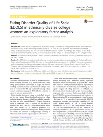
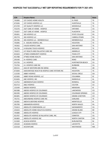
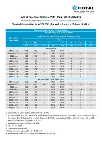
Orlova et al. BMC Biotechnology 2014, age 4 of 11Figure 1 Map of the p1.1 plasmid vector and the cloning scheme for p1.1-based plasmids. A. Plasmid map. UFR: upstream flanking regionof the EEF1A gene; DFR: downstream flanking region; PL: polylinker region; pA: polyadenylation signal; bla – ampicillin resistance gene; blaprom – promoter of the ampicillin resistance gene. B. Cloning scheme for p1.1-based plasmids. Generation of cloning inserts by PCR withadaptor primers is depicted by dashed lines; generation of cloning inserts by restriction is depicted by solid lines. EBV F1-6: correspondingsynthetic fragments of the EBVTR element. 5CH F1-6: corresponding fragments of the upstream flanking region of the EEF1A gene; 3CH F1-6:corresponding fragments of the downstream flanking region of the EEF1A gene.cultivated for 5–10 additional days until the first 10% ofthe wells containing colonies became confluent.To generate stably transfected cell populations usingp1.1eGFP and p1.1(EBVTR-)eGFP plasmids, transientlytransfected cultures were transferred to OptiCHO medium(Invitrogen) lacking HT, and thereafter cultivated in shaking flasks with medium exchange every 3 days until the cellviability increased to 85% (approximately 22–27 days ofcultivation).MTX-driven target gene amplification in culture plateswas performed by seeding the cells from stably transfectedcell populations into 96-well culture plates at a density of5000 cells/well in the CHO-A culture medium, supplemented with 0, 50, 100, 200, 400 or 800 nM MTX. Threeplates were used for each concentration of MTX. The cellswere grown undisturbed for 14 days, after which the plateswere inspected by microscopy and the culture mediumwas changed every 4 days until the first 10% of wells ineach plate became confluent. Plates were screened again byfluorescence microscopy, and cells from the 16 brightestwells from each plate were transferred into a 48-well plate,grown to confluence, and then transferred into 24-wellFigure 2 Map of the p1.2-Hygro plasmid vector and the cloning scheme for p1.2-based plasmids. A. Plasmid map. UFR: upstream flankingregion of the EEF1A gene; DFR: downstream flanking region; Pl: polylinker region; SV40 prom and SV40 PA: promoter and polyadenylation signalfrom the SV40 virus. B. Cloning scheme for p1.2-based plasmids.
Orlova et al. BMC Biotechnology 2014, lates. Colonies lacking normal proliferation speeds or attached to the surface of the plates too tightly for dislodgingby pipetting were discarded. Cells from the eight brightestwells for each MTX concentration were dislodged fromtheir plates, lysed as described below, and then used to determine eGFP levels. Six randomly picked colonies, obtained in the presence of 400 and 800 nM MTX, weretransferred into a 6-well plate and grown with shaking inOptiCHO medium with passages made every three daysfor 60 days. Samples for eGFP level determination werecollected every second passage.Target gene amplification for polyclonal cell populations was performed for the suspension culture of CHODG44 cells, stably transfected by the p1.1eGFP plasmidin presence of 50 nM MTX, as described above. Concentration of the MTX in the culture medium was increasedby two-fold steps, each after two consecutive passages,until the cell viability decreased below 85%. Resultingculture, obtained in presence of 0.8 μM MTX, was splitinto four flasks, supplemented by 0.8; 1.6; 3.2; 6.4 μMMTX and cultured until the cell viability returned to atleast 85% (7–12 days).Generation of polyclonal cell populations involvingtransfected p1.2 plasmids were performed by seedingtransiently transfected cells in 6-well culture plates,using 1 million of viable cells per well in 5 ml of DG44medium, supplemented with the corresponding antibiotic, or 5 ml of OptiCHO medium with 200 nM MTXfor control transfections using p1.1 plasmids. The concentrations of the antibiotics used are shown in Figure 3.Plates were cultivated with shaking until the cell viabilityreturned to at least 85% (20 days), after which themedium was changed every 4 days.Determination of eGFP concentrations in cell lysatesCell culture samples containing approximately one millionof cells were centrifuged and the cell pellets were resuspended in phosphate buffered saline and recentrifuged.The washed cell pellets were resuspended in 0.1 ml of lysissolution containing 150 mM NaCl, 50 mM Tris–HCl atpH 7.5, 1% Triton X-100, a protease inhibitor cocktail(Sigma, St. Louis, MO), and then incubated for 30 min onice with stirring. Cell debris was removed by centrifugation. The concentration of eGFP in the lysate fromthe H-4 cell population (Figure 3) was measured by spectrophotometry at a wavelength of 488 nm using a molarextinction coefficient of 55,000 M 1 · cm 1 and an eGFPmolecular weight of 32.7 kDa [15]. The fluorescence intensity of eGFP in all of the lysates was measured alongwith the serially diluted calibration samples, which wereprepared from the H-4 lysate containing a known concentration of eGFP. Total protein concentration in the lysates was measured by the Bradford method with bovineserum albumin as a standard.Page 5 of 11FACS analysis and quantitative PCRUndiluted cell culture samples were subject to FACS FC500 (Beckman Coulter, Krefeld, Germany) analysis at anemission at 488 nm and detection through a 530/40-nmbandpass filter. At least 10,000 individual cells werecounted for each sample analysed. Quantitative PCRanalysis of the expression plasmid copy numbers in thegenomes of stably transfected cells was performed usingan iCycler iQ thermocycler (Bio-Rad) and qPCRmix-HSSYBR (Evrogen) reaction mixture with the primersshown in Additional file 1: Table S2. The highly purifiedp1.1eGFP plasmid was used as a quantity calibratorusing five different concentrations for each determination performed in triplicate. PCR was performed threetimes with three to four replicates for each sample. Genomic DNA was extracted from cells with a GenomicDNA Purification Kit (Fermentas) and quantified using aQubit fluorometer (Invitrogen) and the dsDNA HS kit(Invitrogen). Quantity calibrator plasmid was used as theexternal standard for the quantification of genomic DNAsamples by fluorometry.Results and discussionConstruction of expression plasmidsSince the transfection efficiency and, probably, the genome integration rate of an expression plasmid is inversely proportional to its size [16], we made a minimalbackbone plasmid by eliminating most of the unnecessary elements from the pUC18 plasmid. The resultingplasmid, pBL-2, lacks the f1 origin of replication, andthe bacterial promoter of the LacZ gene along with theLacZ ORF itself and some flanking DNA regions. Overall, the resulting plasmid length decreased some 600 bpfrom 2686 to 2032 bp.The upstream and downstream regions of the EEF1Agene were obtained from CHO DG44 cell genomic DNAusing the modular assembly cloning technique describedpreviously [13]. A concatemer of terminal repeats fromthe Epstein-Barr virus (EBVTR) [3,4] was assembledfrom synthetic oligonucleotides using the same technique and was inserted along with the IRES from the encephalomyocarditis virus and the murine DHFR openreading frame into the pBL-2 vector. Cloning the upstream and downstream flanking areas of the EEF1Agene into the pBL-2-ID-EBV plasmid resulted in the expression vector p1.1 (Figure 1). A control vector, lackingthe EBVTR fragment, was assembled similarly and isdenoted here as p1.1(EBVTR-). The p1.1 plasmid wasapproximately 1.5 kbp shorter than the original EEF1Abased plasmid, pDEF38, despite addition of the EBVTRfragment. The eGFP ORF with the synthetic consensusKozak sequence [14] was cloned into both vectors andthe resulting plasmids p1.1eGFP and p1.1(EBVTR-)eGFPwere used for CHO cell transfections.
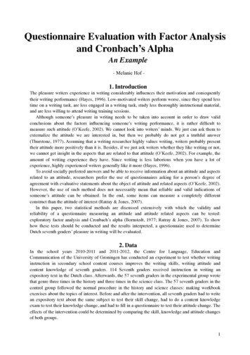
Orlova et al. BMC Biotechnology 2014, age 6 of 11Figure 3 Properties of the cell populations stably transfected by p1.2-based plasmids under various drug selection stringencies. DG44:untransfected CHO DG44 cells; p1.1: cells stably transfected by the p1.1eGFP plasmid and selected in the presence of 200 nM MTX; p1.1(EBVTR-):transfection by the p1.1(EBVTR-)eGFP plasmid using the same conditions. A. Level of intracellular eGFP in cell populations. Error bars indicatethe standard deviation, n 2. B. Proportion of eGFP-negative cell populations measured by FACS. C. Number of copies of genome-integratedplasmids measured by Q-PCR. Amplicons are located inside the eGFP ORF and one representative value experiment from three independentmeasurements is shown. Error bars represents standard deviations, n 3-4. The apparent level of the eGFP ORF DNA for the untransfectedCHO DG44 cells is below 0.1 copies per one haploid genome. D. Codes for the different cell populations and the concentrations ofantibiotics employed.Generation of stably transfected colonies usingp1.1-based plasmidsTransient transfection of the DHFR-deficient CHO DG44cells resulted in significantly decreased transfection efficiencies for both of the EEF1A-based plasmids relativeto the cytomegalovirus (CMV)- promoter-based 4700 bppEGFP-N2 plasmid, and approximately the same transfection efficiencies and eGFP expression levels for plasmidswith or without the EBVTR element (Table 1).At the same time, stable integration rate (or rate ofestablishment of stable episomal maintenance) of thep1.1eGFP plasmid was 24 times higher than that ofthe p1.1(EBVTR-)eGFP control plasmid in the selection medium lacking both HT and MTX (Table 2),clearly indicating that the EBVTR element was activein the very large expression plasmid. Transfection andselection of stably transformed CHO DG44 cells bythe p1.1eGFP plasmid was repeated with the selection medium supplemented with 50 nM MTX. In thiscase, the eGFP expression level increased twice forthe ten most productive wells (Figure 4A). Thus, thep1.1 plasmid is suitable for creation of stably transfected cell clones or populations under variable selectionstringencies.
Orlova et al. BMC Biotechnology 2014, age 7 of 11Table 1 Properties of the transiently transfected cells usedin this studyPlasmid g cells22.8%5.8%6.0%Fluorescence intensity,RFU/50 cells11435.332.0Viable cells86%83%84%MTX-driven target gene amplificationSince the EBVTR element was effective at increasingthe incidence of stable transfection, we tested its abilityto speed up the transgene amplification process. Polyclonal populations of CHO DG44 cells, transfected byp1.1eGFP and p1.1(EBVTR-)eGFP plasmids and selectedfor stable integration by suspension cultivation in theabsence of MTX and HT, were seeded in the 96-wellculture plates in the presence of 0–800 nM MTX andgrown undisturbed until visible colonies developed. Forthe p1.1(EBVTR-)eGFP plasmid lacking the EBVTRelement, no viable cell colonies were obtained in thepresence of 200, 400 and 800 nM MTX. The eGFP expression levels of the highest expressing colonies, obtained in the presence of 0 and 100 nM MTX, was inthe same range as the colonies obtained by direct platingof transiently transfected cells in the absence of MTX(data not shown). In the case of the p1.1-eGFP plasmid,multiple colonies were obtained for all of the concentrations of MTX tested. After visual screening by fluorescence microscopy and expansion, the eight brightestcolonies for each concentration of MTX employed weregrown to confluency in 24-well culture plates. The relative eGFP expression levels for these colonies (Figure 4B)was approximately 8 times higher when cultivated with800 nM MTX, and approximately 2 times higher whencultivated with 400 nM MTX, compared with cultivationwithout MTX. Six randomly chosen colonies, obtainedin the presence of 400 and 800 nM MTX, were scaledup, re-adapted to suspension culture and cultivated for60 days. No substantial decay in the eGFP expressionlevel was detected for each colony (data not shown).Thus, the p1.1 vector is suitable for target gene amplification in the presence of MTX. The resulting cell clonesTable 2 Colony formation efficiency for p1.1eGFP andp1.1(EBVTR-)eGFP plasmidsPlasmid namep1.1eGFPp1.1(EBVTR-)eGFPTotal number of colonies in 10culture plates234295eGFP-expressing colonies in 10culture plates and their proportions2093 (89.4%)52 (54.7%)Fluorescence intensity of thebrightest well, RFU/50 cells21045.1are sufficiently stable for cell bank generation and subsequent large-scale cultivation.Target gene amplification process was also tested forpolyclonal cell population, obtained by the primary selection in the presence of 50 nM MTX. Sequentialaddition of MTX from 100 nM to 400 nM gave no decrease in cell viability, eGFP level was also constant (datanot shown). Further addition of 0.8 – 6.4 μM of MTX,performed in one step for multiple culture flasks, resulted in the concentration-dependent increase of eGFPcontent (Figure 4C), peaking at 9% of the total proteinin the case of 6.4 μM of MTX. Analysis of the copynumbers of the integrated plasmids using quantitativePCR (Figure 4D) showed that higher MTX level andhigher eGFP content correspond to higher copy numberof the integrated plasmid. Hereby, the vector p1.1 is suitable for obtaining highly productive cell populations orclones by direct clone selection in culture plates in thepresence of MTX or by the multi-step target gene amplification in the suspension culture.Polyclonal cell populations stably transfected by p1.2plasmidsHeterologous expression of a functionally active targetprotein often requires co-expression of a small numberof protein processing enzymes. For example, the bloodclotting factor IX expression systems used with CHO orBHK cells rely on co-expression of the signal proteasePACE/furin [17] and the vitamin-K recharging enzyme,VKORC1 [18]. Generally, the expression levels of such“helper” proteins should be lower than that of the targetprotein, but of comparable magnitude. If the target protein is coded by a plasmid bearing a DHFR selectionmarker, helper proteins may be coded by plasmids withthe same structure, but bearing antibiotic resistancemarkers. We have tested resistance markers for threewidely used antibiotics, G418 (a neomycin analogue),zeocin, and hygromycin, in the EEF1A-based expressionvector, which was modified by removing the IRES fragment and the DHFR open reading frame from the p1.1plasmid, and insertion of the corresponding antibioticresistance genes outside of the EEF1A gene flankingareas and controlled by a separate SV40 promoter. Theresulting vectors, denoted p1.2-Neo, p1.2-Zeo, and p1.2Hygro, were used for insertion of the eGFP protein ORF.All three resulting plasmids showed similar transienttransfection efficiencies in CHO DG44 cells (19–24% byelectroporation), and the resulting cell populations wereused to generate stably transfected cell populations inthe suspension culture under variable selection pressuresfor each antibiotic used. The control
1Laboratory of Mammalian Cell Bioengineering, Centre "Bioengineering", Russian Academy of Sciences, 60-letija Oktyabrya 7, Moscow 117312, Russia . (EBVTR) [3,4] in the expression vectors increase the rate of stably transfected colonies forma-tion by five to ten fold [5]. The molecular mechanism of this effect is poorly understood. It is .