Transcription
Wang et al. Journal of Hematology & 2019) 12:31RESEARCHOpen AccessEarly myeloid-derived suppressor cells(HLA-DR /lowCD33 CD16 ) expanded bygranulocyte colony-stimulating factorprevent acute graft-versus-host disease(GVHD) in humanized mouse and mightcontribute to lower GVHD in patientspost allo-HSCTKe Wang1,2†, Meng Lv1†, Ying-Jun Chang1, Xiang-Yu Zhao1, Xiao-Su Zhao1, Yuan-Yuan Zhang1, Yu-Qian Sun1,Zhi-Dong Wang1, Pan Suo1, Yang Zhou1, Dan Liu1, Shu-Zhen Zhai1, Yan Hong1, Yu Wang1, Xiao-Hui Zhang1,Lan-Ping Xu1, Kai-Yan Liu1 and Xiao-Jun Huang1,2*AbstractIntroduction: Myeloid-derived suppressor cells (MDSCs) are proposed to control graft-versus-host disease (GVHD)in allogeneic hematopoietic stem cell transplantation (allo-HSCT). However, the definition of human MDSCs has notyet reached consensus, and the mechanism of MDSCs to control GVHD remains unclear.Methods: Immature myeloid cells (HLA-DR /lowCD33 CD16 ) were tested before and after granulocyte colonystimulating factor (G-CSF) administration in healthy donor and isolated for suppression assays and co-culturewith T cells in vitro. Isolated cells were infused in humanized mice for a xenogeneic model of acute GVHD.One hundred allo-HSCT recipients were enrolled prospectively to assess the role of HLA-DR /lowCD33 CD16 cells ingrafts on the occurrence of acute GVHD.(Continued on next page)* Correspondence: xjhrm@medmail.com.cnPart of this study was presented orally at the 60th Annual Meeting of theAmerican Society of Hematology (ASH) on Dec 1, 2018.†Ke Wang and Meng Lv contributed equally to this work.1Peking University People’s Hospital, Peking University Institute ofHematology,Beijing Key Laboratory of Hematopoietic Stem CellTransplantation, National Clinical Research Center for Hematologic Disease,No 11 Xizhimen South Street, Beijing 100044, China2Center for Life Sciences, Academy for Advanced Interdisciplinary Studies,Peking University, No.5 Yiheyuan Road, Beijing 100871, China The Author(s). 2019 Open Access This article is distributed under the terms of the Creative Commons Attribution 4.0International License (http://creativecommons.org/licenses/by/4.0/), which permits unrestricted use, distribution, andreproduction in any medium, provided you give appropriate credit to the original author(s) and the source, provide a link tothe Creative Commons license, and indicate if changes were made. The Creative Commons Public Domain Dedication o/1.0/) applies to the data made available in this article, unless otherwise stated.
Wang et al. Journal of Hematology & Oncology(2019) 12:31Page 2 of 16(Continued from previous page)Results: In the present study, G-CSF mobilized HLA-DR /lowCD33 CD16 cells with immunosuppressive properties indonor peripheral blood. These cells contained more interleukin-10 and transforming growth factor-beta (TGF-β) cellsafter G-CSF administration and inhibited the proliferation of autologous donor T cells in a TGF-β-dependent manner.Meanwhile, these immature myeloid cells promoted regulatory T cell expansion and induced Th2 differentiation.Importantly, these cells prevented acute GVHD in a humanized mouse model. Moreover, clinical cohort results showedthat the number of HLA-DR /lowCD33 CD16 cells in the donor graft was the only independent risk factor inverselycorrelated with the incidence of grade II–IV acute GVHD in the recipients (HR 0.388, 95% CI 0.158–0.954, p 0.039).Conclusion: HLA-DR /lowCD33 CD16 cells represent functional MDSCs that may control acute GVHD in allo-HSCT.Keywords: Myeloid-derived suppressor cells, Granulocyte colony-stimulating factor, Acute graft-versus-host disease,Humanized mouse, Allogeneic hematopoietic stem cell transplantationIntroductionAllogeneic hematopoietic stem cell transplantation(allo-HSCT) is an effective treatment for patients withmost hematological diseases [1]. However, the applications of allo-HSCT are limited by lethal complications,including graft-versus-host disease (GVHD). The increased understanding of the regulatory cells and molecular pathways involved in limiting pathogenicimmune responses offers the opportunity for promotingGVHD-relapse-free survival (GRFS), aiming to replacenon-specific GVHD control with pharmacological immune suppression [2–5].Extensive research has shown that myeloid-derivedsuppressor cells (MDSCs) have emerged as major immunosuppressive cells in cancer, infection, chronic inflammation, and autoimmune disease, which suggestthat MDSCs may help to control GVHD [6, 7]. However,the phenotypic, morphological, and functional heterogeneity of MDSCs generates confusion in the investigation and analysis of their roles in immune responses asMDSCs represent a heterogeneous population of immature myeloid cells and are defined by their ability to suppress T cells in vitro [8, 9]. In humans, MDSCs mostcommonly express the common myeloid marker, CD33[10], but lack the expression of markers of mature myeloid and lymphoid cells as well as the MHC class-II molecule, HLA-DR [11, 12]. Lin (including CD3, CD19,and CD56) CD14-CD15-HLA-DR CD33 cells containmixed groups of MDSCs comprising more immatureprogenitors (also named early MDSCs, eMDSCs). Furthermore, two other subpopulations with more differentiated surface makers, namely, CD15 or CD14 , aredivided into two groups as follows: G-MDSCs(PMN-MDSCs) and M-MDSCs [8]. However, the definitions of these subsets have not yet reached consensus,and it remains unclear if MDSCs control GVHD in vivo.Though granulocyte colony-stimulating factor (G-CSF)has been routinely used to mobilize stem cells to theperipheral blood from healthy donors, it is alsorecognized as a novel mediator of T cell tolerance [13,14]. Increasing evidences suggest that G-CSF affects different immune cells [13, 15], which modulate T cell responsedirectly by inducing Th2 differentiation [16, 17] or mobilizing functional regulatory T cells [18], or indirectly throughmonocytes [19–21], DC [22], and neutrophils [23] to prevent GVHD. Previous results have suggested thatG-CSF-mobilized donor cells also contain MDSCs, whichare closely associated with GVHD in allo-HSCT [24]. Antonio et al. reported that systemic treatment with G-CSF induces expansion of myeloid cells displaying the phenotypeof M-MDSCs (Linlow/negHLA-DR CD11b CD33 CD14 ),which is the only graft parameter to predict acuteGVHD (aGVHD) [25]. Lv showed that monocyticMDSCs(Lin HLA-DR /lowCD33 CD11b CD14 CD1dim 5 CD16 ) and promyelocytic MDSCs (Lin HLA-DR /lowCD33 CD11b /lowCD14 CD15 CD16 ) inthe graft are correlated with the incidence of cGVHDwithout significant influence on relapse and survival[26]. Fan demonstrated that higher frequency ofLinlow/negHLA-DR CD33 CD11b MDSCs in theG-CSF-primed bone marrow (G-BM) than in theG-CSF peripheral blood stem cell (G-PBSC) harvestgrafts leads to a better GRFS and less GVHD [27]. Incontrast, the frequency of CD14 HLA-DRlow/negM-MDSCs is significantly increased in the peripheral blood after allo-HSCT, especially in patients withacute graft-versus-host disease [28]. Though clinicalrelevance between MDSCs and GVHD has been observed in humans, there is no data available to identify if human MDSCs prevent GVHD in vivo andthere was limited data about the eMDSCs inallo-HSCT. In addition, as positive CD16 representsthe relatively mature marker of myeloid cells, whethernegative CD16 could better define MDSCs remains tobe explored [26, 29].Based on a humanized mouse model and a prospectivecohort study, the present study demonstrated thatHLA-DR /lowCD33 CD16 cells expanded by G-CSF
Wang et al. Journal of Hematology & Oncology(2019) 12:31Page 3 of 16represent subsets of more immature progenitors and immune regulatory properties in donor PBSC have the immunosuppressive potential of eMDSCs, which suppressaGVHD in the allo-HSCT settings.Materials and methodsExperimental modelNOD-SCID-IL-2Rg / mice (NSG mice) were purchased from Vitalstar. All mice were bred and housed ina specific pathogen-free facility in microisolator cagesand used at 8 to 12 weeks of age in protocols approvedby the local Ethical Committee. Each experimentalgroup included six to eight mice, and the experiment isrepeated twice.Donor PB and BM samplesThis study was approved by the Ethics Committee of Peking University People’s Hospital, Beijing, in accordancewith the Declaration of Helsinki. Healthy donors were enrolled from the related donors of patients who had undergone either haplo-identical or HLA-matched allogeneicblood and marrow transplantation at the Peking University People’s Hospital between April 2017 and May 2018.Peripheral blood and BM samples (10 ml in EDTA tube)from recombinant human G-CSF (rhG-CSF)-non-treatedor recombinant human G-CSF-treated (filgrastim, 5 μg/kg/day, five consecutive days) healthy allogeneic donors(randomly selected at Peking University People’s Hospitalin Beijing) were collected after informed consent wasobtained.Flow cytometryDifferent MDSC types in donor PB and BM sampleswere analyzed using flow cytometry (FACS Canto II, BDBiosciences) with the following antibodies: CD45, CD3,CD19, CD56, HLA-DR, CD11b, CD33, CD14, CD15,and CD16 (eBioscience or BD Bioscience) (Table 1).Human HLA-DR /lowCD33 CD16 isolationHLA-DR /lowCD33 CD16 cells were isolated fromGPBSC using fluorescence-activated cell sorting(FACS)—sorted (FACS Aria II, BD Biosciences) withantibodies of HLA-DR, CD33, and CD16 (eBioscience orBD Bioscience) according to the manufacturer’s instructions, which included one step of positive selection forTable 1 Definition of MDSCsHuman MDSCsPhenotypeTotal MDSCLin HLA-DR /lowCD33 /low PMN-MDSC (G-MDSC)Lin HLA-DRM-MDSCLin HLA-DR /lowCD33 CD11b CD14 CD15 CD33 CD11b CD14 CD15eMDSCsLin HLA-DR /lowCD33 CD16 LinCD3, CD19, CD56CD33 cells and two steps of negative selection forHLA-DR and CD16 cells.MorphologyHLA-DR /lowCD33 CD16 morphology was studied oncytospins after May-Grünwald-Giemsa staining. Photographs were taken using 1000 magnification with asmart V350Df digital camera mounted on an OLYMPUSCX31 microscope.T cell proliferation and suppression assaysT cells from GPBSC were purified by positive selectionusing CD3 cell isolation via flow cytometry. Purified CD3 T lymphocytes were labeled with 5 μM 5,6-carboxy-fluorescein diacetate succinimidyl ester (CFSE) (eBioscience)and thereafter were co-cultured with isolated HLA-DR /lowCD33 CD16 at a HLA-DR /lowCD33 CD16 to CD3 T cell ratio of 0.25:1 to 2:1 in the presence of anti-CD3/CD28 beads (Thermo) in RPMI 1640 (Biological Industries) supplemented with 10% FBS, penicillin/streptomycin, and 2 mM L-glutamine. 105 T cells were seeded in96-well U-bottom plates. After 4 days, cells were harvestedfor flow cytometry analysis. To neutralize IL-10 andTGF-β, blocking antibodies were used at 10 to 20 μg/mlconcentrations (blocking antibody for IL-10 and TGF-βfrom R&D Systems). To inhibit arginase activity,nor-NOHA (Calbiochem) was used at a concentration of300 μM. For the inhibition of iNOS, L-NMMA (Sigma)was used at a concentration of 300 μM. To inhibit IDO,indoximod (NLG8189) (Selleck) was used at a concentration of 500 μM. To inhibit Cox2, NS398 (Selleck) wasused at a concentration of 10 μM. All the in vitro experiment was repeated at least three times.T helper cells detected after co-culture with MDSCsT cells from G-PBSC were sorted via flow cytometry.Purified CD3 T lymphocytes were co-cultured with isolated HLA-DR /lowCD33 CD16 at a HLA-DR /lowCD33 CD16 to CD3 T cell ratio of 1:1 in the presence of anti-CD3/CD28 beads (Thermo) in RPMI 1640(Biological Industries) supplemented with 10% FBS,penicillin/streptomycin, and 2 mM L-glutamine. Fourdays later, the percentages of Th1 (CD4 IFNγ ), Th2(CD4 IL-4 ), and Th17 (CD4 IL-17A ) were detectedvia flow cytometry, then the fold changes of Th2/Th1and Th2/(Th1 Th17) were calculated. Human Th1/Th2/Th17 Phenotyping Kit (BD Pharmingen) was used.Regulatory T cell (Treg) detected after co-culture withMDSCsT cells from G-PBSC were sorted via flow cytometry. Purified CD3 T lymphocytes were co-cultured with isolatedHLA-DR /lowCD33 CD16 at a ratio of 1:1 in RPMI 1640(Biological Industries) supplemented with 10% FBS,
Wang et al. Journal of Hematology & Oncology(2019) 12:31penicillin/streptomycin, and 2 mM L-glutamine. Four dayslater, the percentage of Treg (CD4 CD25 FoxP3 ) was detected via flow cytometry.Xenogeneic model of graft-versus-host diseasePage 4 of 16ALL, and MDS is as follows: intravenous cytarabine(4 g/m2/day) on days 10 to 9 and intravenous ATG (2.5mg/kg/day; Sang Stat, Lyon, France) on days 5 to 2,while the application of busulfan, cyclophosphamide,Me-CCNU, and post-transplant GVHD prophylaxis wassimilar to allo-HSCT from MSDs [30–32].The conditioning therapy for patients receivingallo-HSCT from MSDs for severe aplastic anemia (SAA)is as follows: intravenous ATG (2.5 mg/kg/day; Sang Stat,Lyon, France) on days 5 to 2 and intravenous cyclophosphamide (50 mg/kg/day) on days 5 to 2. Intravenous busulfan (3.2 mg/kg/day) on days 7 to 6 wasadded for haplo-HSCT recipients of SAA. Post-transplantGVHD prophylaxis was similar to allo-HSCT for acuteleukemia [33, 34].NSG mice were sublethally irradiated using 180-cGy totalbody irradiation by X-ray on day 1, followed by theintravenous infusion in the caudal vein of 5 106 GPBSCand different dose of HLA-DR /lowCD33 CD16 on day 0.Control groups were transplanted with GPBSC alone. Engraftment of human white blood cell was detected fromperipheral blood samples of mice at 7 days, 14 days, and21 days after transplantation. Engraftment of humaneMDSC was detected in the peripheral blood, spleen, andbone marrow of humanized mice at 14 days and 21 daysafter transplantation. Human Treg (CD4 CD25 Foxp3 ),Th1 (CD4 T-bet ), Th2 (CD4 GATA3 ), Th17(CD4 RORγt ), and the cytokine levels were detectedfrom peripheral blood samples of mice at 21 days aftertransplantation. Levels of cytokines were assessed withpre-designed sets of Luminex immunoassays fromeBioscience (for all cytokines measured in supernatants[Human ProcartaPlex Panel #EPX180-12165-901] according to the recommended protocols, using Magpix detection platform (Luminex Corp., Austin, TX) and xPonentsoftware (Luminex). Mice were monitored for survival,weight, and acute GVHD score three times a week. Theclinical scoring system was based on six parameters:weight loss, posture, activity, fur texture, skin integrity,and diarrhea. A severity scale of 0 to 1 was used for eachparameter, with a maximum score of 6. Mice that had diedor had been sacrificed at 1.5 months after HSCT and thetissues were kept in 4% paraformaldehyde. Tissues fromGVHD target organs (liver, intestine, and skin) were embedded in paraffin, sectioned, and stained withhematoxylin, eosin, and Safran. Photographs were takenusing 200 or 400 magnifications with Basler Microscopy ace 2.3 MP Mono camera mounted on an OLYMPUS BX43 microscope.The risk stratification for recipients of hematological malignancies is followed by Disease Risk Index (DRI) [35].The survival functions were estimated by theKaplan-Meier method using the log-rank test with asymmetric 95% confidence intervals, and the cumulative incidences of aGVHD or cGvHD and TRM/Relapse werecalculated using competing risks with 95% confidence intervals. Diagnosis of GVHD was also evaluated with classic and the National Institutes of Health (NIH) consensuscriteria [36, 37]. Univariate probabilities of survival werecalculated using a left-truncated approach. A bivariableanalysis was applied to check with two-way interactionsbetween variables (Spearman correlation for non-normallydistributed values; Pearson correlation for normally distributed values). All variables included in the multivariableanalysis were tested with time-dependent covariates andconstructed in a proportional hazards (Cox) model if PH 0.05 or a Cox w/Time-Dep Cov model if PH 0.05, whichincluded patient age, sex, donor-patient sex mismatch,graft components, etc. Variables included in the multivariate analysis were selected by backward elimination processwith a criterion of p 0.10 for retention.Transplant procedure of allo-HSCTResultsThe conditioning therapy for patients receiving allo-HSCTfrom matched sibling donors (MSDs) for acute myeloidleukemia (AML), acute lymphoid leukemia (ALL), and myelodysplastic syndromes (MDS) is as follows: intravenouscytarabine (2 g/m2/day) on day 9, intravenous busulfan(3.2 mg/kg/day) on days 8 to 6, intravenous cyclophosphamide (1.8 g/m2/day) on days 5 to 4, and oral methylchloride hexamethylene urea nitrate (Me-CCNU; 250 mg/m2/day) on day 3. The GVHD prophylaxis regimen consisted post-transplant cyclosporine A, mycophenolate mofetil, and short-term methotrexate [30–32].The conditioning therapy for patients receivingallo-HSCT from HLA haplo-identical donors for AML,Survival analysis of allo-HSCT recipientsHLA-DR /lowCD33 CD16 cells expanded in G-CSFmobilized PBSC suppressed T lymphocyte proliferationTo investigate the types of MDSCs or other myeloidcells expanded in the peripheral blood after G-CSFtreatment in donors, the frequency and cell number ofdifferent MDSCs were analyzed. Total MDSCs(HLA-DR /lowCD11b CD33 ), M-MDSCs (HLA-DR /lowCD11b CD33 CD14 CD15 ), G-MDSCs (HLA-DR /lowCD11b CD33 CD14 CD15 ), CD11b CD14 CD15 cells (alternative definition of G-MSCSs), HLA-DR /lowCD14 cells (alternative definition of M-MSCSs),and HLA-DR /lowCD33 CD16 cells in the peripheralblood from ten healthy donors before (homeostasis
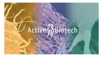
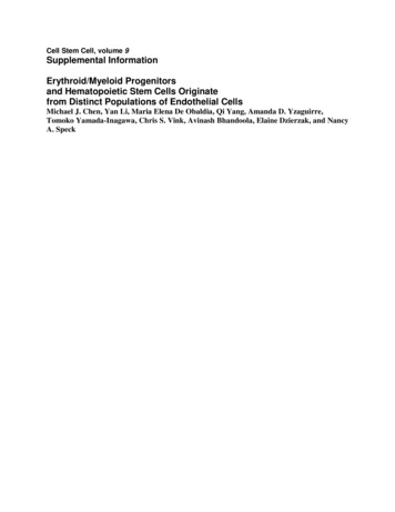
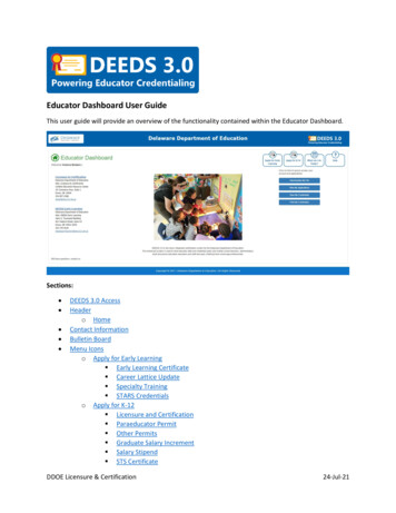
Wang et al. Journal of Hematology & Oncology(2019) 12:31peripheral blood, H-PB) and after G-CSF mobilization(G-CSF-mobilized peripheral blood, G-PB), as well asG-PBSC, were analyzed. The cell numbers and percentage of all MDSCs were significantly increased in G-PBand G-PBSC. M-MDSC population increased in bothfrequency and absolute number in G-PBSC. HLA-DR /lowCD33 CD16 population increased in both frequency and absolute number in G-PB and G-PBSC(Fig. 1a, b, d).May-Grünwald-Giemsa cytospin results showed that themorphological features of HLA-DR /lowCD33 CD16 cellswere similar to those of immature monocyte-like cells(Fig. 1e). The in vitro immune-suppressive activity of theHLA-DR /lowCD33 CD16 population identified amongthe G-PBSC was tested. HLA-DR /lowCD33 CD16 andautologous CD3 T cells were sorted from the G-PBSC ofhealthy donors using FACS. HLA-DR /lowCD33 CD16 cells were co-cultured for 4 days with autologous T cells atdifferent ratios (HLA-DR /lowCD33 CD16 : T 0:1 to 1:2)in the presence of T cell stimulators (anti-CD3/CD28beads). Flow cytometry analysis demonstrated that theHLA-DR /lowCD33 CD16 population had a strong immunosuppressive effect on T cell proliferation in a celldose-dependent manner (Fig. 1f, g). Moreover, HLA-DR /lowCD33 CD16 population demonstrated superiorimmune-suppressive activity compared with CD33 andHLA-DR /lowCD33 CD16 fraction (Additional file 1:Figure S1). Meanwhile, the immune-suppressive activity ofHLA-DR /lowCD33 CD16 cells was inferior to M-MDSCsand G-MDSCs population (Additional file 1: Figure S2).HLA-DR /lowCD33 CD16 cell-induced inhibition of T cellproliferation in vitro depends on TGF-β productionTo investigate the mechanisms underlying the inhibitionof T cell proliferation, the IL-10- and TGF-β-suppressivecytokines expressed by G-CSF-mobilized HLA-DR /lowCD33 CD16 cells were detected. The CD33 fractionand non-mobilized HLA-DR /lowCD33 CD16 cells servedas controls. The median percentage of IL-10 or TGF-βpositive cells among HLA-DR /lowCD33 CD16 population in the G-PBSC was significantly higher than those ofH-PB (IL-10, 10.1% vs. 0.62%, p 0.002; TGF-β, 60.24% vs.10.39%, p 0.0003). Notably, the TGF-β cells among theHLA-DR /lowCD33 CD16 cells significantly increasedcompared to the CD33 fraction or non-mobilizedHLA-DR /lowCD33 CD16 cells (Fig. 2a, b).Subsequently, the inhibition of CD3 T cell proliferation by HLA-DR /lowCD33 CD16 cells was markedlyreduced in the presence of an anti-TGF-β antibody butnot an anti-IL-10 antibody, indicating that the immunosuppressive activity relied more on the presence ofTGF-β (Fig. 2c, d).Other immunosuppressive mechanisms (arginase, iNOS,IDO, and COX2) were excluded by adding selectivePage 5 of 16inhibitors of arginase (nor-NOHA (N(w)-hydroxy-norL-arginine)), iNOS (L-NMMA), IDO (NLG8189) to theco-cultures. However, no reverse effect on T cell proliferation was observed (Additional file 1: Figure S3).HLA-DR /lowCD33 CD16 cells modulate Th cell balanceand induce Treg generationThe present study demonstrated that CD4 CD25 Foxp3 Treg cells increased in the HLA-DR /lowCD33 CD16 cell co-culture group (Fig. 3a, c). Thcell subsets were analyzed after co-culture with or without HLA-DR /lowCD33 CD16 cells. Though the expression of IFN-γ and IL-17A increased, co-culture withHLA-DR /lowCD33 CD16 cells led to a significantlyhigher ratio of Th2/Th1 and Th2/Th1 Th17 cells thanthe control group (Fig. 3b, d, e).HLA-DR /lowCD33 CD16 MDSCs prevent aGVHD inhumanized mouse modelBecause G-CSF expanded HLA-DR /lowCD33 CD16 cells into the peripheral blood and exerted a strongregulatory effect on T cell immune response, we next investigated if such population prevents aGVHD in vivo.To mimic aGVHD after allo-HSCT in humans, a humanized xenogeneic aGVHD model was established byinjection of 5 106 donor G-PBSC into irradiatednon-obese diabetic (NOD)-severe combined immune deficient (SCID)-IL-2Rg / mice (NSG mice) [38, 39](Fig. 4a). Human white blood cell engraftment was detected by CD45 cells at 7, 14, and 21 days after transplantation, and there was no significant difference ofhuman cells engraftment intragroup (14 days, p 0.486;21 days, p 0.4012) (Additional file 1: Figure S4). Wefailed to detect the engraftment of HLA-DR /lowCD33 CD16 MDSCs in PB as these cells could bedetected in the spleen and bone marrow at 14 days aftertransplantation; however, they decreased quickly and almost could not be detected at 21 days afterco-transplantation (Additional file 1: Figure S5). AcuteGVHD occurred in the humanized mice receiving donorG-PBSC during 2 to 4 weeks after transplantation as evidenced by weight loss, disease score (hunching, activity,ruffling, and diarrhea), and death. Similar to humans[40, 41], humanized mice with aGVHD showed severeinflammation, leukocyte infiltration, necrosis, and tissuedamage in target organs, such as the skin, liver, and gutin the G-PBSC group (Fig. 4e). On the base of thismodel, a prevention model was established by one-timeco-infusion of 2.5 106, 1 106, and 5 105 humanG-CSF-mobilized HLA-DR /lowCD33 CD16 cells sortedfrom the same donor with graft at the same day. All ofthese cell doses reduced weight loss (Fig. 4b), improvedoverall survival (Fig. 4c), improved pathological score(Fig. 4d), and lessened tissue damage in target organs in
Wang et al. Journal of Hematology & Oncology(2019) 12:31Page 6 of 16Fig. 1 HLA-DR /lowCD33 CD16 myeloid cells expanded in PB and PBSC after G-CSF administration and exerted a strong immunosuppressive effecton T cell proliferation. a, b The frequency and cell number of different kind of MDSCs and HLA-DR /lowCD33 CD16 population in ten donors (H-PB,G-PB, G-PBSC) were analyzed by flow cytometry. c Gate strategies of HLA-DR /lowCD33 CD16 : FSC, forward scatter; SSC, side scatter. d The proportionof HLA-DR /lowCD33 CD16 in the CD45 fraction from the ten healthy donor’s HPB, GPB, and GPBSC (medians of the different groups were 4.6%,6.5%, and 15.5% of total CD45 cells, Mann-Whitney t test). e May-Grünwald-Giemsa cytospin preparations show morphological features of HLA-DR /lowCD33 CD16 . f T cell proliferation was examined using CFSE dilution. HLA-DR /lowCD33 CD16 and CD3 T cells from the same donor G-PBSC wereco-cultured at different ratios for 4 days with anti-CD3/CD28 beads. T cell proliferation was evaluated using CFSE labeling. Unstimulating T cells werenegative control. The picture shows the representative results. g The percentage of T cells in suppression was shown in different groups. Data wascompared using unpaired t test (ns, not significant)medium and high doses of HLA-DR /lowCD33 CD16 cells compared to mice grafted without or withlow-dose human HLA-DR /lowCD33 CD16 cells(Fig. 4e). For human Treg detected at 21 days aftertransplantation, a significant difference was found between the high-dose HLA-DR /lowCD33 CD16 cellsand GPBSC control (p 0.0498); there was no disparitybetween medium dose vs. control (p 0.6825) and lowdose vs. control (p 0.9) (Fig. 4a, Additional file 1:Figure S6); there were no disparity of Th2/(Th1 Th17)ratio between the different groups (Fig. 4b,Additional file 1: Figure S6). For cytokines detected at 7,14, and 21 days after transplantation, there were significantly less INF-γ and IL-6 at days 7, 14, and 21 and
Wang et al. Journal of Hematology & Oncology(2019) 12:31Page 7 of 16Fig. 2 The percentage of IL-10 and TGF-β cells increased in HLA-DR /lowCD33 CD16 cells in GPBSC and HLA-DR /lowCD33 CD16 cellssuppressed T cell proliferation in a TGF-β-dependent manner. a, b IL-10 and TGF-β were analyzed by cytometry in HPB and GPBSC from eighthealthy donors. IL-10 and TGF-β cells were gated on HLA-DR /lowCD33 CD16 cells. c HLA-DR /lowCD33 CD16 cells sorted from GPBSC andco-cultured with autologous CD3 T cells, with or without anti-IL-10 antibody or anti-TGF-β antibody in culture system in the presence of antiCD3/CD28 beads. The ratio of HLA-DR /lowCD33 CD16 to CD3 T cells was 1:1. Co-cultured for 4 days, T cell proliferation was evaluated usingCFSE labeling. Unstimulated T cells were used as a negative control. Stimulated T cells were used as a positive control. d The percentage of T cellsuppression was shown in a different group. Data were compared using Student’s unpaired t test (ns, not significant; *p 0.05). d Percentages ofT cell suppression in different groups. Data were compared using Student’s unpaired t test (ns, not significant; *p 0.05)IL-17A and TNF-α at days 14 and 21 (high dose ofHLA-DR /lowCD33 CD16 cells vs. control) and lessINF-γ and TNF-α at days 7 and 21, IL-17A at days 7and 14, and IL-6 at day 21 (medium dose of HLA-DR /lowCD33 CD16 cells vs. control) (Additional file 1:Figure S7). IL-4/INF-γ and IL-4/IL-17A ratios werehigher in the high-dose HLA-DR /lowCD33 CD16 cellsgroup vs. control at 7, 14, and 21 days after transplantation; by comparison with medium-dose HLA-DR /lowCD33 CD16 cells group vs. control, IL-4/INF-γ
Wang et al. Journal of Hematology & Oncology(2019) 12:31Page 8 of 16Fig. 3 HLA-DR /lowCD33 CD16 cells could affect the balance of Th subsets induce Treg generation. a Treg (CD4 CD25 Foxp3 ) generation wasdetected through flow cytometry. CD3 T cells were co-cultured with or without HLA-DR /lowCD33 CD16 cells for 4 days. b The percentage ofTreg in CD4 T cell, group HLA-DR /lowCD33 CD16 vs. control, p 0.0048. c Flow cytometry analysis of Th1 (CD4 IFN-γ ), Th2 (CD4 IL-4 ), Th17(CD4 IL-17a ). CD3 T cells were co-cultured with or without HLA-DR /lowCD33 CD16 cells sorted from GPBSC in the presence of CD3/CD28 for4 days. Red for CD4 cell fraction and blue for CD4-negative part. d Th2/Th1 ratio fold change compared with the control group, p 0.0159.e Th2/(Th1 Th17) ratio fold change compared with the control group, p 0.0095ratios were higher at days 7 and 14, while IL-4/IL-17Aratio was higher at day 21 (Fig. 5c, d). These results confirmed that immune-suppressive activity of HLA-DR /lowCD33 CD16 cells which might contribute to alleviating aGVHD in NSG model.Negative correlations between the frequency ofHLA-DR /lowCD33 CD16 cells among donor PBSCsand the development of aGVHD in recipientsIn a prospective study (registered at www.chictr.org asChiCTR-OCH-10000940), 100 allo-HSCT recipients wereenrolled to assess the role of HLA-DR /lowCD33 CD16 in graft on the occurrence of aGVHD. Of these patients,76 patients received a combination of G-BM and G-PBSCgrafts from haplo-donors, and 24 patients received graftsfrom MSDs. The indications for allo-HSCT were as follows: AML (n 39), ALL (n 29), MDS (n 8), SAA (n 14), and lymphoma (n 10). Ninety-nine (99%) patientsreached full donor chimerism before day 30 after transplantation as assessed by the analysis of a variable numberof tandem repeats on the peripheral blood. Platelet recovery (count 20,000/ml) was achieved at a median of day32 post-transplantation, whereas neutrophils reachedvalues greater than 500/ml on day 12. Only 1 patient dieddue to transplant-related mortality at day 11 post-HSCT.The median follow-up period of the enrolled patients was11 months. The cell components in the graft were listed inAdditional file 1: Table S1. Patients were divided into 2groups according to the median absolute counts ofHLA-DR /lowCD33 CD16 MDSCs in grafts (1.88 107/
Wang et al. Journal of Hematology & Oncology(2019) 12:31Page 9 of 16Fig. 4 HLA-DR /lowCD33 CD16 cells could inhibit the development of aGVHD in humanized mouse model. a Protocol for the establishment ofaGVHD model through injecting human GPBSC. Sublethally irradiated NSG mice received 5 106 human GPBSC with or without 2.5 106, 1 106,and 5 105 G-CSF-mobilized HLA-DR /lowCD33 CD16 sorted from GPBSC. b Weight Loss, c median survival, and d pathological score in xenograftedmice were shown. Mice co-transplanted with HLA-DR /lowCD33 CD16 (pink, green, and blue, eight mice each group) had improved survival aftertransplant as compared with GPBSC-injected group (red). For survival, weight change, and disease score: GPBSC high dose versus GPBSC, GPBSC medium dose versus GPBSC, and GPBSC low dose versus GPBSC, p 0.05. e Representative histology of the target organs harvested on day 22 aftertransplantation. All results were derived from two pooled independent experim
grafts leads to a better GRFS and less GVHD [27]. In contrast, the frequency of CD14 HLA-DRlow/ negM-MDSCs is significantly increased in the periph-eral blood after allo-HSCT, especially in patients with acute graft-versus-host disease [28]. Though clinical relevance between MDSCs and GVHD has been ob-served in humans, there is no data .