Transcription
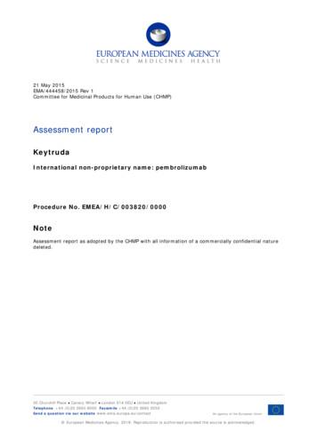
Watanabe et al. World Journal of Surgical Oncology (2015) 13:274DOI 10.1186/s12957-015-0695-2WORLD JOURNAL OFSURGICAL ONCOLOGYCASE REPORTOpen AccessPrimary pulmonary melanoma: a report oftwo casesMototsugu Watanabe1, Hiromasa Yamamoto1, Shinsuke Hashida1,2, Junichi Soh1,3, Seiichiro Sugimoto1,Shinichi Toyooka1,2,3* and Shinichiro Miyoshi1AbstractMalignant melanoma is a refractory malignancy with a dismal prognosis. It generally arises from the skin in mostcases, and cases of primary pulmonary malignant melanoma are rare and often behave aggressively. We havetreated two cases of localized primary pulmonary malignant melanoma using surgical resection. Pulmonarymalignant melanomas often metastasize to the brain and liver; one of our cases exhibited metastasis to the cecumat about 8 months after surgery. Because cutaneous melanomas often carry activating mutations in the BRAF gene(V600E), we performed a BRAF mutational analysis using direct sequencing for both of these tumors arising fromthe lung. However, no BRAF mutations were detected. We detected a p53 mutation, which was thought to be apotential somatic mutation, in one of the two cases using a sequencing panel targeting 20 lung cancer-relatedgenes. Although we also checked the expression of programmed death ligand 1 (PD-L1) on the surface of thetumor cells by immunohistochemical testing, neither of our two cases expressed PD-L1. Further molecular analysesmay uncover the characteristics of primary pulmonary malignant melanomas.Keywords: Primary pulmonary malignant melanoma, Target sequencing, Surgical resectionBackgroundMalignant melanoma is an often fatal cutaneous neoplasm.As a primary tumor, malignant melanoma of the lung israre, accounting for only 0.01 % of all primary lung tumors[1]. To diagnose malignant melanoma as a primary tumorarising from the lung, the absence of primary melanomalesions at cutaneous or nonpulmonary extracutaneous sitesmust be demonstrated [2].Approximately 40 to 60 % of cutaneous melanomascarry mutations in the BRAF gene (V600E), which leadsto the constitutive activation of downstream signalingthrough the MAPK pathway [3, 4]. At present, whetherpulmonary malignant melanomas carry BRAF mutationshas not been reported. Additionally, immunotherapy toinhibit programmed death 1 (PD-1)/programmed deathligand 1 (PD-L1) pathway that has a role for immune escape* Correspondence: toyooka@md.okayama-u.ac.jp1Department of Thoracic, Breast and Endocrinological Surgery, OkayamaUniversity Graduate School of Medicine, Dentistry and PharmaceuticalSciences, Okayama 700-8558, Japan2Department of Clinical Genomic Medicine, Okayama University GraduateSchool of Medicine, Dentistry and Pharmaceutical Sciences, 2-5-1Shikata-cho, Kita-ku, Okayama 700-8558, JapanFull list of author information is available at the end of the articleis developed for malignant melanoma [5, 6]. It is reportedthat the efficacy of anti-PD-1 antibody was related to theexpression of PD-L1 in tumor cells [6]. Here, we describetwo cases of possible primary pulmonary malignant melanoma with wild-type BRAF and no expression of PD-L1.Case presentationCase 1A 66-year-old man with hemoptysis was referred to ourhospital because of a pulmonary malignant tumor. Abronchoscopy revealed a tumor arising from the rightmiddle lobe bronchus (Fig. 1a), and a histological diagnosis based on a bronchial biopsy revealed a malignantmelanoma. A positron emission tomography-computedtomography (PET-CT) scan revealed no other lesionsthat could have represented a primary site (Fig. 1b). Aright middle lobectomy with mediastinal and hilar lymphadenectomy was performed. Some tumor cells had melaninin the cytoplasm (Fig. 1c). Immunohistochemical analysisshowed positive cytoplasmic staining of the tumor cells forHMB-45, Tyrosinase, and MART1, although the expression of TTF-1 in tumor cells was negative. The postoperative course was uneventful. Combination chemotherapy of 2015 Watanabe et al. Open Access This article is distributed under the terms of the Creative Commons Attribution 4.0International License (http://creativecommons.org/licenses/by/4.0/), which permits unrestricted use, distribution, andreproduction in any medium, provided you give appropriate credit to the original author(s) and the source, provide a link tothe Creative Commons license, and indicate if changes were made. The Creative Commons Public Domain Dedication o/1.0/) applies to the data made available in this article, unless otherwise stated.
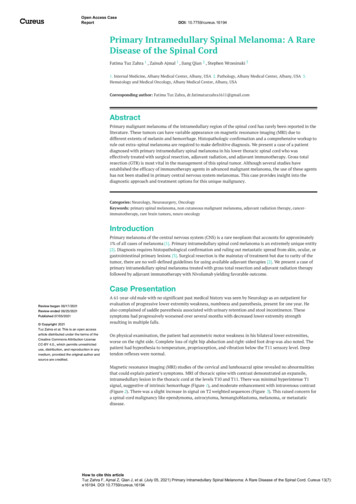
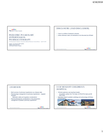
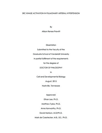
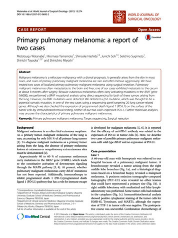
Watanabe et al. World Journal of Surgical Oncology (2015) 13:274Page 2 of 5Fig. 1 Diagnostic images and pathological findings for case 1. a A bronchoscopy revealed a tumor arising from the right middle lobe bronchus.b PET-CT scan revealed no other lesions that could have represented a primary site. c Hematoxylin-eosin staining of the resected tumor, 40(scale bar, 100 μm). d Immunohistochemistry showed negative for PD-L1 staining, 40 (scale bar, 100 μm)dacarbazine, nimustine, and vincristine was performed. Atthe time of the 22 months after surgery, he was alive without disease recurrence.that the tumor was considered to be the recurrence of amelanoma.Molecular profilingCase 2A 46-year-old woman was found to have an abnormalshadow on a chest X-ray during an annual check-up. ACT and PET-CT scan showed a tumor in the left lowerlobe of the lung (Fig. 2a, b). Pathological examination ofa specimen obtained during a transbronchial lung biopsyrevealed that the tumor was a malignant melanoma(Fig. 2c). Because further examinations did not reveal anyother tumors that could have been the primary lesion, wediagnosed the lung tumor as a primary pulmonary malignant melanoma. A left lower lobectomy with mediastinaland hilar lymph node dissection was performed. Immunohistochemical staining was negative for S-100 protein,chromogranin, synaptophysin, keratin, and TTF-1 butpositive for HMB-45 and vimentin. The postoperativecourse was uneventful. She refused to receive adjuvantchemotherapy. Eight months after the pulmonary resection, a PET-CT scan showed the uptake of fluorodeoxyglucose in the cecum (Fig. 2e). An ileocecal resection wasperformed, and the pathological examination indicatedWe determined the BRAF mutational status (exons 11 and15) using direct sequencing in the two tumors. The detailedmethods have been described previously [7]. However, wedid not detect any BRAF mutations. Next, we performed atarget sequencing analysis using the Human Lung CancerPanel (Qiagen, Hilden, Germany), which targets 20 lungcancer-related genes including most of the exons in BRAF,using the same samples. Although various variants weredetected using this analysis, only the p53 mutation (P72R)in case 1 was considered to be a potential somatic mutationafter restricting the variants using GeneRead Software(Qiagen, Venlo, Netherlands) as shown in Table 1. P72Rmutation has been already reported in bladder and gastriccancers, but there are no reports in lung cancer or malignant melanoma ([8], accessed on August 14, 2015).Furthermore, we also checked the expression of PD-L1on the surface of the tumor cells by immunohistochemicaltesting in formalin-fixed, paraffin-embedded tumor specimens with the use of a rabbit monoclonal antihuman PDL1 antibody. In the previous reports, PD-L1 positivity wasdefined as at least 5 % of tumor cells showing cell-surface
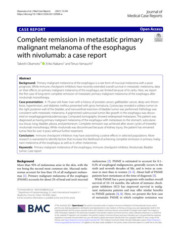
Watanabe et al. World Journal of Surgical Oncology (2015) 13:274Page 3 of 5Fig. 2 Diagnostic images and pathological findings for case 2. a A CT scan shows a large tumor in the left lower lobe of the lung. b Preoperative PET-CTscan revealed no other lesions that could have represented a primary site. c Hematoxylin-eosin staining of the resected tumor, 40 (scale bar, 100 μm). dImmunohistochemistry showed negative for PD-L1 staining, 40 (scale bar, 100 μm). e Eight months after the pulmonary resection, PET-CT scan showedthe uptake of fluorodeoxyglucose in the cecumPD-L1 staining of any intensity in a section containing atleast 100 tumor cells that could be evaluated [9, 10].According to the definition of PD-L1 positivity by them,neither of our two cases was considered to express PD-L1(Figs. 1d and 2d).DiscussionThe total number of operations in general thoracic surgeryis increasing year by year in Japan [11]. However, there area few reports about primary pulmonary melanoma [2].There are four clinical criteria that should be satisfied forthe diagnosis of primary melanoma of the lung: (1) no previously removed skin tumor, unless the pathology examination did not show malignancy and the slides are stillavailable for reevaluation; (2) no excised ocular tumor; (3) asolitary tumor in the surgical specimen from the lung; and(4) no demonstrable melanoma in other organs at the timeof surgery [1, 12]. Both of our cases fulfill the abovementioned diagnostic criteria.Regarding treatment, surgery is necessary for a cure forlocalized pulmonary malignant melanoma because theeffect of chemotherapy on malignant melanoma is limited[13]. Even among patients who undergo surgery, the prognosis of patients with malignant melanoma of the lung isgenerally poor because the lesion often metastasizes soonafter surgery. Regarding metastatic spread, primary pulmonary melanomas often metastasize to the brain and theliver [2, 14–16], similar to other types of primary lung cancer. Intriguingly, one of our cases exhibited metastasis tothe cecum. To the best of our knowledge, this is the firstreport describing a malignant melanoma of the lung metastasizing to the cecum.BRAF-activating mutation is a characteristic of malignantmelanoma, and molecular-targeted therapy for BRAF hasbeen developed [17]. However, our two cases did notexhibit activating mutations in BRAF. Considering the factthat the presence of BRAF-activating mutations has notbeen reported in pulmonary malignant melanoma, theirfeatures may differ from those of primary skin melanoma.A target sequencing analysis did not detect any possiblesomatic driver mutations commonly observed in lungcancer [18], such as EGFR, HER2, KRAS, ALK, MET, orPIK3CA, suggesting that primary pulmonary malignantmelanomas may have different oncogenic pathwaysfrom primary lung cancer from the viewpoint of driveroncogenes.In these days, there are many reports about the efficacyof anti-PD-1 antibody for cutaneous melanoma patients
Watanabe et al. World Journal of Surgical Oncology (2015) 13:274Table 1 Mutational status of 20 genes in cases 1 and 2Gene nameCase 1Case WTERBB2WTWTSTK11WTWTWT wild typeand a potential association between the occurrence of aresponse and expression of PD-L1 in patients receivingnivolumab [10]. On the other hand, Robert et al. reportedthat regardless of PD-L1 status, nivolumab-treatedpatients had improved overall survival, as comparedwith dacarbazine-treated patients [19]. Although ourfindings did not show the positivity of PD-L1 in thetwo cases, there is still the possibility of the efficacy ofimmunotherapy for primary pulmonary melanoma, asthe role of PD-1/PD-L1 pathway in primary pulmonary melanoma is yet to be investigated.ConclusionsIn conclusion, we encountered two cases of malignant melanoma of the lung that did not carry activating mutationsin the BRAF gene. Further molecular analyses may uncoverthe characteristics of primary malignant melanoma.ConsentWritten informed consent was obtained from the patientsfor publication of this case report and any accompanyingimages. A copy of the written consent is available for reviewby the editor in chief of this journal.AbbreviationsPD-1: Programmed death 1; PD-L1: Programmed death ligand 1;PET-CT: Positron emission tomography-computed tomography.Page 4 of 5Competing interestsThe authors declare that they have no competing interests.Authors’ contributionsMW, HY, ST, and SM conceived the project. MW and SH performed theexperiments. MW, HY, JS, SS, and ST collected the data and assisted with theexperiments. MW, HY, and SH analyzed the data. MW, HY, SH, JS, and STprepared the manuscript with input from the other authors. ST and SMsupervised the project. All authors read and approved the final manuscript.AcknowledgementsWe thank Dr. Takehiro Matsubara (Biobank of Okayama UniversityHospital, Okayama, Japan) for his technical support and Dr. NoriyukiTanaka (Department of Pathology, Okayama University Hospital) forproviding advice.Author details1Department of Thoracic, Breast and Endocrinological Surgery, OkayamaUniversity Graduate School of Medicine, Dentistry and PharmaceuticalSciences, Okayama 700-8558, Japan. 2Department of Clinical GenomicMedicine, Okayama University Graduate School of Medicine, Dentistry andPharmaceutical Sciences, 2-5-1 Shikata-cho, Kita-ku, Okayama 700-8558,Japan. 3Biobank of Okayama University Hospital, Okayama 700-8558, Japan.Received: 27 April 2015 Accepted: 7 September 2015References1. Wilson RW, Moran CA. Primary melanoma of the lung: a clinicopathologicand immunohistochemical study of eight cases. Am J Surg Pathol.1997;21:1196–202.2. Maeda R, Isowa N, Onuma H, Miura H, Tokuyasu H, Kawasaki Y.Primary malignant melanoma of the lung with rapid progression.Gen Thorac Cardiovasc Surg. 2009;57:671–4.3. Davies H, Bignell GR, Cox C, Stephens P, Edkins S, Clegg S, et al.Mutations of the BRAF gene in human cancer. Nature. 2002;417:949–54.4. Menzies AMLT, Wilmott JS, Hyman J, Kefford RF, Thompson JF, O’Toole S,et al. Intrapatient homogeneity of BRAFV600E expression in melanoma.Am J Surg Pathol. 2014;38:377–82.5. Schreiber RD, Old LJ, Smyth MJ. Cancer immunoediting: integratingimmunity’s roles in cancer suppression and promotion. Science.2011;331:1565–70.6. Topalian SL, Hodi FS, Brahmer JR, Gettinger SN, Smith DC, McDermott DF,et al. Safety, activity, and immune correlates of anti–PD-1 antibody incancer. N Engl J Med. 2012;366:2443–54.7. Yamamoto H, Shigematsu H, Nomura M, Lockwood WW, Sato M, Okumura N,et al. PIK3CA mutations and copy number gains in human lung cancers.Cancer Res. 2008;68:6913–21.8. cBioPortal for Cancer Genomics. [http://www.cbioportal.org/].9. Taube JM, Anders RA, Young GD, Xu H, Sharma R, McMiller TL, et al.Colocalization of inflammatory response with B7-H1 expression in humanmelanocytic lesions supports an adaptive resistance mechanism of immuneescape. Sci Transl Med. 2012;4:27–37.10. Wolchok JD, Kluger H, Callahan MK, Postow MA, Rizvi NA, Lesokhin AM,et al. Nivolumab plus ipilimumab in advanced melanoma. N Engl J Med.2013;369:122–33.11. Committee for Scientific Affairs TJAfTS, Masuda M, Kuwano H, Okumura M,Amano J, Arai H, et al. Thoracic and cardiovascular surgery in Japan during2012 : annual report by the Japanese association for thoracic surgery.Gen Thorac Cardiovasc Surg. 2014;62:734–64.12. Jensen OAEJ. Primary malignant melanoma. Scand J Respir Dis.1967;48:127–35.13. Bajetta E, Del Vecchio M, Bernard-Marty C, Vitali M, Buzzoni R, Rixe O, et al.Metastatic melanoma: chemotherapy. Semin Oncol. 2002;29:427–45.14. Neri S, Komatsu T, Kitamura J, Otsuka K, Katakami N, Takahashi Y.Malignant melanoma of the lung: report of two cases. Ann ThoracCardiovasc Surg. 2011;17:170–3.15. Gong L, Liu X-Y, Zhang W-D, Zhu S-J, Yao L, Han X-J, et al. Primarypulmonary malignant melanoma. A clinicopathologic study of two cases.Diagn Pathol. 2012;7:123.
Watanabe et al. World Journal of Surgical Oncology (2015) 13:274Page 5 of 516. Ouarssani A, Atoini F, Reda R, Lhou FA, Rguibi MI. Malignant melanoma ofthe lung: a case report. Pan Afr Med J. 2012;11:68.17. Chapman PB, Hauschild A, Robert C, Haanen JB, Ascierto P, Larkin J, et al.Improved survival with vemurafenib in melanoma with BRAF V600Emutation. N Engl J Med. 2011;364:2507–16.18. Toyooka S, Mitsudomi T, Soh J, Aokage K, Yamane M, Oto T, et al.Molecular oncology of lung cancer. Gen Thorac Cardiovasc Surg.2011;59:527–37.19. Robert C, Long GV, Brady B, Dutriaux C, Maio M, Mortier L, et al.Nivolumab in previously untreated melanoma without BRAF mutation.N Engl J Med. 2015;372:320–30.Submit your next manuscript to BioMed Centraland take full advantage of: Convenient online submission Thorough peer review No space constraints or color figure charges Immediate publication on acceptance Inclusion in PubMed, CAS, Scopus and Google Scholar Research which is freely available for redistributionSubmit your manuscript atwww.biomedcentral.com/submit
from primary lung cancer from the viewpoint of driver oncogenes. In these days, there are many reports about the efficacy of anti-PD-1 antibody for cutaneous melanoma patients Fig. 2 Diagnostic images and pathological findings for case 2. a A CT scan shows a large tumor in the left lower lobe of the lung. b Preoperative PET-CT