Transcription
Molecular docking and ADMET based study to identify potential phytochemicalinhibitors for papain-like protease of SARS-CoV-2Padmika Madushanka Wadanambia*, Uthpali Mannapperumab, Nimanthi JayathilakacaIndependent Researcher, Wadduwa-12560, Sri Lanka.ORCID iD: https://orcid.org/0000-0002-4742-1553bDepartment of Pharmacology, Faculty of Medicine, University of Colombo, Sri Lanka.ORCID iD: https://orcid.org/0000-0002-7433-2855cDepartment of Chemistry, Faculty of Science, University of Kelaniya, Kelaniya, SriLanka.ORCID iD: https://orcid.org/0000-0001-8741-3075Corresponding author: Padmika Madushanka Wadanambi, Independent Researcher,Wadduwa-12560, Sri Lanka.E-mail: pwadanambi@gmail.comAbstractThe severe acute respiratory syndrome coronavirus 2 (SARS-CoV-2) is the causativepathogen for the novel COVID-19 disease. SARS-CoV-2 papain-like protease (PLpro) isresponsible for viral replication and host innate immunity suppression. Thus, this studyaimed to explore potential phytochemical inhibitors against this dual therapeutic targetusing virtual screening methods. Thirty-one phytochemicals with reported anti-SARS-1
CoV-1 PLpro activity were used to construct the phytochemical library along with twopositive controls. Molecular docking using AutoDock 4.2 was employed to calculatebinding affinity and inhibition constant of each compound within the S3/S4 bindingpocket of SARS-CoV-2 PLpro. Based on the docking results, twelve compounds weresubjected to non-covalent interaction analysis utilizing the Discovery Studio Visualizersoftware. Further, their physicochemical, pharmacokinetics and toxicity descriptors wereevaluated using molinspiration and pkCSM web servers, respectively. Hirsutenone fromAlnus japonica and broussoflavan A from Broussonetia papyrifera, displayed thestrongest binding affinity (-8.23 kcal/mol and -8.13 kcal/mol), lowest inhibition constant(920.39 nM and 1.1 μM) and highest ligand efficiency (0.34 and 0.26) among allphytochemicals towards the binding pocket of SARS-CoV-2 PLpro, demonstratingsuperiority to PLpro inhibitors, 3k and GRL0617 which were used as positive controls.Additionally, hirsutenone, broussoflavan A and broussochalcone A (from Broussonetiapapyrifera) possessed favorable physicochemical properties for oral drug development,satisfying Lipinski’s and Veber’s rules. Furthermore, in silico pharmacokinetics andtoxicity predictions revealed that the three aforementioned phytochemicals are watersoluble, non-mutagenic, non-hepatotoxic and biologically safe. Hence these leadcompounds might be exploited to accelerate the drug discovery process against theongoing COVID-19 infection.Keywords: SARS-CoV-2, COVID-19, papain-like protease, PLpro inhibitors, virtualscreening, pharmacokinetics, molecular dockingIntroduction2
The COVID-19 pandemic has continued unabated across the world since 2019.According to the latest data available through the World Health Organization (WHO),168,040,871laboratory-confirmed cases of COVID-19 have been reported resulting in3,494,758 fatalities by May 27, 2021(WHO, 2021). However, COVID-19 shows anoverall low mortality rate (2.08%) compared to the mortality rate of the previouslyreported Middle East respiratory syndrome coronavirus (MERS-CoV) –(9.6%) and themortality rate of the severe acute respiratory syndrome coronavirus (SARS-CoV-1)(34.4%) diseases irrespective of its rapid dissemination across the world (KordzadehKermani et al., 2020). The highly contagious virus is transmitted through respiratorydroplets of an infected individual or fomites. The elderly and individuals withcomorbidities are more susceptible to developing fatal complications due to COVID-19(Petushkova and Zamyatnin, 2020). Thus, it has become a life threatening disease.The novel SARS-CoV-2 belonging to the beta coronavirus family is the causative agentof COVID-19. The virion contains positive sense, single stranded RNA genetic materialcomprised of 30 000 nucleotides (Arya et al., 2021). Inside the host cell, the viralgenome encodes two cysteine proteases, main protease or 3-chymotrypsin likeprotease (Mpro or 3CLpro) and papain-like protease (PLpro). Proteolysis mediated by3CLpro and PLpro, slices large polyproteins into smaller components for packaging newvirions to promote the spread of infection. PLpro can cleave translated largepolypeptides (pp1a and pp1ab) into non-structural, functional protein subunits (NSP1-3).Later, these non-structural proteins assemble into the viral replicase-transcriptasecomplex (RTC) to initiate viral replication leading to proliferation of the virus (Arya et al.,2021). Besides the protease activity, PLpro possesses two more functions such as3
deubiquitination and deISGylation which can effectively remove post translationalmodifications such as ubiquitin and Interferon-stimulated gene 15 (ISG-15) from thehost cell proteins. Therefore, inhibition of SARS-CoV-2 PLpro may inhibit viralreplication and also promote antiviral immunity mechanisms inside the host cell (Shin etal., 2020). In addition to SARS-CoV-2 structural proteins, other non-structural proteinsand human host proteins, SARS-CoV-2 PLpro is an attractive dual therapeutic target fornovel drug development (Zhou et al., 2020).To date, no effective drug has been discovered to eradicate the COVID-19 from theworld. The first ever repurposed drug of COVID-19, remdesivir approved by UnitedStates Food and Drug Administration (U.S. FDA), did not effectively allow recovery fromCOVID-19 infection (Office of the Commissioner, & Food and Drug Administration, U.S., 2020). Therefore, current clinical management of COVID-19 consists of infectionprevention and supportive care, including supplemental oxygen and mechanicalventilatory support for severe cases. A number of new treatments, such as monoclonalantibodies, are available under emergency use authorization (EUA), with ongoing trialsto evaluate their potential efficacy (Deb et al., 2021).Although, several vaccines including Pfizer-BioNTech, Moderna, AstraZeneca,Covishield, Janssen, Sinopharm, Sinovac and Sputnik V COVID-19 vaccines had beenauthorized for emergency use by 2021, widespread application of these vaccines islimited by several factors such as availability, limited infrastructure for storage anddistribution, economic restraints and consumer acceptability as well as limited researchand clinical studies supporting the widespread use for all demographics (WHO, 2021b).4
However, the efficacy of these vaccines against new mutant variants of SARS-CoV-2 isstill under investigation at the time of drafting this article. To date no ideal drug or avaccine has been discovered by scientists to eradicate this fatal COVID-19 forever.Therefore, it is critical to develop effective drugs to control the COVID-19 infection.The whole genome of novel SARS-CoV-2 shares 80% sequence similarity with thepreviously determined SARS-CoV-1 genome. Moreover, amino acid sequence ofSARS-CoV-2 PLpro shows 83% sequence similarity to SARS-CoV-1 PLpro.Interestingly, the trio of catalytic site residues Cys-His-Asp were conserved in both,SARS-CoV-2 (C111- H272-D286) and in SARS-CoV-1 (C112-H273-D287) in viralPLpro proteins.The active site in the palm subdomain of SARS-CoV-2 PLpro was made up of largerS3/S4 pockets rather than the more constrained S1/S2 pockets near the catalyticresidues (Arya et al., 2020). The S3/S4 binding pocket residues in the substrate bindingcleft within the ubiquitin-specific protease (USP) domain of the two PLpro proteins inSARS-CoV-1 and SARS-CoV-2 were discovered to be identical. When small moleculesbind within the S3/S4 pockets, flexible blocking loop 2 (BL2) transforms to a closedconformation, restricting substrate access and thereby reducing protease activity (Gaoet al., 2021). As a result, targeting S3/S4 binding sites present a potential therapeuticstrategy for developing possible SARS-CoV-2 PLpro inhibitors.Researchers have conducted numerous computational and laboratory studies to identifypotential SARS-CoV-2 PLpro inhibitors using medicines currently in clinical use(Kandeel et al., 2020; Kouznetsova et al., 2020; Klemm et al., 2020), natural5
compounds (Naidoo et al., 2020; Baildya et al., 2021;Alfaro et al., 2020) and syntheticcompounds (Thurakkal et al., 2021; Freitas et al., 2020). Bioactive phytochemicalsderived from medicinal plants have received widespread attention due to theirtherapeutic effects in treating a variety of health conditions with no or minimal healthrisks. Given the sequence and structural similarities between SARS-CoV-1 and SARSCoV-2 PLpro, we constructed a phytochemical library using phytochemicals withinhibitory activity against SARS CoV-1 PLpro and conducted molecular docking and insilico absorption, distribution, metabolism, excretion, and toxicity (ADMET) predictionsto assess pharmacological activity and inhibitory potential on SARS-CoV-2 PLpro toaccelerate drug discovery against COVID 19 infection.Materials and MethodsConstruction of anti SARS-CoV-2 PLpro phytochemical libraryPhytochemicals from four different medicinal plants, that were proven potent inhibitorsbased on their half maximal inhibitory concentration [IC50] against SARS-CoV-1 PLprowere selected by literature search to construct the phytochemical library for virtualscreening. The selected medicinal plants included Paulownia tomentosa (Cho et al.,2013), Alnus japonica (Park et al., 2012), Broussonetia papyrifera (Park et al., 2017)and Psoralea corylifolia (Kim et al., 2014) which are traditionally used in Chinese herbalmedicine. The 2D chemical structures of the selected phytochemicals and twonaphthalene based PLpro inhibitor compounds, GRL0617 (Ratia et al., 2008) and 3k(Báez-Santos et al., 2014) (used as positive controls) were drawn usingACD/ChemSketch Freeware (ACD/ChemSketch, 2021).6
SARS-CoV-2 PLpro protein structure preparationThe crystal structure of the viral target protein, SARS-CoV-2 PLpro was retrieved fromthe Research Collaboratory for Structural Bioinformatics (RCSB) Protein Data Bank(PDB) database (https://www.rcsb.org/) (Berman et al., 2002) bearing PDB ID: 6WX4 ata resolution of 1.66 Å(R-Value Free: 0.196,R-Value Work: 0.170). The heteroatomsincluding peptide inhibitor (VIR251), unnecessary ligands and crystallographic watermolecules were removed using UCSF Chimera v.1.12 (Pettersen et al., 2004). Thenative zinc ion was retained. Hydrogen atoms were added to the protein at physiologicalpH 7.4 using Avogadro v.1.2.0 (Hanwell et al., 2012) and the protein was converted topdb format.Ligand structures preparationAll selected phytochemicals were retrieved into UCSF Chimera by uploading theirrespective PubChem CIDs (https://pubchem.ncbi.nlm.nih.gov/). Two naphthalene basedinhibitors were used as positive controls. Both inhibitors were taken from theirrespective PDB entries. Then, hydrogen atoms were added to each ligand at pH 7.4(physiological pH) using Avogadro v.1.2.0 software. Further, Gastieger charges wereadded to all ligands. Except the two reported inhibitors, all phytochemicals weresubjected to energy minimization with 100 steepest descent steps (step size 0.02 Å)followed by 10 conjugate gradient steps (step size 0.02 Å). Finally, all ligands includingthe two naphthalene-based inhibitors were converted to pdb format.Selection of binding site residues in the SARS-CoV-2 PLpro7
The binding site residues were selected by analyzing the co-crystalized structure ofSARS-CoV-2 PLpro (PDB ID: 6WX4) (Rut et al., 2020). Further, residues that fall withinthe 5 Å of the binding site of the leading naphthalene-based inhibitor, 3k, adjacent to thecatalytic triad were selected based on the ligand bound structure of SARS-CoV-1PLpro (PDB ID: 4OW0) after superimposition with the SARS-CoV-2 PLpro dockingprotein template (PDB ID: 6WX4). The binding sites for the docking were defined byplacing a grid box of suitable dimensions centered above binding site residues in theSARS-CoV-2 PLpro structure (PDB ID: 6WX4) followed by docking execution of 3k.Site-specific molecular docking studyThe grid box size was designed to cover all the important binding site residues of theS3/S4 substrate binding pocket and giving sufficient space to occupy each ligand duringthe molecular docking experiment. The optimum grid box size was set as 38, 39, 45 inX, Y, Z directions whereas center grid box parameters were positioned at 8.050, (28.623), (-40.224) in X, Y, Z axes. These grid box settings were fixed by spacing at0.375 Å. All pdb files of ligands and protein were converted to pdbqt format usingAutoDock 1.5.6. The Kollman charges were added to the protein during the dockingprocess. The site-specific molecular docking was executed using AutoDock 4.2software (Morris et al., 2009). Molecular docking calculations were performed inLamarckian Genetic Algorithm (LGA) retaining all other parameters as default. Rigidprotein structure and flexible ligands were used throughout the molecular dockingprocess. For each ligand, ligand efficiency was calculated by dividing the binding affinity(- G) by the number of non-hydrogen atoms in the ligand (n-NHA).8
Non-covalent interaction analysisThe best docking pose of each ligand based on binding affinity was converted to thepdb format and visualized using Discovery Studio Visualizer Client 2020 to analyze noncovalent interactions belonging to favorable hydrogen bond, hydrophobic, electrostaticand other categories (BIOVIA, 2020).The detailed two dimensional (2D) and threedimensional (3D) interaction plots were generated using BIOVIA Discovery StudioVisualizer Client 2020 and UCSF Chimera v.1.12.Calculation of physicochemical properties and oral bioavailabilityMolinspiration web server (Molinspiration Cheminformatics, 2021) was employed tocalculate the various physicochemical properties of the top twelve compounds includingoctanol/water partition-coefficient (LogP), topological polar surface area (TPSA),number of non-hydrogen atoms (n-NHA), molecular weight (MW), number of hydrogenbond acceptors (n-HBA), number of hydrogen bond donors (n-HBD), number ofrotatable bonds (n-RotB) and the volume of the molecule. The SMILE (SimplifiedMolecular Input Line Entry System) notation of top ranked compounds was uploadedinto molinspiration web server for predictions. Lipinski’s rule of five (LRo5) (Lipinski etal., 1997) and Veber rule (Veber et al., 2002) were applied to predict the oralbioavailability of each top ligand.According to the LRo5, a compound must have MW 500 Dalton, LogP 5, n-HBA 10 and n-HBD 5 to be selected as a drug candidate for oral administration. In addition,the Veber rule suggests two more descriptors, TPSA and n-RotB which must be equal9
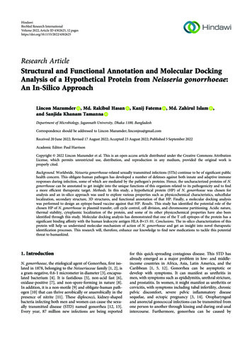
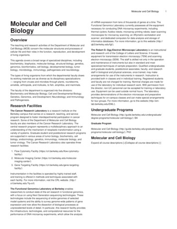
to or less than 140 Å and 10 respectively to be eligible as a drug candidate for oraladministration.Evaluation of in silico pharmacokinetics and toxicityThe pkCSM web tool was used to predict different ADMET attributes such as watersolubility (WS), human intestinal absorption (HIA), steady state volume of distribution(VDss) , blood-brain barrier permeability (BBBp), fraction unbound (FU), substrate ofcytochrome P450 isoenzymes (S), inhibitor of cytochrome P450 isoenzymes (I), totalclearance (TC), Ames toxicity (AMES) and hepatotoxicity (HEP) (Pires et al., 2015).ResultsConstruction of anti-SARS-CoV-2 PLpro phytochemical libraryPhytochemical data were collected from four medicinal plants namely Broussonetiapapyrifera (9), Paulownia tomentosa (11), Alnus japonica (6) and Psoralea corylifolia (5)based on their in vitro SARS-CoV-1 PLpro antiviral activity assays. To build thephytochemical library for molecular docking experiments, thirty one phytochemicalswere chosen based on the lowest IC50 values. Additionally, 3k and GRL0617 whichreported SARS-CoV PLpro inhibition were chosen as the positive controls throughoutthe computational study. All phytochemicals belonged to two major classes of naturalproducts – i.e. flavonoids (25 compounds) and diarylheptanoids (6 compounds). Detailsof all the phytochemicals used in this study are shown in Table 1. Figure 1 depicts the2D chemical structures of the compounds used in this analysis.Table 1: List of SARS-CoV-1 PLpro inhibitors collected from literature10
No LigandIC50* cone B11.6 0.7FlavonoidChalconeA202Broussochalcone A9.2 .4 11.3FlavonoidChalconeA204Papyriflavonol A3.7 1.6FlavonoidFlavonolA205Kazinol A66.2 6.8FlavonoidFlavaneA206Kazinol B31.4 2.9FlavonoidFlavaneA207Broussoflavan A30.4 5.5FlavonoidFlavaneA208Kazinol F27.8 B18pane9Kazinol J15.2 1.6FlavonoidDiphenylpropane10Tomentin A6.2 0.04FlavonoidC-geranylatedflavanone11Tomentin B6.1 0.02FlavonoidC-geranylatedflavanone12Tomentin C11.6 0.13FlavonoidC-geranylatedflavanone13Tomentin D12.5 0.22FlavonoidC-geranylatedflavanone14Tomentin E5.0 diplacol9.5 diplacol9.2 yldiplacone13.2 0.14FlavonoidC-geranylated
flavanone184’-O-methyldiplacone12.7 C19flavanone19Mimulone14.4 0.27FlavonoidC-geranylatedflavanone20Diplacone10.4 1 0.3DiarylheptanoidLinear diarylhePtanoid22Hirsutanonol7.8 1.7DiarylheptanoidLinear diarylhePtanoid23Oregonin20.1 2.2DiarylheptanoidLinear diarylheptanoid24Rubranol12.3 0.9DiarylheptanoidLinear diarylheptanoid25Rubranoside B8.0 0.2DiarylheptanoidLinear diarylheptanoid26Rubranoside A9.1 1.0DiarylheptanoidLinear diarylheptanoid27Bavachinin38.4 2.4FlavonoidFlavanoneD2128Neobavaisoflavone18.3 1.1FlavonoidIsoflavoneD2129Isobavachalcone7.3 10.1 1.2FlavonoidChalconeD2131Corylifol A32.3 3.2FlavonoidIsoflavoneD21323k (control 1)0.15 0.01N/AN/AN/A2533GRL0617 (control 2 )0.6 0.1N/AN/AN/A2412#
*The half maximal inhibitory concentration (IC50) values are shown as mean standard deviation based onexperiments repeated three times.#Control 2 reported IC50 value of 2.4 0.2μM against SARS-CoV-2 PLproN/A – Not applicableA- Broussonetia papyrifera, B- Paulownia tomentosa, C- Alnus japonica, D- Psoralea corylifolia20 – Park et al., 2012 / 18 – Freitas et al., 2020 / 19 – Cho et al., 2013 / 21 – Park et al., 2017 /24 – Ratia et al., 2008 / 25 - Báez-Santos et al., 201413
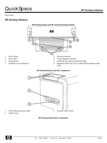
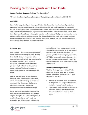
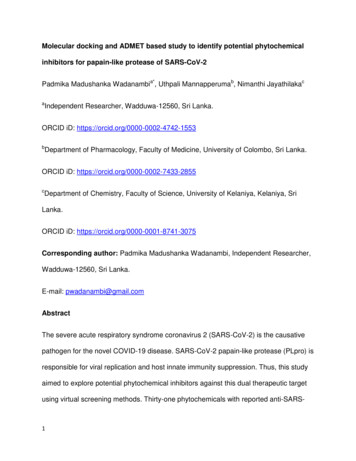
Figure1. 2D chemical structures of phytochemicals and reported inhibitorsSelection of binding site residues in the SARS-CoV-2 PLproThe substrate binding site residues on SARS-CoV-2 PLpro identified based on theregion that lays 5 Å of the binding site of the naphthalene-based inhibitor, 3k, adjacentto the catalytic triad on the ligand bound structure of SARS-CoV-1 PLpro, are shown inFigure 2. The crucial binding site residues (12 residues) comprised of polar amino acids(Tyr264/ Asn267/ Tyr268/ Gln269/ Tyr273/ Thr301), non-polar amino acids (Leu162/Gly163/ Met208/ Pro247/ Pro248) and negatively charged amino acid (Asp164).14
C-terminal(A)N-TerminalFigure 2: (A) Structure of SARS-CoV-2 PLpro representing (B) Zinc binding site, (C)Catalytic triad and (D) S3/S4 binding pockets. Zinc ion is represented in blue sphere.BL2 loop is shown in cyan color.15
Site-specific molecular docking studyOut of thirty three compounds, hirsutenone displayed the lowest binding energy (highestdocking score) of -8.23 kcal/mol with an inhibition constant (Ki) of 920.39 nM.Interestingly, it showed the highest ligand efficiency (LE) value of 0.34 among selectedcompounds. The phytochemical, rubranoside A exhibited the weakest binding energyvalue of -4.09 kcal/mol (Ki of 1.01 mM) with the lowest LE (0.12) among thecompounds. All compounds including phytochemicals and positive controls exhibitedbinding energies ranging from -8.23 kcal/mol to -4.09 kcal/mol with Ki values rangingfrom 920.39 nM to 1.01 mM. The positive controls, 3k and GRL0617 were found to havebinding energy values of -7.89 kcal/mol (Ki of 1.63 μM) and -7.02 kcal/mol (Ki of 7.18μM) respectively. Both hirsutenone and broussoflavan A (-8.13 kcal/mol and Ki of 1.1μM) showed strong affinity in terms of binding energies towards the SARS-CoV-2 PLprobinding pocket compared to the two reported inhibitors used in this study whereasbroussochalcone A (-7.78 kcal/mol and Ki of 1.97 μM), kazinol B (-7.67 kcal/mol and Kiof 2.39 μM), papyriflavonol A(-7.46 kcal/mol and Ki of 3.41 μM), tomentin E (-7.29kcal/mol and Ki of 4.5 μM), tomentin A(-7.22 kcal/mol and Ki of 5.08 μM), corylifol A (7.18 kcal/mol and Ki of 5.41 μM), 4’-O-Methyldiplacone(-7.07 kcal/mol and Ki of 6.56μM) and 3’-O-methyldiplacone (-7.06 kcal/mol and Ki of 6.67 μM) ranked better than theGRL0617. Ten phytochemicals along with the two positive controls were selected basedon their binding energies to gain further insights into intermolecular interactions betweeneach ligand and the protein.16
The molecular docking results of compounds including the binding energy, inhibitionconstant and ligand efficiency are given in Table 2.Table 2: Docking results of phytochemicals and reported inhibitors, used for the studyNo LigandPubChem CID FormulaLBE* 3920.39nM0.342Broussoflavan A44257178C25H30O6-8.131.1μM0.2633k (control ne A6438825C20H20O5-7.781.97μM0.315Kazinol B480869C25H28O4-7.672.39μM0.266Papyriflavonol A10343070C25H26O7-7.463.41μM0.237Tomentin E71659767C26H32O8-7.294.5μM0.218Tomentin A71659627C25H30O7-7.225.08μM0.239Corylifol A25056407C25H26O4-7.185.41μM0.2510 4’-O-methyldiplacone 24854122C26H30O6-7.076.56μM0.2211 3’-O-methyldiplacone 14539951C26H30O6-7.066.67μM0.2212 GRL0617 (control 2)24941262C20H20N2O-7.027.18μM0.3113 Tomentin D71659766C27H34O8-6.958.08μM0.2014 Bavachinin10337211C21H22O4-6.928.53μM0.2815 Kazinol A442414C25H30O4-6.908.72μM0.2416 Tomentin B71659628C26H32O7-6.879.25μM0.2117 8 Mimulone5716903C25H28O5-6.7311.72μM0.2219 820 Tomentin C71659765C27H34O8-6.6313.73μM0.1921 0.20carpin17
22 Kazinol J21637732C26H34O4-6.4917.49μM0.2223 24 Diplacone14539948C25H28O6-6.4120.12μM0.2125 Kazinol F184311C25H32O4-6.1530.85μM0.2126 Isobavachalcone5281255C20H20O4-5.8353.63μM0.2427 0.1828 Rubranol10088141C19H24O5-5.7660.41μM0.2429 Broussochalcone B6450879C20H20O4-5.6473.23μM0.2430 Rubranoside B24011643C24H32O9-5.4798.03μM0.1731 Hirsutanonol9928190C19H22O6-4.92246.38μM0.2032 Oregonin14707658C24H30O10-4.83289.90μM0.1433 Rubranoside A10097263C25H34O10-4.091.01mM0.12chalconeLBE*- Ligand Binding Energy, EIC**- Estimated Inhibition Constant, LE***- Ligand EfficiencyNon-covalent interaction analysis of all ligands with the binding site residues ofSARS-CoV-2 PLproThese compounds were stabilized through a network of interactions. They are hydrogenbond interactions (based on conventional hydrogen bond, carbon-hydrogen bond andsalt bridge), electrostatic interactions (based on pi-anion), hydrophobic interactions(based on pi-sigma, alkyl, pi-alkyl, pi-pi t shaped, amide-pi stacked, pi-pi stacked) andother interactions such as pi-lone pair and pi-sulfur in the docking site. Out of twelvebinding site amino acid residues, excluding Met208, Pro247, Asn267 and Gln269, eightresidues participated in forming both hydrogen bonds and other interaction bond types18
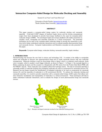
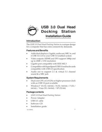
with the thirty-three compounds. The residues, Met208, Pro247 and Asn267 formedother interaction bond types except conventional hydrogen bonds while Gln269 wasinvolved only in conventional hydrogen bonding. Apart from twelve binding siteresidues, eight more amino acids were involved in protein-ligand interactions. Out ofeight residues, Lys157, Glu167, Gly271 and Asp302 were observed to form onlyconventional hydrogen bonds while the remaining Cys111, Ala246 and Gly 266 formonly other interaction bond types. At distances of 4.37 and 4.28 Å, respectively, 3'-Omethyldiplacone and Mimulone formed weak hydrophobic (Alkyl) interactions with oneof the catalytic residues, Cys111. The amino acid residue, Arg166 showed bothconventional hydrogen bond interactions as well as other interaction bond types. Almostall compounds formed hydrogen bonds varying from 1 to 6 with the binding siteexcluding the phytochemical, 4’-O-methylbavachalcone. It was observed that the sameamino acid residue may form multiple bonds through the same or different types ofinteractions with the ligand. In such cases, the residue was counted only once, eventhough there may be several interactions between the residue and the ligand. The mostfrequent hydrogen bonding amino acid residues were Asp164 (11/33), Tyr273 (11/33),Arg166 (10/33), Gly163 (9/33), Tyr268 (6/33) and Asp302 (6/33) while Asp164 (25/33),Leu162 (22/33), Pro248 (20/33), Pro247 (19/33) and Tyr264 (15/33) were morecommon in forming different types of interactions other than conventional hydrogenbonds. The number of ligands that forms conventional hydrogen bond with amino acidresidues is demonstrated in Figure 3a. Figure 3b shows the number of ligands that formdifferent interactions with amino acid residues other than conventional hydrogenbonding. Supplementary figure S1a and S1b show the amino acid contribution towards19
forming each type of interaction with the compounds separately, except conventionalhydrogen bonding. Non-covalent interaction analysis of all compounds is given in Table3 and S1.8(a)Number of u167Arg166Gly163Asp164Leu162Lys1570Amino Acids
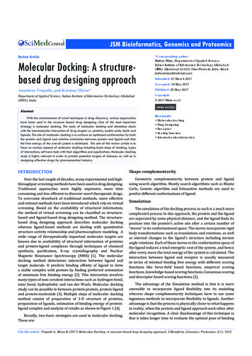
18Number of Ligands161412108Compounds (1-12)6Compounds Amino AcidsFigure 3: Intermolecular interaction analysis between ligands and amino acid residues of S3/S4 bindingpocket. (a) Analysis of conventional hydrogen bond interactions between ligands and amino acidresidues. (b) Analysis of overall interactions (except conventional hydrogen bonds) between ligands andamino acid residues.21
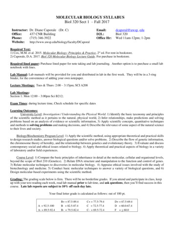
(a)109Number of o AcidsCarbon hydrogen bondElectrostatic (Pi-Anion)Hydrophobic (Pi-Sigma)Hydrophobic (Alkyl)Hydrophobic (Pi-Alkyl)Hydrophobic (Pi-Pi T Shaped)Hydrophobic (Amide-Pi Stacked)Other (Pi-Sulfur)22Hydrophobic (Pi-Pi Stacked)Other (Pi-Lone Pair)
(b)1614Number of 1Met2080Amino AcidsCarbon hydrogen bondElectrostatic (Pi-Anion)Hydrophobic (Pi-Alkyl)Hydrophobic (Pi-Pi T Shaped)Hydrophobic (Pi-Pi Stacked)Other (Pi-Lone Pair)Hydrophobic (Alkyl)Hydrophobic (Amide-Pi Stacked)Other (Pi-Sulfur)Figure S1: Intermolecular interaction analysis between ligands and amino acid residues of S3/S4 bindingpocket. (a) Analysis of different bond types (except conventional hydrogen bonds) between top 12 ligandsand amino acid residues. (b) Analysis of different bond types (except conventional hydrogen bonds)between compounds (13-33) and amino acid residues.Table 3. Non-covalent interaction analysis of best docking complexesDocked gand(A )D: Lys157: HZUNK1:O52.46344Electrostatic (Pi-Anion) – Asp164D: Arg166: HNUNK1:O42.30243Hydrophobic (Pi-Sigma) – Asp164/ (Pi-Alkyl) -
Broussoflavan AReported Inhibitor 1Broussochalcone AKazinol B42*41D: Glu167: OE1UNK1: H1.77988Arg166D: Glu167: OE1UNK1: H2.023Other (Pi-Sulfur) – Met208D: Asp302: OD1UNK1: H1.84894D: Asp302: OD1UNK1: H1.93373D: Tyr268: OUNK1: H2.01975Electrostatic (Pi-Anion) – Asp164/ HydrophobicD: Gly163: OUNK1: H2.6726(Pi-Sigma) – Tyr264/ (Alkyl) – Pro247, Pro248,D: Tyr268: OUNK1: H2.27659Arg166/ (Pi-Alkyl) – Pro247, Pro248D: Pro248: OUNK1: H2.61912D: Tyr268: OS88902: H2.17461Hydrophobic (Pi-Alkyl) – Leu162, Pro247,D: Asp164: OD2S88902: H1.80559Pro248D: Tyr273: HHUNK1: O42.2497Carbon hydrogen bond – Arg166/ ElectrostaticD: Asp302: OD1UNK1: H1.91536(Pi-Anion) – Asp164/ Other (Pi-Sulfur) – Met208D: Asp302: OD1UNK1: H2.13925/ Hydrophobic (Amide-Pi Stacked) – Ala246,D: Gly163: OUNK1: H2.14603Pro247/ (Pi-Alkyl) – Tyr268, Arg166D: Gly163: OUNK1: H2.35927Electrostatic (Pi-Anion) – Asp164/ Hydrophobic(Pi-Pi Stacked) – Tyr264/ (Alkyl) – Pro247,Pro248, Arg166, Met208/ (Pi-Alkyl) – Pro247,Pro248Papyriflavonol ATomentin E2434D: Gln269: OUNK1: H2.00773Electrostatic (Pi-Anion) – Asp164/ HydrophobicD: Gly163: OUNK1: H1.95379(Pi-Sigma) – Leu162/ (Alkyl) – Arg166D: Tyr273: OHUNK1: H2.44521D: Arg166: HHUNK1: O32.01321Electrostatic (Pi-Anion) – Asp164/ HydrophobicD: Asp164: OD2UNK1: H2.0845(Pi-Sigma) – Leu162/ (Alkyl) – Pro247, Pro248
Tomentin ACorylifol A32D: Tyr273: OHUNK1: H1.96507D: Gly163: OUNK1: H2.17545D: Arg166: HEUNK1: O31.84603Electrostatic (Pi-Anion) – Asp164/ HydrophobicD: Asp164: OD2UNK1: H1.95492(Alkyl) – Arg166, Met208D: Gly163: OUNK1: H2.15016D: Thr301: OG1UNK1: H2.28093Carbon hydrogen bond – Pro248, Tyr264,D: Leu162: OUNK1: H2.6581Tyr268/ Hydrophobic (Pi-Pi Stacked) – Tyr264,(Amide-Pi Stacked) – Leu162, Gly163, Asp164/(Alkyl) – Pro247, Pro2484’-O-Methyldiplacone3D: Lys157: HZUNK1: O42.52056Carbon hydrogen bond – Pro248/ ElectrostaticD: Thr301: OG1UNK1: H1.97597(Pi-Anion) – Asp164/ Hydrophobic (Pi-Pi T shapD: Glu167: OE1UNK1: H2.15726-ed) – Tyr264/ (Amide-Pi Stacked) – Gly163,Asp164/ (Alkyl) – Met208, Pro247, Pro2483’-O-Methyldiplacone2D: Gly271: OUNK1: H2.29941Other (Pi-Lone
therapeutic effects in treating a variety of health conditions with no or minimal health risks. Given the sequence and structural similarities between SARS-CoV-1 and SARS-CoV-2 PLpro, we constructed a phytochemical library using phytochemicals with inhibitory activity against SARS CoV-1 PLpro and conducted molecular docking and in