Transcription
REPRODUCTIONRESEARCHFANCD2 is required for the repression of germlinetransposable elementsYan Nie1, Andrew F Wilson1, Tony DeFalco2,3, Amom Ruhikanta Meetei1,3,Satoshi H Namekawa2,3 and Qishen Pang1,31Division of Experimental Hematology and Cancer Biology, Cincinnati, Ohio, USA, 2Division of ReproductiveSciences, Cincinnati Children’s Hospital Medical Center, Cincinnati, Ohio, USA and 3Department of Pediatrics,University of Cincinnati College of Medicine, Cincinnati, OhioCorrespondence should be addressed to Q Pang; Email: Qishen.pang@cchmc.orgAbstractThe Fanconi anemia (FA) DNA damage response (DDR) pathway regulate important cellular processes such as DNA replication, cellcycle control, and DNA damage repair. Here we show that FANCD2, a key member of the FA DDR pathway, interacts with severalimportant components of the germ-cell-specific Prmt5/piRNA pathways that orchestrate the repression of transposable elements(TEs). By using the Pou5f1-eGFP reporter mice, which marks pure populations of primordial germ cells (PGCs), we demonstrate thatFA deficiency results in de-repression of TEs, depletion of PGCs, and defective spermatogenesis and oogenesis. Fancd2-KO PGCsexhibited excessive DNA damage and exacerbated apoptosis. Mechanistically, we observed a significant reduction of PRMT5catalyzed H2A/H4R3me2s marks on the LINE1 TEs in E10.5 PGCs of Fancd2-KO; Pou5f1-eGFP and Fanca-KO;Pou5f1-eGFP embryos.Furthermore, we utilized the Fancd2-KI model to show that FANCD2 and PRMT5 co-occupied the promoter of LINE1 in WT PGCsand that this co-occupancy was lost in FA-deficient (Fanca-KO) PGCs. These results suggest that the FA pathway takes part in TErepression in early PGCs, likely through a mechanism involving FANCD2-facilitated, PRMT5-catalyzed repressive H2A/H4R3me2smarks on TEs.Reproduction (2020) 159 659–668IntroductionFanconi anemia (FA) is a genetic disease associatedwith bone marrow failure, increased cancer, and severegermline defects, and the FA pathway is known toplay a major role in the DNA damage response (DDR)network (Bagby 2003, 2018, Tischkowitz & Hodgson2003). FA is genetically heterogeneous, with at least 22complementation groups (FANCA-FANCW) identifiedthus far (Dong et al. 2015, Sawyer et al. 2015, Knieset al. 2017). Eight of the FA proteins (FANCA, B, C, E, F,G, L, and M) form the FA core complex that functions asan ubiquitin ligase. In response to DNA damage or DNAreplication stress, the FA core complex monoubiquitinatestwo downstream FA proteins, FANCD2 and FANCI,which then recruit the downstream FA proteins andother DNA repair factors, to nuclear loci containingdamaged DNA and consequently influence importantcellular processes such as DNA replication, cell-cyclecontrol, and DNA damage response and repair (Deans& West 2011, Kottemann & Smogorzewska 2013).DDR comprises an elaborate network of signalingpathways that enable cells to execute biologicalresponses to genotoxic stress (Jackson & Bartek 2009).Defects in DDR pathways have been shown to be 2020 Society for Reproduction and FertilityISSN 1470–1626 (paper) 1741–7899 (online) associated with infertility, gamete aneuploidy, anddiseased risk in offspring . As a major DDR pathway,FA has been subjected to extensive study for its role ingermline genomic maintenance. In addition to genomeinstability and cancer susceptibility, FA deficiency inmice has been shown to compromise male and femalefertility. Specifically, deletion of the genes for the murineFA proteins Fanca, Fancb, Fancc, Fancl, Fancm, andFancp causes primordial germ cell (PGC) depletion,meiotic defects, and ultimately hypogonadism andinfertility (Nadler & Braun 2000, Agoulnik et al. 2002,Wong et al. 2003, Crossan et al. 2011, Kato et al. 2015,Alavattam et al. 2016). More recently, it was shownthat hypogonadism of Fancm mutant mice was a resultof reduced proliferation of PGCs, likely resulting fromelevated DNA damage (Luo et al. 2014). These studieshighlight the importance of the FA DDR pathway ingerm cell development and function. A recent studydemonstrated that the FA pathway is critical for DNAcrosslinking repair to safeguard genomic stability ofPGCs (Hill & Crossan 2019). However, the mechanismby which the FA pathway functions in genomemaintenance in the germline, particularly in PGCs,remains largely e version via https://rep.bioscientifica.comDownloaded from Bioscientifica.com at 08/12/2022 12:01:05PMvia free access
660Y Nie and othersTransposable elements (TEs) induce DNA doublestrand breaks (DSBs), insertional mutations, andchromosome rearrangements, leading to genomicinstability (Goodier & Kazazian 2008). This geneticconsequence is particularly dangerous in germ cells, asgenome changes induced by TEs will be transmitted tothe next generation. Thus, TE silencing is considered anessential mechanism for defending germline integrity andfertility. There are two key mechanisms for the repressionof germline TEs: the H2A/H4R3me2s modificationcatalyzed by the protein arginine methyltransferase 5(PRMT5) in early (E8.5–E11.5) PGCs and the de novoDNA methylation orchestrated by the Piwi-interactingsmall RNA (piRNA) pathway initiating at approximatelyE14.5 (Aravin et al. 2008, Kim et al. 2014). Consequently,mice deficient in genes encoding Prmt5 or factors in thepiRNA pathway exhibit overexpression of TEs, loss ofgerm cells, and ultimately male and/or female sterility(Saito & Siomi 2010, Siomi et al. 2011, Kim et al. 2014,Weick & Miska 2014, Berrens & Reik 2015). Despiteextensive investigation, the mechanisms by which germcells control TE activity are largely not known and, inparticular, the DDR pathways functioning in this criticalprocess have not been identified.In the current study, we show that FANCD2, a keymember of the FA DDR pathway, interacts with severalimportant components of the PRMT5/piRNA pathwaysessential for TE silencing in primordial germ cells (PGCs).Loss of FANCD2 results in massive de-repression ofTEs, depletion of PGCs, defective spermatogenesis andoogenesis, and ultimately infertility. Early FA-deficientPGCs show reduced H2A/H4R3me2s marks on LINE1transposons. Further, we reveal the co-occupancy ofFANCD2 and PRMT5 at the promoter of LINE1 TEs inearly PGCs.reporter mice were dissected, and single PGCs from individualgenotyped embryos were sorted by GFP expression or bySSEA1-APC (R&D Systems; Cat.# FAB2155A) positive cellsusing a MoFlo XDP high speed flow sorter (Beckman Coulter).For PGC culture, STO feeder cells (American Type CultureCollection (ATCC), Cat.# CRL-1503) were seeded in a 96-wellplate coated with 1% gelatin (Sigma-Aldrich, Cat.# G1393) andcultured in DMEM (Gibco, Cat. # 11-960-044) supplementedwith 10% FBS and 5% P/S. Upon confluency, STO cells wereincubated with mitomycin C (Sigma-Aldrich, Cat.# M4287) atthe concentration of 10 µg/mL for 4 h. Then the cells werewashed three times with 10 mL 1 PBS. The sorted PGCswere plated onto the STO feeder layer in PGC culture medium(DMEM supplemented with 1% P/S, 10% FBS, 100 ng/mL SCF(Peprotech, Cat.# 250-03), 25 nM bFGF (Peprotech, Cat.# 10018B), and 25 nM LiF (Peprotech, Cat.# 250-02)).Proliferation assayA cell suspension containing GFP-positive PGCs obtained fromE10.5 embryos of WT and Fancd2-/- females expressing thePou5f1-eGFP reporter gene was prepared as described. PGCswere subjected to pulse labeling with Bromodeoxyuridine(BrdU; final concentration 10 µM) for 60 min. We used thereagents from the APC-BrdU Flow Kit (BD Biosciences,Cat.#552598), according to the manufacturer’s instructions.Analysis of apoptosisCell apoptosis was measured by Annexin V (BD Pharmingen,Cat. # 550474) and DAPI (BD Pharmingen, Cat. # 564907)staining using flow cytometry, according to the manufacturer’sinstructions.Quantitative PCR analysis of TE expressionFancd2-KI mice were generated in our laboratory (Zhang et al.2017). Fancd2 /- and Fanca /- mice were provided by Dr MarkusGrompe (Oregon Health & Sciences University) (Houghtalinget al. 2003) and Dr Madeleine Carreau (Laval University) (Wonget al. 2003), respectively. The Fancd2-KI;Pou5f1-eGFP, FancaKO;Pou5f1-eGFP, or Fancd2-KO; Pou5f1-eGFP mice weregenerated by crossing the Fancd2-KI, Fanca-KO, or Fancd2-KOmice to the Pou5f1-eGFP reporter mice (B6;CBA-Tg(Pou5f1EGFP)2Mnn/J; JAX Stock #:004654), respectively. Mice weremaintained on C57BL/6J background in the animal barrier facilityat Cincinnati Children’s Hospital Medical Center. Animals werekept in accordance with the protocol approved by the CCHMCInstitutional Animal Care and Use Committee.Total RNA from FACS-sorted (GFP SSEA1 ) PGCsisolated from E11.5 embryos of WT, Fanca-/-, and Fancd2/- females expressing the Pou5f1-eGFP reporter gene wasprepared using the RNeasy kit (Qiagen) following themanufacturer’s protocol. RT was performed with randomhexamers and Superscript II RT (Invitrogen) and was carriedout at 42 C for 60 min and stopped at 95 C for 5 min.First-strand cDNA was used for qPCR using the followingprimers: L15UTR-F 5′-GGCGAAAGGCAAACGTAAGA-3′,L15UTR-R 5′-GGAGTGCTGCGTTCTGATGA-3; mpleswerenormalized to the level of WT controls, and relative expressionlevels were determined by the standard curve method.Isolation and culture of primordial germ cells (PGCs)Preparation of protein extracts and immunoblottingFor isolation of PGCs, genital ridges of E10.5, E11.5, or E13.5embryos from timed crosses of FA mice and Pou5f1-eGFPTo prepare protein lysates, testes from 8- to 10-week-old miceor in vitro expanded PGCs isolated from E13.5 embryos wereMaterials and methodsMiceReproduction (2020) 159 659–668 https://rep.bioscientifica.comDownloaded from Bioscientifica.com at 08/12/2022 12:01:05PMvia free access
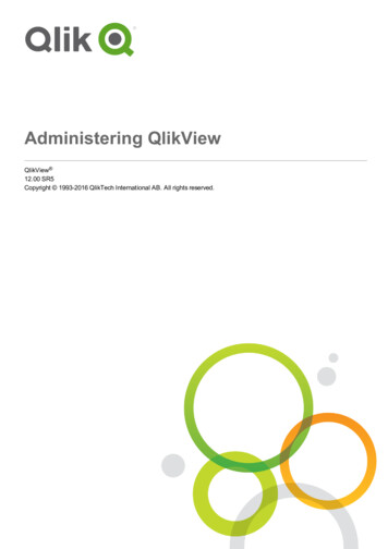
Fancd2 in germline TE silencinglysed using a homogenizer on ice in three volumes of ice-chilledLysis buffer with Roche Complete proteinase inhibitor (Roche,Cat. # 04693116001). Lysates were cleared by centrifugation(40,000 g, 20 min, at 4 C). Protein concentration was quantifiedby using Bio-Rad reagent and resolved on SDS-PAGE andtransferred onto nitrocellulose membranes. Immunoblots werethen incubated with primary antibodies specific for MILI (CellSignaling, Cat. #2071), PRMT5 (Sigma-Aldrich, Cat. # 07-405),MAEL (Santa Cruz, Cat. # sc-398925), TDRD1 (R&D Systems,Cat. # AF320), TDRD6 (Sigma-Aldrich, Cat. # ABE231), TDRD8/STK31 (Abcam, Cat. # ab155172), FLAG (Sigma-Aldrich, Cat. #F3165), and β-actin (Sigma-Aldrich, Cat. # A5441).μChIP analysisSmall-scale ChIP (μChIP) experiments were conducted asdescribed previously (Dahl & Collas 2008). Briefly, FACSsorted (GFP SSEA1 ) PGCs isolated from E10.5 embryosof WT, Fanca-KO, or Fancd2-KO females expressing bothFancd2-KI transgene and the Pou5f1-eGFP reporter werecrosslinked with 0.5% paraformaldehyde, lysed in radioimmunoprecipitation assay (RIPA) buffer containing proteaseinhibitors and sonicated to achieve a mean DNA fragmentsize of around 200–400 base pairs. After centrifugation, thechromatin extracts were then precleared with Dynal MagneticBeads (Invitrogen) (4 C, 1 h) followed by centrifugation(425 g, 30 min). Supernatant (precleared chromatin) wasimmunoprecipitated overnight with Dynal Magnetic Beadscoupled with anti-H4R3me2s (1 µg per ChIP, ab5823, Abcam),anti-FLAG (Sigma-Aldrich, Cat. # F3165), anti-Prmt5 (SigmaAldrich, Cat. # 07-405), or normal rabbit serum. After reversecrosslinking (with protease K at 42 C for 2 h and 68 C for 6h), DNA was purified (phenol-chloroform extraction) and usedfor qPCR analysis. Primers used were as follows: LINE1 5UTRForward 5′-GGCGAAAGGCAAACGTAAGA-3′, LINE1 5UTRReverse 5′-GGAGTGCTGCGTTCTGATGA-3′.Comet assayComet assay was performed using the Trevigen’s Comet AssayKit, following the manufacturer’s instructions. In brief, the661PGCs suspension was mixed with molten LMAgarose at aratio of 1:10 (v/v) and transferred onto slides. After incubationin the lysis buffer and alkaline treatment in electrophoresisbuffer, electrophoresis (20 min, 25 V, and approximately 300mA) and neutralization were performed. Subsequently, slideswere dehydrated with ethanol. For evaluation, the slides werestained with 100 μL of diluted SYBR Gold (1:10,000 in TEbuffer, Invitrogen) and the comets were evaluated by imageanalysis using the software comet IV version 4.2 (PerceptiveInstruments, Haverhill, UK). The percentage of tail DNA (%tail DNA) for each comet was calculated. The experimentswere performed three times (duplicates, 50 cells per technicalreplicate).Statistical analysisStudent’s t-test was performed using GraphPad Prism v6(GrapPad software). Comparison of more than two groups wasanalyzed by one-way ANOVA test. Values of P 0.05 wereconsidered statistically significant. Results are presented asmean s.d. * indicates P 0.05; **, P 0.01; ***, P 0.001.ResultsFancd2 interacts with germline-specificPrmt5/piRNA factorsWe recently reported that by using a Fancd2 knock-inmouse model and proteomic approach, we unveileda FANCD2 in vivo interaction network (Zhang et al.2017). Significantly, analysis of FANCD2-containingcomplexes from four different tissues (ES cells, E11.5embryos, testes, and spleen) revealed distinct patternsof FANCD2-associated proteins (Fig. 1A), suggestingthe presence of developmental and tissue-specificdifferences in the repertoire of FANCD2 interactors.Specifically, in testes, we observed several proteins ofthe germ-cell-specific PRMT5/piRNA pathways, whichorchestrate the repression of transposable elements (TEs)(Saito & Siomi 2010, Siomi et al. 2011, Kim et al. 2014,Weick & Miska 2014, Berrens & Reik 2015). We validatedFigure 1 Fancd2 interacts with germline-specific PRMT5/piRNA factors. (A) List of tissue specific FANCD2-associated proteins. The testis-specificPrmt5/piRNA factors are marked by bold. (B) Validation of the testis-specific PRMT5/piRNA factors by anti-FLAG immunoprecipitation (IP) fromtestis lysates and detected by antibodies against the indicated proteins. (C) Western analysis of the indicated PRMT5/piRNA factors in the testesof WT, Fancd2-KI, and Fancd2-KO mice. https://rep.bioscientifica.comReproduction (2020) 159 659–668Downloaded from Bioscientifica.com at 08/12/2022 12:01:05PMvia free access
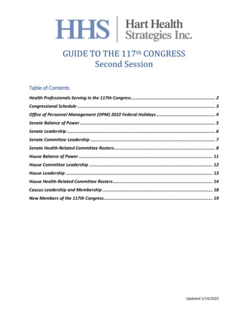
662Y Nie and othersthe interaction between the FLAG-tagged FANCD2 andthese PRMT5/piRNA factors in Fancd2-KI mice (Fig.1B). We also addressed whether the loss of FANCD2affected these PRMT5/piRNA factors. Western analysisfor MAEL and TDRD8 showed that these proteins werestill present in the Fancd2-KO testes (Fig. 1C). However,the levels of PRMT5, MILI, TDRD1, and TDRD6 werereduced in the Fancd2-KO testes compared to those inWT and Fancd2-KI mice (Fig. 1C). These results indicatethat FANCD2 interacts with germline-specific PRMT5/piRNA factors and suggest that FANCD2 may play a rolein the regulation of germline TEs.Loss of Fancd2 leads to defective spermatogenesis andoogenesisThe observation that FANCD2 interacts with PRMT5/piRNA factors prompted us to determine whetherFANCD2 plays a role in germline development.Consistent with previous report (Houghtaling et al. 2003),we found that Fancd2-KO mice were smaller than WTor Fancd2-KI mice at the embryonic stage (d.p.c 13.5)(Fig. 2A). Second, the size of testes and ovary in 8-weekold-adult Fancd2-KO mice were significantly reduced incomparison to age-matched WT or and Fancd2-KI mice(Fig. 2B). Third, H&E stained testis and ovary of 8-weekold WT, Fancd2-KO, and Fancd2-KI mice show severelydefective spermatogenesis and oogenesis in Fancd2-KOmice, as compared to WT and Fancd2-KI mice (Fig.2C). Consequently, loss of Fancd2 results in completesterility in both males and females (Zhang et al. 2017and data not shown). Thus, these data confirm the pastfinding and suggest that Fancd2 is required for germlinemaintenance.Loss of Fancd2 results in depletion of PGCs duringembryonic developmentIt has been reported that deletion of the mouse FA genesFanca, Fancb, Fancc, Fancl, Fancm, and Fancp causesprimordial germ cell (PGC) depletion, hypogonadism,and ultimately infertility (Nadler & Braun 2000,Agoulnik et al. 2002, Wong et al. 2003, Crossan et al.2011, Kato et al. 2015). In addition, it has been shownthat hypogonadism in Fancm mutant mice is a resultof reduced proliferation of PGCs (Luo et al. 2014).Therefore, we asked whether Fancd2 deficiencywould affect PGC maintenance during embryonicFigure 2 Loss of Fancd2 leads to hypogonadism. (A) Images of embryos (left) of the indicated genotypes at E13.5. Genotypes (right) of theembryos were determined by PCR. (B) Representative photo of the testes and ovary of WT and Fancd2-KO mice. (C) Gonadal defects inFancd2-/- mice. Shown are H&E stained sections of testes and ovaries from 8-week-old WT and Fancd2-/- mice (200 magnification).Reproduction (2020) 159 659–668 https://rep.bioscientifica.comDownloaded from Bioscientifica.com at 08/12/2022 12:01:05PMvia free access
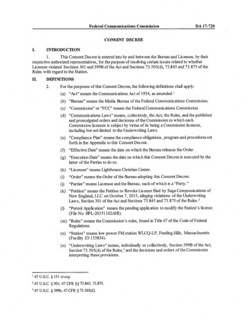
Fancd2 in germline TE silencingdevelopment. To facilitate the purification and analysisof PGCs by flow cytometry, we crossed Fancd2 /- miceto Pou5f1 (Oct4)-eGFP (Pou5f1-eGFP hereafter) reportermice, which have been used to mark and isolate purepopulations of PGCs by FACS (Szabo et al. 2002). Usinganother PGC marker, SSEA1 (Hayashi et al. 2011), weobserved a significant reduction of GFP SSEA1 PGCsin Fancd2-KO mice compared to WT controls at bothE11.5 (WT 384 76 vs Fancd2-KO 147 49, n 5) andE13.5 (WT 634 61 vs Fancd2-KO 283 44, n 6) (Fig.3A and B). These results indicate that loss of Fancd2leads to PGC depletion.Reduced proliferation and increased apoptosis inFancd2-KO PGCsSince we observed a significant reduction of PGCs inFancd2-KO embryos, we sought to determine the effectof Fancd2 deficiency on the proliferation and survivalof mouse PGCs. To this end, GFP PGCs from E11.5embryos were sorted by FACS and cultured on STO feedercells for 15 days (Fig. 4A). We found that Fancd2-KOPGCs proliferated significantly slower than WT PGCsFigure 3 Loss of Fancd2 leads to PGC depletion. (A) Representativedot plots of flow cytometric analysis of GFP SSEA1 PGCs fromE11.5 embryos of WT and Fancd2-/- females expressing thePou5f1-eGFP reporter gene. (B) The number of PGCs at E11.5 andE13.5 embryos of WT and Fancd2-/- females expressing thePou5f1-eGFP reporter gene was determined by flow cytometry forGFP SSEA1 PGCs. Results are presented as mean s.d. of threeindependent experiments. *P 0.05, **P 0.01. https://rep.bioscientifica.com663in stroma-supported culture (Fig. 4B), suggesting thatFancd2 is essential for proliferation of PGCs. Consistentwith these observations, the number of BrdU-positivecells was reduced in Fancd2-KO PGCs (16.2 4.1%,n 5) compared to WT controls (27.8 6.4%, n 5) (Fig.4C), indicating that FANCD2 is required for proliferationof mouse PGCs. To determine whether Fancd2 isdirectly involved in the cell survival of mouse PGCs, weanalyzed apoptosis using Annexin V staining followedby flow cytometry. Notably, Annexin V-positive cellswere up-regulated in Fancd2-KO PGCs (5.40 0.38%,n 5) compared to WT controls (1.73 0.62%, n 5)(Fig. 4D). These data suggest that Fancd2 plays a rolein regulating proliferation and survival of mouse PGCs.FA deficiency leads to de-repression of germline TEsOur proteomic analyses suggested that the FA pathwaymay play a TE silencing function in vivo during germcell development. For this reason, we decided to furtherstudy the link between the FA pathway and germ cellspecific TE silencing machinery. To determine if theloss of Fancd2 results in TE de-repression in PGCs, weisolated GFP SSEA1 PGCs from WT;Pou5f1-eGFP andFancd2-KO;Pou5f1-eGFP mice and determined theexpression of two TEs, LINE1 (long interspersed nuclearelements) and IAP (intracisternal A particle), whoseunrestrained expression has been linked to infertility(Hayashi et al. 2011), by quantitative qPCR. We observeda four- to seven-fold increase in LINE1 and a three- tofive-fold increase in IAP expression in E11.5 Fancd2-KOPGCs compared to WT controls (Fig. 5A). Thus, Fancd2is essential for transcriptional or posttranscriptionalsilencing of two TEs known to cause infertility in mice.To determine whether the depression of LINE1 and IAPTEs in PGCs required the FA core complex, we generatedWT; Pou5f1-eGFP and Fanca-/-;Pou5f1-eGFP mice andanalyzed the levels of LINE1 and IAP TEs in E11.5 PGCs.Consistent with the observation in Fancd2-KO PGCs,Fanca deficiency also resulted in de-repression of LINE1and IAP TEs in PGCs (Fig. 5B). These results indicate thatthe FANCD2/FA pathway is essential for TE repressionduring early germline development.PRMT5-catalyzed repressive H2A/H4R3me2s isessential for TE silencing in PGCs between E8.5–E11.5(Kim et al. 2014), when these actively dividing PGCsare undergoing global DNA demethylation (Hajkovaet al. 2002, Seisenberger et al. 2012). To investigatethe mechanistic link between the FANCD2/FA pathwayand PRMT5-directed TE silencing in early PGCs, wegenerated three mouse models: (1.) Fancd2-KO;Pou5f1eGFP for the effect of Fancd2 deficiency on PRMT5mediated H2A/H4R3me2s repressive chromatinmodifications on LINE1 TEs; (2.) Fanca-KO;Pou5f1eGFP for the requirement of the FA core complex forthese PRMT5-mediated H2A/H4R3me2s marks onLINE1; and (3.) Fancd2-KI;Pou5f1-eGFP in a WT orReproduction (2020) 159 659–668Downloaded from Bioscientifica.com at 08/12/2022 12:01:05PMvia free access
664Y Nie and othersFigure 4 Reduced proliferation and increased apoptosis in Fancd2-KO PGCs. (A) Reduced proliferation in Fancd2-KO PGCs. GFP PGCs fromE11.5 embryos of WT and Fancd2-/- females expressing the Pou5f1-eGFP reporter gene were sorted by FACS and cultured on STO feeder cellsfor 15 days. Images were taken at the indicated time points. (B) Cumulative growth curve of PGCs during PGC culture initiation. GFP PGCsfrom E11.5 embryos of WT and Fancd2-/- females expressing the Pou5f1-eGFP reporter gene were sorted by FACS and cultured on STO feedercells for the indicated days. Values indicate mean s.d. of three independent experiments. *P 0.05, **P 0.01. (C) Diminished BrdUincorporation in Fancd2-KO PGCs. GFP PGCs from E11.5 embryos of WT and Fancd2-/- females expressing the Pou5f1-eGFP reporter genewere sorted by FACS and subjected to pulse labeling with BrdU followed by flow cytometric analysis for BrdU-positive cells. Values indicatemean s.d. of three independent experiments. *P 0.05. (D) Increased apoptosis in Fancd2-KO PGCs. GFP PGCs from E11.5 embryos of WTand Fancd2-/- females expressing the Pou5f1-eGFP reporter gene were subjected to flow cytometric analysis for Annexin V-positive, DAPInegative apoptotic cells. Results are presented as mean s.d. of three independent experiments. **P 0.01.Fanca-KO background for interaction between FANCD2and PRMT5 on sites of H2A/H4R3me2s marks in LINE1chromatin. We chose E10.5 PGCs to investigate theFANCD2-PRMT5 interaction in TE silencing becausePRMT5-mediated H2A/H4R3me2s marks on LINE1 areenriched at E10.5–E11.5, which are responsible for TErepression during this specific stage of developmentwhen PGCs undergo global demethylation of DNA(Hajkova et al. 2002, Seisenberger et al. 2012). SinceLINE1 TEs are silenced via 5′-UTR promoter methylation(Smith et al. 2012), we compared repressive H2A/H4R3me2s marks on the LINE1 5′-UTR promoter inE10.5 PGCs from WT;Pou5f1-eGFP, Fancd2-KO;Pou5f1eGFP, and Fanca-KO; Pou5f1-eGFP embryos by microchromatin immunoprecipitation (µChIP) assays (Dahl& Collas 2008). We observed that, at E10.5, the LINE1promoter was strongly enriched with H2A/H4R3me2smarks, indicative of a PRMT5- repressed chromatinReproduction (2020) 159 659–668 state, in WT; Pou5f1-eGFP PGCs (Fig. 5C). In contrast,these PRMT5-mediated H2A/H4R3me2s marks weresignificantly decreased in Fancd2-KO; Pou5f1-eGFP andFanca-KO;Pou5f1-eGFP PGCs (Fig. 5C), consistent withLINE1 transcriptional activation.Next, we examined the recruitment of FANCD2 andPRMT5 to the promoter of LINE1 in E10.5 PGCs fromWT (Fanca / ;Fancd2-KI) and FA-deficient (Fanca-/;Fancd2-KI) embryos by µChIP assays using agaroseconjugated antibodies against FLAG (tagged FANCD2)and PRMT5 coupled with protein A magnetic beads.We found that FANCD2 and PRMT5 co-occupiedthe promoter of LINE1 in WT PGCs and that thisco-occupancy was lost in Fanca-/-;Fancd2-KI PGCs (Fig.5D). Notably, Fanca-KO caused not only complete loss ofFLAG-FANCD2 occupancy but also significant reductionof PRMT5 recruitment to the LINE1 promoter, indicatinga pattern of FANCD2-dependent PRMT5 occupancy.https://rep.bioscientifica.comDownloaded from Bioscientifica.com at 08/12/2022 12:01:05PMvia free access
Fancd2 in germline TE silencing665Figure 5 FA deficiency leads to de-repression of germline TEs. (A) Fancd2 is essential for TE repression in PGCs. qPCR analysis of TEs inFACS-sorted GFP SSEA1 PGCs from E11.5 embryos of WT and Fancd2-/- females expressing the Pou5f1-eGFP reporter gene, using the primersets described in Materials and methods. Results are presented as mean s.d. of three independent experiments. *P 0.05, **P 0.01. (B)De-repression of germline TEs in Fanca-deficient PGCs. qPCR analysis of TEs in FACS-sorted GFP SSEA1 PGCs from E11.5 embryos of WTand Fanca-/- females expressing the Pou5f1-eGFP reporter gene, using the primer sets described in Materials and methods. Results are presentedas mean s.d. of three independent experiments. *P 0.05, **P 0.01. (C) µChIP analysis for H2A/H4R3me2s at the promoter region of LINE1TEs in FACS-sorted GFP SSEA1 PGCs from E10.5 embryos of WT, Fanca-/-, and Fancd2-/- females expressing the Pou5f1-eGFP reporter gene.Percentages of anti-H2A/H4R3me2s immunoprecipitated DNA compared to input are shown. w/o, without primary antibodies. Results arepresented as mean s.d. of three independent experiments. *P 0.05. (D) µChIP analysis for Fancd2 (FLAG) and Prmt5 at the promoter region ofLINE1 TEs in FACS-sorted GFP SSEA1 PGCs from E10.5 embryos of WT, Fanca-/-, and Fancd2-/- females expressing the Pou5f1-eGFP reportergene and the Fancd2KI/KI transgene. Percentages of anti-FLAG and anti-Prmt5 immunoprecipitated DNA compared to input are shown. w/o,without primary antibodies. Results are presented as mean s.d. of three independent experiments. *P 0.05. (E) Western analysis of the Prmt5and Fancd2 proteins in the in vitro expanded PGCs from E13.5 WT (WT;Fancd2-KI) and Fanca-deficient (Fanca-/-;Fancd2-KI) embryos, usingantibodies against the indicated proteins. (E) Western analysis of the Prmt5 and Fancd2 proteins in the E13.5 PGCs from WT (WT;Fancd2-KI)and Fanca-deficient (Fanca-/-;Fancd2-KI) mice, using antibodies against the indicated proteins. (F) Increased accumulation of DNA damage inFancd2-deficient PGCs. Genomic DNA was stained with SYBR Gold and imaged after single cell electrophoresis (left). Quantification for thepercent of DNA in the comet tail is shown (right). Results are presented as mean s.d. of three independent experiments. *P 0.05.Together, these results suggest that FANCD2 recruitsPRMT5 to the LINE1 promoter to support TE repression.The requirement of the FA core component FANCAfor repression of LINE1 and IAP TEs, the establishmentof the PRMT5-mediated H2A/H4R3me2s marks, andthe recruitment of FANCD2 and PRMT5 to the LINE1promoter in PGCs prompted us to analyze the levelsof PRMT5 and FANCD2 proteins in in vitro cultureexpanded PGCs from WT (WT;Fancd2-KI) and Fancadeficient (Fanca-/-;Fancd2-KI) mice. We did not observesignificant alteration in the protein levels of either PRMT5or FANCD2 in Fanca-/-;Fancd2-KI mice compared to theWT;Fancd2-KI controls (Fig. 5E). These results indicatethat, while FANCA is not required for the stability ofthe FANCD2 and PRMT5 proteins, a functional FAcore complex is essential for FANCD2-dependent andPRMT5-mediated germline TE repression. https://rep.bioscientifica.comWe also investigated whether the observed increasein the expression of TEs could lead to an increase inthe accumulation of DNA damage in Fancd2-deficientPGCs. To this end, we performed the Comet assay andrevealed that the Fancd2 / PGCs exhibited significantlymore DNA damage (% comet tail DNA) (Fig. 5F) thantheir wild‐type counterparts. This result demonstratesexcessive DNA damage in the Fancd2 / PGCs andsuggests that de-repression of germline TEs could causeapoptosis of germ cells resulting from DNA damage.DiscussionThe FA DDR pathway plays critical roles in themaintenance of germline integrity; however, theunderlying mechanisms are largely unknown. In thisstudy, we show that (1.) FANCD2 interacts with severalReproduction (2020) 159 659–668Downloaded from Bioscientifica.com at 08/12/2022 12:01:05PMvia free access
666Y Nie and othersimportant PRMT5/piRNA factors essential for germline TEsilencing; (2.) loss of FANCD2 results in de-repression ofTEs, depletion of PGCs, defective spermatogenesis andoogenesis, and infertility; (3.) FA deficiency (Fancd2-KOand Fanca-KO) causes a significant reduction of PRMT5catalyzed H2A/H4R3me2s marks on LINE1 TEs inearly PGCs; and (4.) FANCD2 and PRMT5 co-occupythe promoter of LINE1 in PGCs, which is lost inFA-deficient (Fanca-KO) PGCs. These results indicatea mechanistic link between the FA DDR pathway andgermline maintenance, likely through the regulation ofTE expression in early PGCs.De-regulated TE expression causes germ cell lossleading to infertility and induces germline genomicinstability compromising the viability and healthof offspring. Despite intensive investigation, themechanisms by which germ cells control TE activity arelargely not known and the DDR and repair pathway(s)functioning in this critical process have not beenidentified. The conventional model suggests that, asa major DDR pathway involved in germl
using a MoFlo XDP high speed flow sorter (Beckman Coulter). For PGC culture, STO feeder cells (American Type Culture Collection (ATCC), Cat.# CRL-1503) were seeded in a 96-well plate coated with 1% gelatin (Sigma-Aldrich, Cat.# G1393) and cultured in DMEM (Gibco, Cat. # 11-960-044) supplemented with 10% FBS and 5% P/S. Upon confluency, STO .