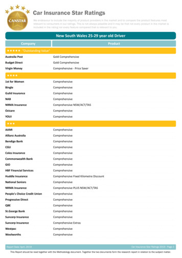
Transcription
Chiu et al. Immunity & Ageing(2018) ARCHOpen AccessA comprehensive characterization ofaggravated aging-related changes in Tlymphocytes and monocytes in end-stagerenal disease: the iESRD studyYen-Ling Chiu1,2,3, Kai-Hsiang Shu1,4, Feng-Jung Yang2,5, Tzu-Ying Chou1, Ping-Min Chen1, Fang-Yun Lay1,Szu-Yu Pan1, Cheng-Jui Lin6, Nicolle H R Litjens7, Michiel G H Betjes7, Selma Bermudez8, Kung-Chi Kao4,Jean-San Chia4, George Wang9, Yu-Sen Peng1 and Yi-Fang Chuang8,10,11*AbstractBackground: Patients with end-stage renal disease (ESRD) exhibit a premature aging phenotype of the immunesystem. Nevertheless, the etiology and impact of these changes in ESRD patients remain unknown.Results: Compared to healthy individuals, ESRD patients exhibit accelerated immunosenescence in both T cell andmonocyte compartments, characterized by a dramatic reduction in naïve CD4 and CD8 T cell numbers butincrease in CD8 TEMRA cell and proinflammatory monocyte numbers. Notably, within ESRD patients, aging-relatedimmune changes positively correlated not only with increasing age but also with longer dialysis vintage. Inmultivariable-adjusted logistic regression models, the combination of high terminally differentiated CD8 T celllevel and high intermediate monocyte level, as a composite predictive immunophenotype, was independentlyassociated with prevalent coronary artery disease as well as cardiovascular disease, after adjustment for age, sex,systemic inflammation and presence of diabetes. Levels of terminally differentiated CD8 T cells also positivelycorrelated with the level of uremic toxin p-cresyl sulfate.Conclusions: Aging-associated adaptive and innate immune changes are aggravated in ESRD and are associatedwith cardiovascular diseases. For the first time, our study demonstrates the potential link betweenimmunosenescence in ESRD and duration of exposure to the uremic milieu.Keywords: Immunosenescence, Aging, CVD, Inflammation, ESRDBackgroundPatients with end-stage renal disease (ESRD) exhibitmany physiological changes reminiscent of acceleratedaging processes and have an increased mortality and susceptibility to diseases when compared to chronologicalage-matched individuals [1]. Impaired physical functions,muscle wasting, cognitive function decline, acceleratedvascular disease and increased risks of death are amongthe many aging-related complications increased in* Correspondence: chuangy@ym.edu.tw8International Health Program, National Yang Ming University School ofPublic Health, Taipei, Taiwan10Institute of Public Health, National Yang Ming University School of PublicHealth, Taipei, TaiwanFull list of author information is available at the end of the articlefrequency in ESRD [2]. The immune system of ESRD patients also exhibits significant changes from that of healthyindividuals. For example, while a low grade-inflammationcan be observed during normal aging [3], it is significantlyenhanced by uremia [4]. Accompanying low-grade inflammation, immune cells develop different phenotypicmarkers and functions during normal aging. Thesechanges are are collectively called “immunosenescence”and are considered to contribute to various aging-relatedmorbidities, including increased risks for infectious eventsand cardiovascular diseases [5–7].During normal aging, lymphocytes and monocytes experience dramatic changes. The subset distribution inthe CD8 T cell compartment is different between young The Author(s). 2018 Open Access This article is distributed under the terms of the Creative Commons Attribution 4.0International License (http://creativecommons.org/licenses/by/4.0/), which permits unrestricted use, distribution, andreproduction in any medium, provided you give appropriate credit to the original author(s) and the source, provide a link tothe Creative Commons license, and indicate if changes were made. The Creative Commons Public Domain Dedication o/1.0/) applies to the data made available in this article, unless otherwise stated.
Chiu et al. Immunity & Ageing(2018) 15:27and old people; with progressive terminal differentiation [8],loss of co-stimulatory molecules, shortening of telomeresand impaired response toward infectious pathogens and vaccinations [9, 10] occur during aging. CD4 T cells also exhibit aging-related changes. For example, naïve CD4 T cellsfrom aged animals show reduced IL-2 production, proliferation, helper function, effector generation and memory function [11]. Premature aging of the T cell compartment hasbeen observed in ESRD patients, characterized by decreasedthymic output of naïve cells and increased susceptibility toward apoptosis [12]. We had previously reported that higherlevels of CD4 CD28- cells can be found in ESRD patients[13] and CD4 T cells activation is affected in ESRD patientsin an age-dependent manner [14]. Recently, it has been reported that elderly kidney transplant patients also exhibitmore advanced T cell differentiation compared to youngerpatients [15]. Besides lymphocytes, CD14 CD16 intermediate monocytes as well as the CD14 CD16 non-classical monocytes also increase in numbers duringaging [16] and are further increased in ESRD patients [17].CD14 CD16 intermediate monocytes are of particularinterest because these cells produce high levels of TNF-αand IL-6 upon activation and are involved in many infectiousand pathogenic inflammatory diseases [18, 19].As a result, enhanced aging-related immune changescan be considered as one characteristic of the prematureaging phenotype of renal failure. However, it remainsunclear how mechanistically the immune system suffersfrom these enhanced aging-related changes in renal failure patients. In addition, previous studies attempted tocharacterize the immune system of ESRD patients werefrequently based on small numbers of patient and didnot include both monocyte and lymphocyte panels atthe same time. We hypothesize that if ESRD patientshave accelerated aging, exposure of immune cells to theuremic milieu will also have an impact on immunosenescence, independent of chronological age. In addition,profiling both adaptive and innate immune subsetsshould be performed simultaneously to better understand the overall effects of uremia on aging-related immune responses.We thus initiated the Immunity in ESRD study, or theiESRD study to comprehensively characterize the immune changes in ESRD. The iESRD study is an on-goinglongitudinal cohort study with the ultimate goal to investigate if immune changes are associated withlong-term clinical outcomes. Here, we present the findings from analyzing the baseline data of this study basedon 412 hemodialysis patients.MethodsStudy participantsThe immunity in ESRD study (iESRD) is a multicenterstudy which recruited ESRD patients undergoing regularPage 2 of 10hemodialysis with age 20 years at two academic teaching hospitals in Taiwan: the Far Eastern Memorial Hospital and the National Taiwan University Hospital YunLin branch. A total 432 patients signed informed consent and were screened for eligibility. Those with recenthospitalization within three months, active infection, incomplete blood test results or poor blood samples quality were excluded, making only 412 patients included inthe final study (198 from Far Eastern Memorial Hospitaland 214 from National Taiwan University Hospital).CMV serostatus in all individuals were determinedusing the Roche Elecsys assay. All methods were carriedout in accordance with relevant guidelines and regulations. All experimental protocols were approved by theResearch Ethics Committee of both institutions (FEMH103084-E and NTUYL 201511092 RINA). Informedconsent was obtained from all participants and/or theirlegal guardians.Data collectionBiochemical data were collected on the same day of peripheral blood mononuclear cells (PBMCs) sampling.Blood samples were collected before start of ahemodialysis session in the middle of week. Diagnosis ofcoronary artery disease (CAD) was defined as either 1) 50% stenosis of at least one coronary artery on coronary angiography or 2) documented reperfusion defect onstressed nuclear medicine scan. Peripheral arterial occlusive disease and stroke were based on medical chart review. Cardiovascular disease (CVD) is defined by themedical history of either CAD, peripheral arterial occlusive disease or stroke.T cell and monocyte differentiation panelOn the day of blood sampling, PBMCs were isolated byFicoll-Paque PLUS gradient centrifugation following themanufacturer’s protocol (GE Healthcare). Freshly isolatedPBMCs were immediately stained with antibody cocktailsand processed for flow cytometer reading and analysis aspreviously described [12, 17]. The gating strategy is shown inAdditional file 1: Figure S1. Briefly, singlets were identifiedby forward scatter area and height. Lymphocytes were subsequently gated by forward and side scatter characteristics, andanti-CD3-AF700 (clone UCHT1, Biolegend) was used toidentify CD3 T cells. CD4 and CD8 T cells, determinedby anti-CD4-PerCP-Cy5.5 (clone OKT4, Biolegend) andanti-CD8-APC-Cy7 (clone SK1, Biolegend), were furtheranalyzed by their surface anti-CCR7-APC (clone G043H7,Biolegend) and anti-CD45RA-Alexa488 (clone HI100, Biolegend) expression to separate into the CCR7 CD45RA TNAIVE subset, the CCR7 CD45RA-TCM subset, theCCR7-CD45RA-TEM subset and the CCR7-CD45RA TEMRA subset. CD28-PE-Cy5 (clone CD28.2, eBioscience)and CD95-PE (clone DX2, eBioscience) were used to further
Chiu et al. Immunity & Ageing(2018) 15:27define stem cell memory T cells (Tscm) from the TNAIVEsubset.Anti-CD86-PE (clone IT2.2, eBioscience) was used togate the CD86 monocytes. By anti-CD14-FITC (cloneM5E2, Biolegend) and anti-CD16-APC (clone 3G8,eBioscience), monocytes were further classified as classical (Mon1, CD14 CD16-), intermediate (Mon2,CD14 CD16 ), and non-classical (Mon3, CD14 CD16 ) monocytes. We used a clinical complete bloodcount (CBC) analyzer Beckman Coulter LH 780 to determine the absolute lymphocyte and monocyte countsfor each sample and subset cell numbers were subsequently enumerated. All the experiments were performed in Far Eastern Memorial Hospital and analyzedusing a Beckman Coulter MoFlo -XDP multicolor flowcytometer.Measurements of uremic toxin p-cresyl sulfate andindoxyl sulfateSerum p-cresyl sulfate (PCS) and indoxyl sulfate (IS)were measured with liquid chromatography-mass spectrometry (4000 QTRAP, USA). In brief, serum sampleswere prepared and deproteinised by heat denaturation.The concentrations of IS and PCS were measured inserum ultrafiltrates, obtained by using Microcon YM-30separators (Millipore, Billerica, MA, USA). HPLC wasperformed at room temperature using a dC18 column(3.0 50 mm, Atlantis, Waters). The sensitivity of thisassay was 1 μg/L for PCS and 1 μg/L for IS.Statistical analysesBaseline characteristics were described as mean standard deviation for continuous variables, and frequency forcategorical variables. Spearman’s correlation was appliedto evaluate the correlation of immunological markerswith age and biochemical data. Partial regression plotswere used to analyze the relationships between immunecell subset percentages and age adjusting for dialysis vintage, or between immune cell subset percentages anddialysis vintage adjusting for age. CAD and CVD wereanalyzed separately as in most cardiovascular outcomestudies.The R corrplot package was used to draw the correlogram to visualize the relationships between immune cellsubsets (Freely available at ation-matrix-using-correlogram). A p value of more than 0.05 was consideredinsignificant and only significant results are shown onthe correlogram.Logistic regression models, adjusted for age, gender, albumin, hemoglobin, diabetes mellitus, and hs-CRP, wereused to evaluate the independent relationship betweenimmunophenotype and the presence of CAD or CVD.All statistical tests were two-tailed, and a p value of lessPage 3 of 10than 0.05 was considered be significant. The statisticalanalyses were performed with STATA version 13.1.ResultsAggravated aging-related immune changes in ESRDpatientsFirst, we compared the immune cell subsets in the peripheral blood between 412 ESRD patients and 57age-matched healthy individuals using multicolor flowcytometry (representative staining, Additional file 1:Figure S1). The demographic and biochemical data ofthe iESRD participants are summarized in Table 1. Maincauses of ESRD were diabetes (37.3%), chronic glomerulonephritis (27.6%), hypertension (14.3%) and others(20.8%). Because cytomegalovirus (CMV) infection profoundly affects human immune system homeostasis, wefirst tested CMV seropositivity frequency among participants. All healthy individuals (n 57; 100% CMV seropositive) were CMV seropositive and only 4 out of 412ESRD patients were seronegative for CMV (99% seropositive). Despite the majority of our study samples wasCMV seropositive, we detected many immune subsetsdifferences between healthy versus ESRD (Table 2). Forboth CD4 and CD8 T cells, ESRD patients demonstrated lower percentages of TNAIVE cells but increasedpercentages of memory stem TSCM cells, which are theconsidered to be the least differentiated memory T cellsin humans and plays an important role in immune protection upon pathogen rechallenge [20]. Interestingly,these antigen-experienced, naïve phenotype T cells recently were reported to increase during aging [21]. CD8 Effector memory TEM and terminally differentiatedTEMRA cells, both memory T cells with higher levels ofdifferentiation, were increased in percentages in ESRDpatients. Besides these distributional changes, ESRDTable 1 Demographic data of iESRD participantsVariableMean (SD)Age (years)61.7 (12.2)Male (%)50.7Diabetes (%)44.6Malignancy (%)12.1Dialysis vintage (years)6.2 (5.1)Albumin (g/dL)4.0 (0.4)Hemoglobin (g/dL)10.9 (1.4)T-cholesterol (mg/dL)152.2 (37.3)Triglyceride (mg/dL)147.1 (95.4)intact-PTH (pg/mL)374.5 (423.6)Calcium (mg/dL)9.3 (0.8)Phosphate (mg/dL)4.9 (1.4)Kt/V (Gotch)1.4 (0.2)The complete demographic data of 412 iESRD participants is shown
Chiu et al. Immunity & Ageing(2018) 15:27Page 4 of 10Table 2 Comparisons of immune cell subsets between ESRDand controlsCell subset percentageHealthy (57)ESRD (412)P valueCD4 T cells62.8 (10.3)56.8 (13.3) 0.001*Naïve T cells41.6 (15.6)28.5 (12.9) 0.001*Stem Memory T cells3.18 (2.01)7.50 (6.24) 0.001*Central Memory T cells30.7 (9.6)41.6 (11.1) 0.001*Effector Memory T cells27.0 (14.7)28.3 (12.9)0.47Terminally Differentiated T cells1.80 (2.24)2.36 (2.72)0.13CD8 T cells26.5 (8.97)29.2 (10.1)0.051Naïve T cells32.9 (16.6)21.8 (16.1) 0.001*Stem Memory T cells4.78 (5.26)7.66 (6.20) 0.002*Central Memory T cells6.30 (3.58)7.02 (7.91)0.50Effector Memory T cells29.1 (11.7)34.1 (16.6) 0.023*Terminally Differentiated T cells32.9 (14.4)38.1 (16.7) 0.025*Classical Monocytes64.1 (12.7)56.9 (11.7) 0.001*Intermediate Monocytes6.25 (4.91)10.1 (6.55) 0.001*Non-Classical Monocytes14.1 (10.8)19.9 (9.7) 0.001*MonocytesAbsolute cell numberHealthy (57)ESRD (412)P valueCD4 T cells530 (307)523 (232) 0.02*Naïve T cells247 (199)164 (112) 0.001*Stem Memory T cells17.2 (15.5)11.5 (9.1) 0.001*Central Memory T cells188 (114)229 (116)0.65Effector Memory T cells89.0 (49.5)120 (86.4) 0.006*Terminally Differentiated T cells5.76 (7.59)9.25 (11.7)0.10277 (270)275 (180) 0.012*Naïve T cells103 (97.7)54.5 (61.9) 0.001*Stem Memory T cells13.7 (17.6)4.63 (5.50) 0.001*CD8 T cellsCentral Memory T cells11.6 (9.22)12.1 (13.9)0.47Effector Memory T cells92.9 (58.0)102 (83.6)0.26Terminally Differentiated T cells70.2 (53.9)105 (95.2) 0.013*Classical Monocytes248 (91.2)264 (141)0.13Intermediate Monocytes19.2 (21.9)40.3 (33.9) 0.001*Non-Classical Monocytes18.4 (12.0)56.3 (38.2) 0.001*MonocytesPercentages and absolute numbers (per μl blood) of naïve (TNAIVE), stem cellmemory (TSCM), central memory (TCM), effector memory (TEM), terminallydifferentiated (TEMRA) subsets and three monocyte subsets (classicalmonocytes, intermediate monocytes, non-classical monocytes) were shown asmean (SD) and were compared between healthy controls and ESRD patients.The inter-group differences were analyzed by Student’s t-test*P value 0.05patients exhibit a dramatic 40–50% reduction in CD4 and CD8 naïve T cell numbers and especially have asignificant increase in their CD8 TEMRA cell numbers.For CD4 T cells, although percentages of TEM and TEMRAsubsets were not significantly increased, the absolute cellnumber of TEM cells was increased in ESRD patients.Significant differences in the monocyte differentiation status were also found (Table 2). ESRD patientsexhibited higher percentages of intermediate andnon-classical monocytes and lower percentages ofclassical monocytes in their peripheral blood. In absolutecell number terms, the intermediate and non-classicalmonocytes were both significantly increased. Similar toTEMRA cells, levels of intermediate monocytes andnon-classical monocytes are known to increase duringaging [16]. Overall, our observations confirmed that manyimmunological changes observed in ESRD are reminiscentof immunosenescence observed during normal aging.We next tested whether T cell compartment changesand monocyte compartment changes are related. Asshown in Additional file 1: Figure S2, we performed correlogram analyses in both healthy and ESRD individualsusing either cell type percentage or absolute cell numbers. We found that monocyte subset distribution and Tcell differentiation are not significantly correlated, butcells of the same lineage tend to be significantly correlated in absolute number.Dialysis vintage positively associates withimmunosenescenceAlthough ESRD patients clearly exhibit aggravated immune aging, the etiology of aggravated immune agingremains unclear. We hypothesize, if the uremia milieuaffects immune cell homeostasis, duration of ESRD ordialysis treatment (dialysis vintage years) should have asignificant impact on the severity of observed agingphenotype, independent from the effect of age. We nextinterrogated the relationship between percentage of eachimmune cell subset with age and dialysis vintage inmultivariable-adjusted regression models. The completeregression analysis results are shown in Table 3 and keyrepresentative plots are shown in Fig. 1. Because longerdialysis vintage was associated with the progressive decrease in total T cell counts (significant for both CD4 and CD8 T cells, data not shown), for this analysis weused subset percentages to reflect premature aging ofeach cell compartment instead of absolute cell counts.As shown in Table 3, age profoundly affected the T celldifferentiation status by decreasing the percentage ofTNAIVE cells and increasing the percentage of TEM andTEMRA cells. Both CD4 and CD8 TNAIVE cells decrease in percentage with aging, but effects of age onTEMRA cells were more pronounced in the CD8 compartment than CD4 cells. Consistent with a previousstudy made in non-renal failure population [16], we alsofound that age positively associated with the percentageof intermediate monocytes. When we further adjustedetiology of ESRD in the model, the relationships betweenage and immune cell subsets did not change (data notshown). Longer dialysis vintage years robustly associated
Chiu et al. Immunity & Ageing(2018) 15:27Page 5 of 10Table 3 Independent effects of age and dialysis vintage onimmune cell agingCell Subset (percentage)AgeDialysis VintageβP valueβP valueNaïve T cells 0.22 0.001*0.200.095Stem Memory T cells0.080.029*0.030.72Central Memory T cells 0.050.24 0.60 0.001*Effector Memory T cells0.26 0.001*0.410.001*Terminally Differentiated T cells0.020.120.020.46Naïve T cells 0.56 0.001*0.140.31Stem Memory T cells0.120.002*0.0010.91Central Memory T cells 0.010.610.110.16Effector Memory T cells0.160.014* 0.67 0.001*Terminally Differentiated T cells0.42 0.001*0.470.002*Classical Monocytes0.020.72 0.320.005*Intermediate Monocytes 0.030.210.27 0.001*Non-Classical Monocytes 0.020.640.060.56CD4 T cellsCD8 T cellsMonocytesTo separate the effects of age from dialysis vintage on immune changes, wetested the independent effects of age and dialysis vintage on cell subsetpercentages. In a multivariable-adjusted regression model (using subsetpercentage as the independent variable), the independent associationsbetween immune cell percentage and age as well as the independentassociations between immune cell percentage and vintage are shown*P value 0.05with higher percentages of CD8 TEMRA cells (β 0.47,p 0.002). Importantly, dialysis vintage also positivelyassociated with percentages of intermediate monocytesand negatively associated with the percentages of classical monocytes. To further confirm the effects of dialysisvintage on immune changes, ESRD patients were separated into tertiles based on vintage years for trend analysisand were also analyzed by robust regression to eliminatethe concern of outliers (Additional file 1: Table S1).CD4 CD28null cells are important aging-related T cellsubset that had been reported to increase during aging.Although CD4 CD28null cells were also increased in dialysis patients, neither percentages nor absolute counts ofCD4 CD28null cells in dialysis patients were correlatedwith dialysis vintage (data not shown). Overall, the dialysisvintage, after statistical adjustment for age, significantlyassociated with both immunosenescent T cell differentiation (especially in CD8 T cells) and higher levels ofintermediate monocytes.Aging-related immune changes correlate withcardiovascular risk factors and systemic inflammationIt is well-known that ESRD patients exhibit a dramaticincreased risk for cardiovascular disease when comparedto age-matched healthy individuals [22]. In the literature,inflammation is responsible for increased risk of atherosclerotic diseases and mortality in ESRD because ESRDpatients also exhibit high level of chronic inflammation[23, 24]. Since immunosenescence contributes to atherosclerotic diseases in the elderly without renal diseases[25], we studied the correlation between parameters ofimmunosenescence with traditional as well asnon-traditional cardiovascular risk factors in the iESRDcohort. We selected CD8 TNAIVE, CD8 TEMRA andintermediate monocytes as key immunosenescence parameters to perform further analysis in the current studybecause both adaptive and innate immunity were implicated in previous studies on atherosclerotic vascular diseases [26] and these subsets were closely associated withage and/or dialysis vintage. As shown in Additional file 1:Table S2, these immune changes were associated withtraditional and non-traditional cardiovascular risk factors.Most importantly, systemic inflammation as measured byhigh-sensitivity C-reactive protein was associated with decreased CD8 TNAIVE and increase in intermediate monocyte numbers. The presence of diabetes, another importantcardiovascular risk factor, has little impact on the extent ofimmunosenescence (Additional file 1: Table S3).ESRD patients with concurrent cardiovascular diseasedisplay more severe immunosenescenceTo test the impact of aging-related immune changes oncardiovascular health, percentages and cell numbers ofCD8 TNAIVE, CD8 TEMRA cells and intermediatemonocytes were further compared between patients withand without coronary artery disease (CAD) and betweenpatients with and without cardiovascular disease (CVD).Among 412 patients, 106 patients had history of coronary artery disease determined by history of myocardialinfarction, positive coronary angiography or positivethallium scan; 132 patients had cardiovascular diseasedefined by the history of either coronary artery diseaseas defined in the method section, stroke, or peripheralarterial occlusive disease. As shown in Additional file 1:Table S4, patients with CAD or CVD had lower percentages of CD8 naive T cells and higher percentages ofCD8 TEMRA cells. Patients with CVD also had significant higher percentages of intermediate monocytes.The high-CD8 TEMRA/high-intermediate monocyteimmunophenotype independently associates withexisting cardiovascular diseasesAlthough patients with concurrent cardiovascular disease had higher percentages of CD8 TEMRA cells andCD14 CD16 intermediate monocytes in their peripheral blood, the differences between groups were relativelysmall regarding a given immune subset. These findingsprompted us to create a composite immunophenotypebased on both cell subsets. To date, no study has studied
Chiu et al. Immunity & Ageing(2018) 15:27Page 6 of 10Fig. 1 Independent associations between immune cell percentages with age and dialysis vintage. Scatter plots and regression linesdemonstrated the relationship between immune cell differentiations with age or dialysis vintage in ESRD patients. Since dialysis vintagepotentially modulates the effects of age on immunophenotype, we used partial regression plots to show the relationship between immune cellsubset percentage and age adjusting for dialysis vintage, or between immune subset percentage and dialysis vintage adjusting for age. Whenindicated, the Y axis presents residuals from regressing immune cell subset percentage against dialysis vintage or age while the X axis presentsresiduals from regressing age against dialysis vintage or dialysis vintage against age. For presentation, the axes were labeled as they are insteadof e(age X) or e(vintage X)both adaptive and innate immune cells simultaneously toinvestigate whether aging-related immune changes inESRD patients (or even individuals with normal renalfunction) are related to atherosclerotic complications. Tocharacterize a clinically useful phenotype based on bothcell types, we tested the use of medium-split of each variable and defined immunophenotypes based on expressionlevels of CD8 TEMRA and intermediate monocyte. Wefound that all the iESRD participants can be separatedinto four groups: high CD8 TEMRA/high intermediatemonocytes, high CD8 TEMRA/low intermediate monocytes, low CD8 TEMRA/high intermediate monocytes andthe low CD8 TEMRA/low intermediate monocytes. Wethen tested the independent associations of immunophenotypes with the concomitant presence of CAD and CVD.We found that the “high CD8 TEMRA/high intermediatemonocyte” phenotype is independently associated withthe presence of CAD and CVD (Table 4). We performedthe likelihood ratio test to compare models with and without immunophenotype in the presence of age, DM, CRPand Hb, and the result was significant (p value 0.0137).This suggests that immunophenotype as a whole resultsin a statistically significant improvement in model fit.When cell percentages instead of cell numbers wereused in the model, the associations between immunophenotype and CAD or CVD remains statistically significant (Additional file 1: Table S5).Uremic toxin p-cresyl sulfate positively correlated withlevels of CD8 TEMRA cellsIn renal failure patients, retention of uremic toxins is akey mechanism underlying the generation of oxidative
Chiu et al. Immunity & Ageing(2018) 15:27Page 7 of 10Table 4 A combinatorial aging-associated immunophenotypeindependently associates with coronary artery disease andcardiovascular diseaseOR (95% CI)P valueVariables in model (independent variable: CAD)ImmunophenotypeHigh CD8 TEMRA High MonINT2.40 (1.18–4.90)0.016*High CD8 TEMRA Low MonINT1.56 (0.74–3.28)0.24Low CD8 TEMRA High MonINT1.01 (0.46–2.16)0.99Low CD8 TEMRA Low MonINTaccumulation of CD8 TEMRA cells, and levels of uremictoxin were not associated with levels of intermediatemonocytes (data not shown).1.00Age1.03 (1.01–1.06)0.003*Gender (Male)1.28 (0.79–2.08)0.31Diabetes3.26 (1.99–5.33) 0.001*Albumin (g/dL)1.21 (0.56–2.21)0.62hs-CRP (mg/dL)1.49 (1.17–1.89)0.001*Hemoglobin (g/dL)1.10 (0.93–1.30)0.28Variables in model (independent variable: CVD)ImmunosenescenceHigh CD8 TEMRA High MonINT2.39 (1.21–4.70)0.012*High CD8 TEMRA Low MonINT1.93 (0.97–3.84)0.06Low CD8 TEMRA High MonINT1.47 (0.72–2.97)0.29Low CD8 TEMRA Low MonINT1.00Age1.03 (1.01–1.06)0.001*Gender (Male)1.28 (0.81–2.01)0.29Diabetes2.92 (1.86–4.60) 0.001*Albumin (g/dL)1.03 (0.51–2.08)0.93hs-CRP (mg/dL)1.40 (1.11–1.77)0.005*Hemoglobin (g/dL)1.04 (0.90–1.22)0.54Multivariable-adjusted logistic regression models were adjusted for: age,gender, albumin, hemoglobin, DM, hs-CRP and immunophenotype group. Theimmunophenotype groups were constructed as a categorical variable basedon the median-split of the absolute number of CD8 TEMRA cells andintermediate monocyte number (MonINT), with the Low MonINT Low CD8 TEMRA group as the reference group. The results were expressed as odds ratio(OR), 95% confidence interval (CI)*P value 0.05stress and inflammation [27]. Others and our previousstudy also indicated that higher levels of uremic toxinsin ESRD patients were related to atherosclerotic complications and mortality [28, 29]. Because aging-related immune changes positively associated with dialysis vintage,we were curious about the relationships between uremictoxins with the level of CD8 TEMRA and intermediatemonocytes. We measured two major uremic toxins,p-cresyl sulfate and indoxyl sulfate, in 100 iESRD participants. As shown in Table 5, we found that levels ofuremic toxin p-cresyl sulfate significantly correlated withhigher levels of CD8 TEMRA cells in both relative percentage and absolute cell number terms. Nevertheless,levels of indoxyl sulfate were not associated with theDiscussionThe immunity in ESRD, or “iESRD study” was designedwith the goal of identifying biomarkers that can accurately assess the health status of ESRD patients undergoing hemodialysis and of investigating the potentialmechanism underlying the aggravated aging-related immune changes that may ultimately also apply to the general population. A longitudinal follow-up of the cohortparticipants is currently be
using a Beckman Coulter MoFlo -XDP multicolor flow cytometer. Measurements of uremic toxin p-cresyl sulfate and indoxyl sulfate Serum p-cresyl sulfate (PCS) and indoxyl sulfate (IS) were measured with liquid chromatography-mass spec-trometry (4000 QTRAP, USA). In brief, serum samples were prepared and deproteinised by heat denaturation.